



Betamethasone
Based on 4 publication(s) in Google Scholar
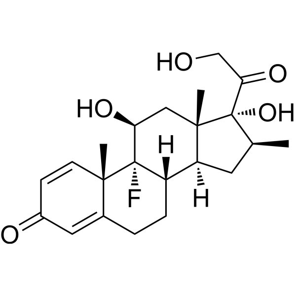
Betamethasone is a synthetic glucocorticoid with anti-inflammatory and immunosuppressive activities. Betamethasone accelerates fetal lung maturation and induces gene expression and apoptosis.
For research use only. We do not sell to patients.
- Purity : 99.95%
- CAS No.: 378-44-9
- Formula: C22H29FO5
- Molecular Weight:392.46
-
Storage:
4°C, protect from light
* In solvent : -80°C, 6 months; -20°C, 1 month (protect from light)

To place orders, for customer services and technical support, please contact: MedChemExpress USA
Tel: 609-228-6898 E-mail: [email protected] [email protected]


-
 Biological Activity
-
Chemical Information
-
Solvent & Solubility
- Protocols
- Purity & Documentation
- References
-
 Help & FAQs


-
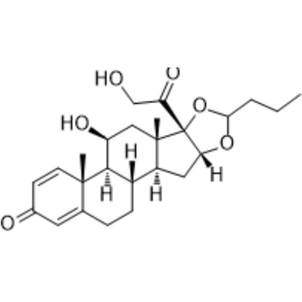
HY-13580
BudesonideGlucocorticoid Receptor Agonist -
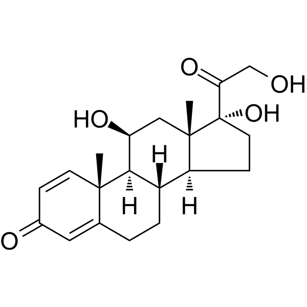
HY-17463
PrednisoloneGlucocorticoid -
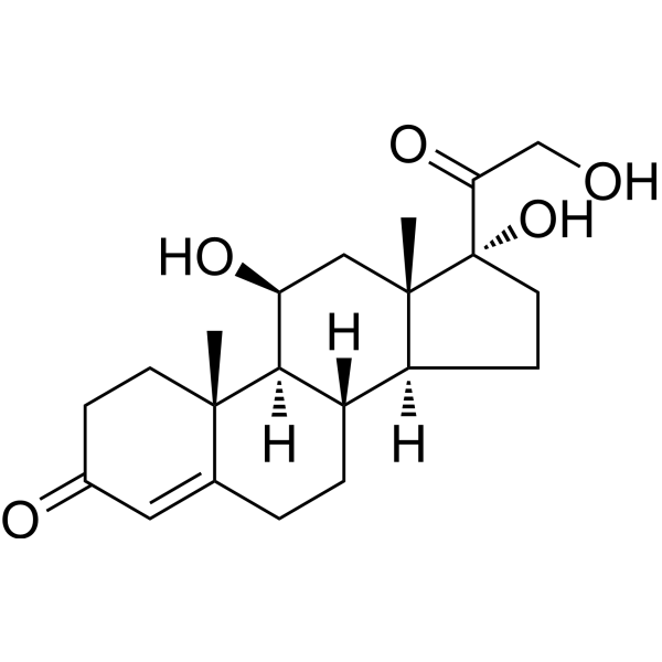
HY-N0583
HydrocortisoneGlucocorticoid Receptor Agonist -

HY-14648
DexamethasoneGR Agonist -
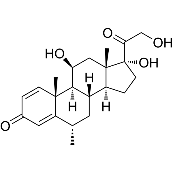
HY-B0260
MethylprednisoloneGlucocorticoid -
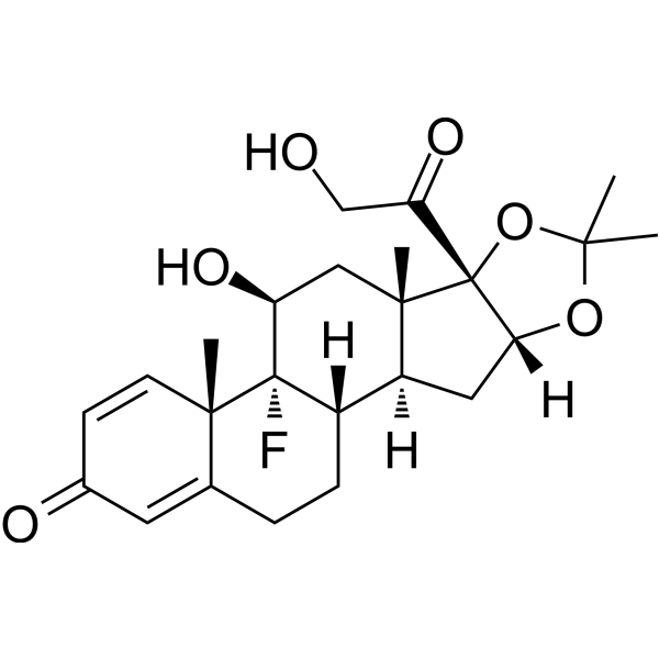
HY-B0636
Triamcinolone acetonideAnti-Inflammatory Agent -
HY-B0154
Fluticasone propionateGlucocorticoid Receptor Agonist -
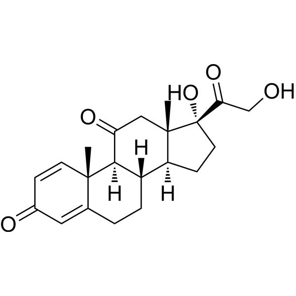
HY-B0214
PrednisoneGlucocorticoid Receptor Agonist -
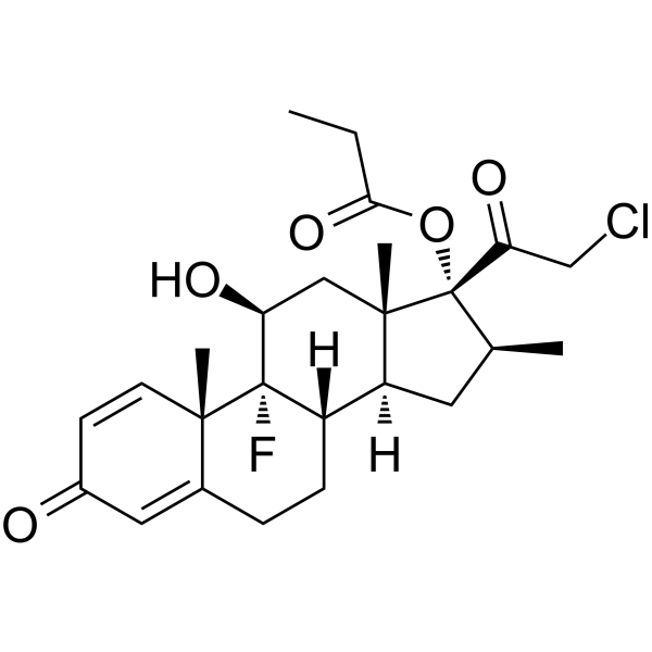
HY-13600
Clobetasol propionateCYP3A5 Inhibitor -
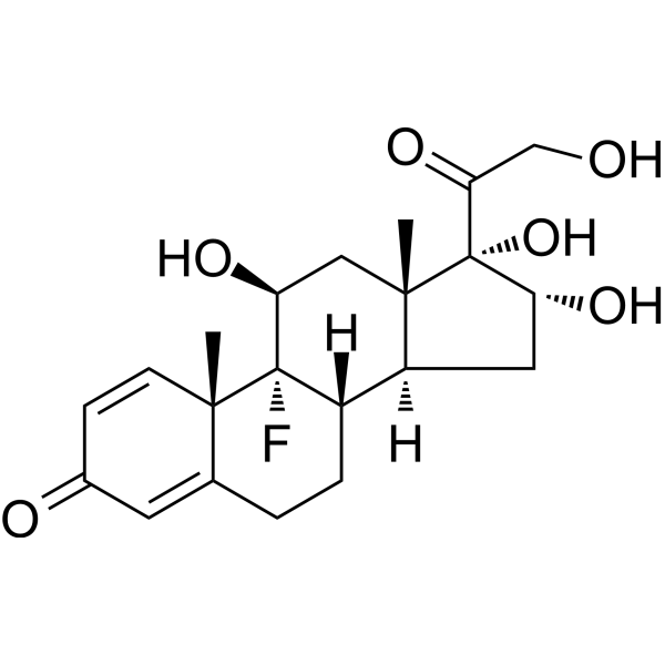
HY-B0328
TriamcinoloneSHRs Agonist -
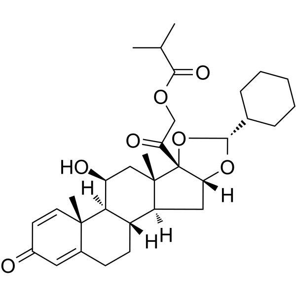
HY-B0625
CiclesonideGlucocorticoid -
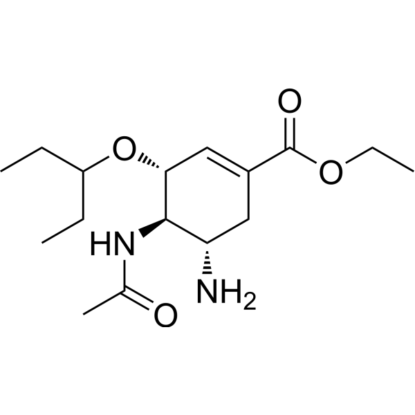
HY-13317
OseltamivirInfluenza Virus Inhibitor
Publications Citing Use of MedChemExpress (MCE) Betamethasone
More Customer Validation & Images

-
WB
-
RT-PCR


Biological Activity
Description
Cellular Effect
|
Cell Line

|
Type | Value | Description | References |
|---|---|---|---|---|
| Keratinocyte | IC50 |
300 nM
Compound: Betamethasone
|
Cytotoxicity against primary human endothelial keratinocytes assessed as inhibition of cell viability preincubated for 2 hrs followed by cytokine addition measured after 72 hrs by PrestoBlue staining based fluorescence assay
Cytotoxicity against primary human endothelial keratinocytes assessed as inhibition of cell viability preincubated for 2 hrs followed by cytokine addition measured after 72 hrs by PrestoBlue staining based fluorescence assay
|
[PMID: 28558967] |
| THP-1 | IC50 |
0.013 μM
Compound: Betamethasone
|
Inhibition of LPS-induced TNFalpha production in human THP1 cells after 24 hrs
Inhibition of LPS-induced TNFalpha production in human THP1 cells after 24 hrs
|
[PMID: 25111439] |
| THP-1 | IC50 |
0.022 μM
Compound: Betamethasone
|
Inhibition of LPS-induced IL6 production in human THP1 cells after 24 hrs
Inhibition of LPS-induced IL6 production in human THP1 cells after 24 hrs
|
[PMID: 25111439] |
In Vitro
In Vivo
MedChemExpress (MCE) has not independently confirmed the accuracy of these methods. They are for reference only.
-
Animal Model:Rambouillet-Colombia ewes bred on a single occasion are received hypercapnic challenges[1]
-
Dosage:0.48 mg
-
Administration:Injected into the fetal jugular vein at a rate of 1 ml/h (10 μg betamethasone/h) and maintained over the next 48 h.
-
Result:Decreased cerebral blood flow (CBF) in all brain regions measured except the hippocampus after 24 h of infusion.
The reduction in CBF was diminished to about 25-30 % after 48 h of infusion.
Clinical Trial
| NCT Number | Sponsor | Condition | Start Date |
Phase
|
|---|---|---|---|---|
| NCT01329991 | Plexxikon| | 2011-05 | PHASE1 |


Chemical Information
-
CAS No. 378-44-9
-
Appearance Solid
-
Molecular Weight 392.46
-
Formula C22H29FO5
-
Color White to off-white
-
SMILES
C[C@@]12[C@](C[C@H](C)[C@]2(O)C(CO)=O)([H])[C@]3([H])CCC4=CC(C=C[C@]4(C)[C@@]3(F)[C@@H](O)C1)=O
-
Shipping
Room temperature in continental US; may vary elsewhere.
-
Storage
4°C, protect from light
* In solvent : -80°C, 6 months; -20°C, 1 month (protect from light)


Publications (4)
-
Journal Impact Factor
-
Most Recent
-
Cell Metab
Sensing steroid hormone 17α-hydroxypregnenolone by GPR56 enables protection from ferroptosis-induced liver injury. [Abstract]2024 Nov 5;36(11):2402-2418.e10. PMID: 39389061 -
Cell Death Differ
Functional genomic screens uncover FERMT2 as a critical regulator of YAP/TAZ-driven tumorigenicity. [Abstract]2026 Mar 6. PMID: 41792242
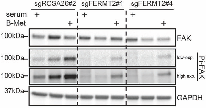
Betamethasone purchased from MedChemExpress. Usage Cited in: Cell Death Differ. 2026 Mar 6. [Abstract]
Western Blotting analysis of MDA-MB-231-Cas9 treated with Betamethasone (B-Met, 1 μM; 24 h). GAPDH was used as the loading control.
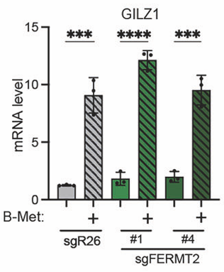
Betamethasone purchased from MedChemExpress. Usage Cited in: Cell Death Differ. 2026 Mar 6. [Abstract]
Betamethasone (B-Met, 1 μM; 24 h). RT-qPCR analysis of a glucocorticoid responsive gene, GILZ1.
-
Proc Natl Acad Sci U S A
2022 Apr 12;119(15):e2117004119. PMID: 35394864 -


Solvent & Solubility
In Vitro:
DMSO : ≥ 50 mg/mL (127.40 mM; Hygroscopic DMSO has a significant impact on the solubility of product, please use newly opened DMSO)
* "≥" means soluble, but saturation unknown.
Please refer to the solubility information to select the appropriate solvent. Once prepared, please aliquot and store the solution to prevent product inactivation from repeated freeze-thaw cycles.
Storage method and period of stock solution: -80°C, 6 months; -20°C, 1 month (protect from light). When stored at -80°C, please use it within 6 months. When stored at -20°C, please use it within 1 month.
Please refer to the solubility information to select the appropriate solvent. Once prepared, please aliquot and store the solution to prevent product inactivation from repeated freeze-thaw cycles.
Storage method and period of stock solution: -80°C, 6 months; -20°C, 1 month (protect from light). When stored at -80°C, please use it within 6 months. When stored at -20°C, please use it within 1 month.
Concentration (start) × Volume (start) = Concentration (final) × Volume (final)
In Vivo:
Select the appropriate dissolution method based on your experimental animal and administration route.
- For the following dissolution methods, please ensure to first prepare a clear stock solution using an In Vitro approach and then sequentially add co-solvents:
- To ensure reliable experimental results, the clarified stock solution can be appropriately stored based on storage conditions. As for the working solution for In Vivo experiments, it is recommended to prepare freshly and use it on the same day.
- The percentages shown for the solvents indicate their volumetric ratio in the final prepared solution. If precipitation or phase separation occurs during preparation, heat and/or sonication can be used to aid dissolution.
Add each solvent one by one: 10% DMSO 40% PEG300 5% Tween-80 45% Saline
Solubility: ≥ 2.5 mg/mL (6.37 mM); Clear solution
This protocol yields a clear solution of ≥ 2.5 mg/mL (saturation unknown).
Taking 1 mL working solution as an example, add 100 μL DMSO stock solution (25.0 mg/mL) to 400 μL PEG300, and mix evenly; then add 50 μL Tween-80 and mix evenly; then add 450 μL Saline to adjust the volume to 1 mL.
Preparation of Saline: Dissolve 0.9 g sodium chloride in ddH₂O and dilute to 100 mL to obtain a clear Saline solution.
Add each solvent one by one: 10% DMSO 90% (20% SBE-β-CD in Saline)
Solubility: ≥ 2.5 mg/mL (6.37 mM); Clear solution
This protocol yields a clear solution of ≥ 2.5 mg/mL (saturation unknown).
Taking 1 mL working solution as an example, add 100 μL DMSO stock solution (25.0 mg/mL) to 900 μL 20% SBE-β-CD in Saline, and mix evenly.
Preparation of 20% SBE-β-CD in Saline (4°C, storage for one week): 2 g SBE-β-CD powder is dissolved in 10 mL Saline, completely dissolve until clear.
In Vivo Dissolution Calculator
Please enter the basic information of animal experiments:
-
-
-
-
Recommended: Prepare an additional quantity of animals to account for potential losses during experiments.
Please enter your animal formula composition:
-
%DMSO +
Recommended: Keep the proportion of DMSO in working solution below 2% if your animal is weak.
-
%+
-
+%Tween-80 + +
-
%Saline +
The co-solvents required include: DMSO, . All of co-solvents are available by MedChemExpress (MCE). , Tween 80. All of co-solvents are available by MedChemExpress (MCE).
Working solution concentration: 0.22 mg/mL
Method for preparing stock solution: mg drug dissolved in μL DMSO. Stock solution concentration: mg/mL. * In solvent : -80°C, 6 months; -20°C, 1 month (protect from light)
1. Take μL DMSO stock solution;
2. Add μL .
μL , mix evenly;
3. Then add μL Tween 80, mix evenly;
4. Then add μL
Please ensure that the stock solution in the first step is dissolved to a clear state, and add co-solvents in sequence. You can use ultrasonic heating (ultrasonic cleaner, recommended frequency 20-40 kHz), vortexing, etc. to assist dissolution.

Protocols
-
RT-PCR
Reverse transcription technology uses RNA as a template to synthesize DNA. RT-PCR is simple, specific and sensitive, and can be used to detect gene expression levels and expression differences in cells; detect RNA virus content; clone cDNA sequences of specific genes.
-
RNA extraction experimental
By lysing cells, releasing RNA, and removing impurities such as proteins and DNA, high-purity RNA products are finally obtained. The commonly used traditional method is the guanidine isothiocyanate/phenol/chloroform method (Trizol), which is suitable for a variety of animal materials including animal tissues, microorganisms, cultured cells, etc., and most plant materials.
-
Apoptosis
Apoptosis, also called programmed cell death, is generally characterized by distinct morphological characteristics.
-
TUNEL staining for apoptotic DNA fragmentation
TUNEL staining detects DNA strand breaks by using terminal deoxynucleotidyl transferase to add labeled nucleotides to exposed 3′-OH DNA termini, generating either microscopic staining in fixed cells or tissue sections, or fluorescence/cytometric signal in cell suspensions. TUNEL positivity reflects DNA fragmentation but should not be interpreted alone as definitive apoptosis, because TUNEL can also label necrotic, autolytic, mechanically damaged, or DNA-repair-associated DNA breaks.
-
Annexin V plus membrane-impermeant dye apoptosis staining
Annexin V-based apoptosis assays rely on the detection of phosphatidylserine (PS) externalization from the inner leaflet of the plasma membrane to the outer leaflet, an early biochemical hallmark of apoptosis. Fluorescently labeled Annexin V binds PS in a calcium-dependent manner, enabling identification of early apoptotic cells by flow cytometry or fluorescence microscopy. When combined with a membrane-impermeant DNA-binding dye (e. g. , propidium iodide), this approach allows discrimination between viable (Annexin V−/dye−), early apoptotic (Annexin V+/dye−), and late apoptotic or necrotic (Annexin V+/dye+) cell populations by assessing membrane integrity and PS exposure.
-
Research Protocol for Endocrine Diseases
Endocrine diseases often arise from disrupted hormone production, hormone signaling, or target-tissue responsiveness; for diabetes-focused endocrine disease models, insulin signaling regulates glucose uptake, hepatic glucose output, lipid metabolism, and β-cell compensation. Type 2 diabetes develops through interacting defects in insulin resistance, β-cell dysfunction, adipose inflammation, hepatic glucose overproduction, altered incretin signaling, and ectopic lipid metabolism. A major unresolved question is whether endocrine dysfunction is driven primarily by target-tissue insulin resistance, intrinsic β-cell failure, immune/inflammatory stress, or combined multi-organ failure that differs by disease stage.
-
Apoptosis Solutions
Apoptosis is a regulated, generally non-lytic cell-death pathway that removes unwanted, damaged, infected, or abnormal cells through coordinated morphological changes, caspase activation, DNA fragmentation, and membrane remodeling. The intrinsic apoptosis pathway is controlled mainly by mitochondrial outer membrane permeabilization, BCL-2 family proteins, cytochrome c release, apoptosome formation, caspase-9 activation, and downstream executioner caspase-3/7 activation. The extrinsic apoptosis pathway is initiated by death receptors such as Fas, TNFR, and TRAIL receptors, which recruit adaptor proteins and activate caspase-8 before engaging executioner caspases or mitochondrial amplification through BID cleavage. Apoptosis is linked to many phenotypes, including cancer cell killing, tissue homeostasis, immune regulation, neurodegeneration, infection response, and treatment-induced cytotoxicity; unresolved questions include how apoptosis interacts with necroptosis, pyroptosis, ferroptos
-
Research Protocol for Inflammation-related Diseases
The NLRP3 inflammasome is a cytosolic innate immune signaling platform that integrates priming signals and danger-signal activation to promote caspase-1 activation, maturation of IL-1β and IL-18, and gasdermin D-mediated pyroptotic cell death. The core experimental logic is to determine whether inflammatory disease phenotypes are driven by increased NLRP3 expression, ASC-containing inflammasome assembly, caspase-1 cleavage, GSDMD cleavage, and extracellular release of IL-1β/IL-18 rather than by nonspecific cell injury alone. The pathway is strongly linked to inflammation-related disease phenotypes because monosodium urate crystals activate NALP3/NLRP3 inflammasome signaling in gout-like crystal inflammation, cholesterol crystals activate NLRP3 inflammasomes in atherogenesis models, and DSS-induced intestinal inflammation has been reported to involve NLRP3 inflammasome activity. However, experimental colitis studies also show context-dependent protective effects of NLRP3 inflammasome co
-
Real Time qPCR (Q-PCR)
Real-time quantitative PCR (qPCR) quantifies an amplifiable nucleic-acid target by monitoring fluorescence during PCR cycling rather than measuring product only after amplification. The increase in fluorescence tracks accumulation of PCR product, and the quantification cycle (Cq; historically also Ct/CP) is related to the initial amount of target: samples containing more starting target generally reach the defined fluorescence threshold in fewer cycles.


Purity & Documentation
-
Data Sheet (282 KB)
-
SDS (418 KB)
- English - EN (418 KB)
- Français - FR (418 KB)
- Deutsch - DE (418 KB)
- Norwegian - NO (418 KB)
- Español - ES (418 KB)
- Swedish - SV (418 KB)
- Italian - IT (418 KB)
- Korean - KR (418 KB)
- Portuguese - PT (418 KB)
-
Handling Instructions (2659 KB)
References
[1]. Schwab M, et, al. Effects of betamethasone administration to the fetal sheep in late gestation on fetal cerebral blood flow. J Physiol. 2000 Nov 1;528(Pt 3):619-32. [Content Brief]
[2]. Xie W, et, al. Betamethasone affects cerebral expressions of NF-kappaB and cytokines that correlate with pain behavior in a rat model of neuropathy. Ann Clin Lab Sci. Winter 2006;36(1):39-46. [Content Brief]
[3]. Kubin ME, et, al. Clinical Efficiency of Topical Calcipotriol/Betamethasone Treatment in Psoriasis Relies on Suppression of the Inflammatory TNFα - IL-23 - IL-17 Axis. Acta Derm Venereol. 2017 Apr 6;97(4):449-455. [Content Brief]
[4]. Hofmann TH, et, al. Various glucocorticoids differ in their ability to induce gene expression, apoptosis and to repress NF-kappaB-dependent transcription. FEBS Lett. 1998 Dec 28;441(3):441-6. [Content Brief]


Complete Stock Solution Preparation Table
Please refer to the solubility information to select the appropriate solvent. Once prepared, please aliquot and store the solution to prevent product inactivation from repeated freeze-thaw cycles.
Storage method and period of stock solution: -80°C, 6 months; -20°C, 1 month (protect from light). When stored at -80°C, please use it within 6 months. When stored at -20°C, please use it within 1 month.
| Optional Solvent | Concentration Solvent Mass | 1 mg | 5 mg | 10 mg | 25 mg |
|---|---|---|---|---|---|
| DMSO | 1 mM | 2.5480 mL | 12.7402 mL | 25.4803 mL | 63.7008 mL |
| 5 mM | 0.5096 mL | 2.5480 mL | 5.0961 mL | 12.7402 mL | |
| 10 mM | 0.2548 mL | 1.2740 mL | 2.5480 mL | 6.3701 mL | |
| 15 mM | 0.1699 mL | 0.8493 mL | 1.6987 mL | 4.2467 mL | |
| 20 mM | 0.1274 mL | 0.6370 mL | 1.2740 mL | 3.1850 mL | |
| 25 mM | 0.1019 mL | 0.5096 mL | 1.0192 mL | 2.5480 mL | |
| 30 mM | 0.0849 mL | 0.4247 mL | 0.8493 mL | 2.1234 mL | |
| 40 mM | 0.0637 mL | 0.3185 mL | 0.6370 mL | 1.5925 mL | |
| 50 mM | 0.0510 mL | 0.2548 mL | 0.5096 mL | 1.2740 mL | |
| 60 mM | 0.0425 mL | 0.2123 mL | 0.4247 mL | 1.0617 mL | |
| 80 mM | 0.0319 mL | 0.1593 mL | 0.3185 mL | 0.7963 mL | |
| 100 mM | 0.0255 mL | 0.1274 mL | 0.2548 mL | 0.6370 mL |


Betamethasone Related Classifications
HY-13570 Related Classifications
 Powered by Bioz