


ER-Tracker Red
Based on 5 publication(s) in Google Scholar
ER-Tracker dye is a derivative of BODIPY series dyes coupled with Glibenclamide (HY-15206), highly selective binding to the endoplasmic reticulum, non-toxic to cells at low concentrations, this type of dye is an environmentally sensitive probe, and formaldehyde treatment can still retain part of the fluorescence, with high fluorescence life, good extinction coefficient and other characteristics. Glibenclamide is an atp-dependent K+ channel blocker (Kir6, KATP) and CFTR Cl-channel blocker that binds in the endoplasmic reticulum. ER-Tracker is not suitable for staining cells after fixation. Ex/Em=587/615 nm.
For research use only. We do not sell to patients.
- Purity: 98.21%
- Formula: C44H42BClF2N6O7S2
- Molecular Weight:915.23
-
Storage:
4°C, protect from light
* In solvent : -80°C, 6 months; -20°C, 1 month (protect from light)

To place orders, for customer services and technical support, please contact: MedChemExpress USA
Tel: 609-228-6898 E-mail: [email protected] [email protected]


-
 Biological Activity
-
Chemical Information
-
Solvent & Solubility
- Purity & Documentation
- References
-
 Help & FAQs


Publications Citing Use of MedChemExpress (MCE) ER-Tracker Red
More Customer Validation & Images

-
Cell Imaging/Staining
-
Cell Imaging/Staining
-
Cell Imaging/Staining
-
Cell Imaging/Staining


Biological Activity
Guide (The following is the experimental plan we recommend. This plan serves only as a reference guide. The specific operations should be adjusted according to your actual needs.)
1. Preparation of ER-Tracker Solution
1.1 Preparation of Storage Solution
Prepare a 1 mM solution using anhydrous DMSO.
1.2 Preparation of working solution
Dilute the stored solution with preheated serum-free cell culture medium or PBS to prepare an ER-Tracker working solution with concentrations ranging from 100 nM to 1 μM.
Note: Please adjust the concentration of the ER-Tracker working solution according to the actual situation, and prepare it as needed.
2. Cell Staining (Suspension Cells)
2.1 Centrifuge to collect the cells, then add PBS for two washes, each for 5 minutes. The cell density is 1×10⁶ per mL.
2.2 Add 1 mL of ER-Tracker working solution and incubate at room temperature for 5 to 30 minutes.
2.3 400 grams, centrifuge for 3-4 minutes, then discard the supernatant.
2.4 Add PBS to wash the cells twice, for 5 minutes each time.
2.5 After resuspending the cells in 1 mL of serum-free medium or PBS, observe them using a fluorescence microscope or a flow cytometer.
3. Cell Staining (Monolayer Cells)
3.1 Place the monolayer cells on a sterile cover slip.
3.2 Remove the cover glass from the culture medium and aspirate away the excess medium.
3.3 Add 100 μL of the dye working solution and gently shake to ensure complete coverage of the cells. Incubate for 5 to 30 minutes.
3.4 Remove the dye working solution and wash with the culture medium 2-3 times, each time for 5 minutes. Observe under a fluorescence microscope.
Note: If flow cytometry is to be used for detection, the cells should first be digested with trypsin, resuspended, and then stained.
MedChemExpress (MCE) has not independently confirmed the accuracy of these methods. They are for reference only.
615
587


Chemical Information
-
Appearance Solid
-
Molecular Weight 915.23
-
Formula C44H42BClF2N6O7S2
-
Color Purple to blue
-
SMILES
O=C(C(C=C(Cl)C=C1NC(COC2=CC=C(C3=[N]4C(C=C3)=CC5=CC=C(C6=CC=CS6)[N-]5[B+3]4([F-])[F-])C=C2)=O)=C1OC)NCCC(C=C7)=CC=C7S(NC(NC8CCCCC8)=O)(=O)=O
-
Shipping
Room temperature in continental US; may vary elsewhere.
-
Storage
4°C, protect from light
* In solvent : -80°C, 6 months; -20°C, 1 month (protect from light)


Publications (5)
-
Journal Impact Factor
-
Most Recent
-
Nat Commun
Impairing the interaction between Erg11 and cytochrome P450 reductase Ncp1 enhances azoles' antifungal activities. [Abstract]2025 Jul 24;16(1):6821. PMID: 40707518
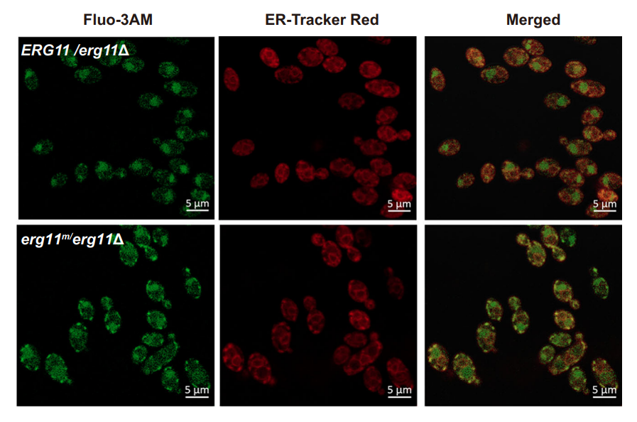
ER-Tracker Red purchased from MedChemExpress. Usage Cited in: Nat Commun. 2025 Jul 24;16(1):6821. [Abstract]
ER-Tracker Red (2 μM; 30 min). Representative images depicting the localization of Ca2+ localization (green) in relation to ER membranes (red) were obtained from ERG11/erg11Δ and erg11m/erg11Δ mutants.
-
Mol Plant Pathol
A fatty acid elongase complex regulates cell membrane integrity and septin-dependent host infection by the rice blast fungus. [Abstract]2024 Jul;25(7):e13494. PMID: 39003585
ER-Tracker Red purchased from MedChemExpress. Usage Cited in: Mol Plant Pathol. 2024 Jul;25(7):e13494. [Abstract]
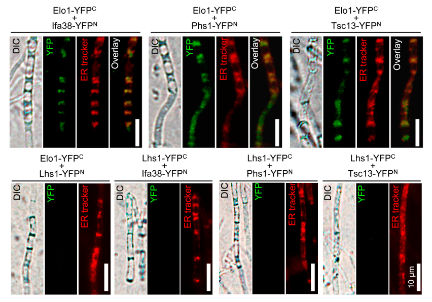
Visualization of protein interaction in the bimolecular fluorescence complementation (BiFC) assay. Vegetative hyphae of each strain expressing a pair of BiFC constructs were stained with ER-Tracker Red (1 μM; 10 min) and visualized under epifluorescence microscopy.
ER-Tracker Red purchased from MedChemExpress. Usage Cited in: Mol Plant Pathol. 2024 Jul;25(7):e13494. [Abstract]
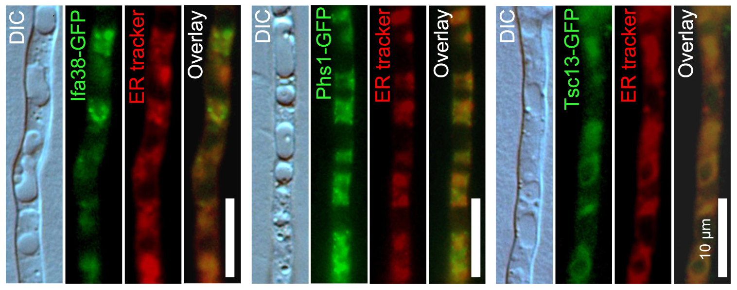
ER-Tracker Red (1 μM; 10 min) was used to examine protein subcellular localization by fusion with green fluorescent protein (GFP).
-
J Biol Chem
Phosphodiesterase 5 expression in photoreceptors rescues retinal degeneration induced by deregulation of membrane guanylyl cyclase. [Abstract]2025 Feb 3:108265. PMID: 39909376
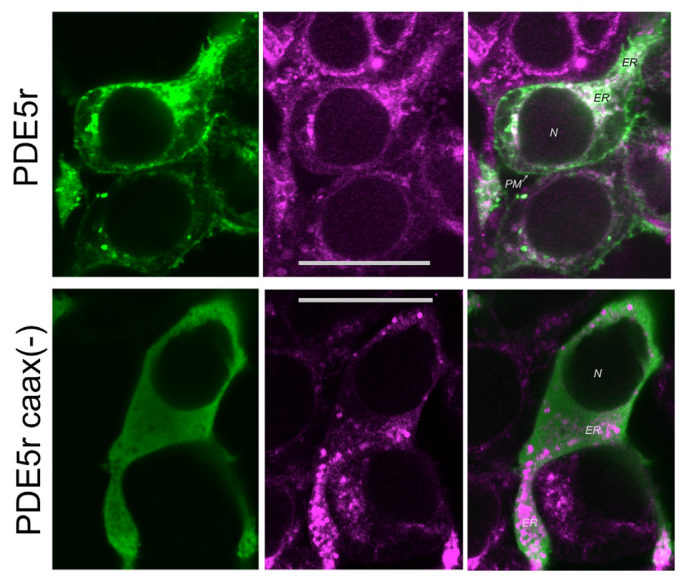
ER-Tracker Red purchased from MedChemExpress. Usage Cited in: J Biol Chem. 2025 Feb 3:108265. [Abstract]
The comparison of the typical cellular distribution of PDE5r and PDE5r lacking the isoprenylation signal ("PDE5r caax-") in HEK293 cells transfected with ER-Tracker Red (1 μM; 30 min) (middle, pseudo-magenta).
-
-


Solvent & Solubility
DMSO : 10 mg/mL (10.93 mM; Need ultrasonic; Hygroscopic DMSO has a significant impact on the solubility of product, please use newly opened DMSO)
Please refer to the solubility information to select the appropriate solvent. Once prepared, please aliquot and store the solution to prevent product inactivation from repeated freeze-thaw cycles.
Storage method and period of stock solution: -80°C, 6 months; -20°C, 1 month (protect from light). When stored at -80°C, please use it within 6 months. When stored at -20°C, please use it within 1 month.
Concentration (start) × Volume (start) = Concentration (final) × Volume (final)


Purity & Documentation
-
Data Sheet (277 KB)
-
SDS (252 KB)
- English - EN (252 KB)
- Français - FR (252 KB)
- Deutsch - DE (252 KB)
- Norwegian - NO (252 KB)
- Español - ES (252 KB)
- Swedish - SV (252 KB)
- Italian - IT (252 KB)
- Korean - KR (252 KB)
- Portuguese - PT (252 KB)
-
Handling Instructions (2659 KB)
References
[1]. Tanuja T Merianda, et al. A functional equivalent of endoplasmic reticulum and Golgi in axons for secretion of locally synthesized proteins. Mol Cell Neurosci. 2009 Feb;40(2):128-42. [Content Brief]
[3]. Adiki Raja Sekhar, et al. A cell-permeant small molecule for the super-resolution imaging of the endoplasmic reticulum in live cells. Org Biomol Chem. 2019 Apr 10;17(15):3732-3736. [Content Brief]


Complete Stock Solution Preparation Table
Please refer to the solubility information to select the appropriate solvent. Once prepared, please aliquot and store the solution to prevent product inactivation from repeated freeze-thaw cycles.
Storage method and period of stock solution: -80°C, 6 months; -20°C, 1 month (protect from light). When stored at -80°C, please use it within 6 months. When stored at -20°C, please use it within 1 month.
| Optional Solvent | Concentration Solvent Mass | 1 mg | 5 mg | 10 mg | 25 mg |
|---|---|---|---|---|---|
| DMSO | 1 mM | 1.0926 mL | 5.4631 mL | 10.9262 mL | 27.3155 mL |
| 5 mM | 0.2185 mL | 1.0926 mL | 2.1852 mL | 5.4631 mL | |
| 10 mM | 0.1093 mL | 0.5463 mL | 1.0926 mL | 2.7316 mL |


 Powered by Bioz