



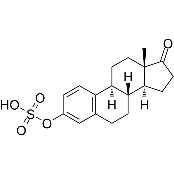
Estrone sulfate
Based on 3 publication(s) in Google Scholar
Estrone sulfate is an inactive endogenous estrogen that can be converted into Estrone (HY-B0234) and Estradiol (HY-B0141). Estrone sulfate is also a substrate of the OATP1B3 transporter. Estrone sulfate can be converted into Estrone and Estradiol in normal mammary parenchymal cells. Estrone sulfate stimulates the growth of nitrosomethylurea-induced mammary tumors in ovariectomized rats and the colony formation of dispersed nitrosomethylurea mammary cells, with conversion into Estrone and Estradiol occurring both in vivo and in vitro during this process. Estrone sulfate is applicable to breast cancer-related research.
For research use only. We do not sell to patients.
- CAS No.: 481-97-0
- Formula: C18H22O5S
- Molecular Weight:350.43
-
Storage:
Please store the product under the recommended conditions in the Certificate of Analysis.

To place orders, for customer services and technical support, please contact: MedChemExpress USA
Tel: 609-228-6898 E-mail: [email protected] [email protected]


-
 Biological Activity
-
Chemical Information
- Protocols
- Purity & Documentation
- References
-
 Help & FAQs


Publications Citing Use of MedChemExpress (MCE) Estrone sulfate
MoreAll Endogenous Metabolite Isoforms
More

Biological Activity
Description
IC50 & Target
|
Human Endogenous Metabolite |
Cellular Effect
|
Cell Line

|
Type | Value | Description | References |
|---|---|---|---|---|
| JEG-3 | IC50 |
7600 nM
Compound: E1S (enzyme substrate)
|
Estrone sulfatase activity against homogenized human JEG-3 cells was determined by measuring the [3H]E1 obtained from [3H]E1S
Estrone sulfatase activity against homogenized human JEG-3 cells was determined by measuring the [3H]E1 obtained from [3H]E1S
|
[PMID: 9873454] |
| JEG-3 | IC50 |
7600 nM
Compound: Estrone sulfate (E1S)
|
Inhibition of steroid sulfatase activity of JEG-3 cells
Inhibition of steroid sulfatase activity of JEG-3 cells
|
[PMID: 11087571] |
| Sf9 | IC50 |
45 μM
Compound: Estrone-3-sulfate
|
TP_TRANSPORTER: inhibition of E217betaG uptake in membrane vesicles from MRP4-expressing Sf9 cells
TP_TRANSPORTER: inhibition of E217betaG uptake in membrane vesicles from MRP4-expressing Sf9 cells
|
[PMID: 12523936] |
| Sf9 | IC50 |
95 μM
Compound: Estrone-3-sulfate
|
TP_TRANSPORTER: inhibition of DHEAS in membrane vesicles from MRP4-expressing Sf9 cells
TP_TRANSPORTER: inhibition of DHEAS in membrane vesicles from MRP4-expressing Sf9 cells
|
[PMID: 12523936] |
In Vitro
Estrone sulfate (1.0 μM; 1-64 min) is taken up at a low rate by dispersed normal breast parenchymal cells, resulting in a cells/medium concentration ratio of 0.20-0.33 when incubated at 37°C[2].
Estrone sulfate (1.0 nM-100 μM) is converted to estrone by dispersed normal breast parenchymal cells with a mean half-time of 628 min per 106 cells (intact cells) or 246 min per 106 cells (homogenized cells)[2].
Estrone sulfate is not produced via conversion of estrone by dispersed normal breast parenchymal cells under conditions where MCF-7 breast cancer cells exhibit sulfotransferase activity[2].
Estrone sulfate stimulates colony formation in dispersed nitrosomethylurea mammary cells and is converted to estrone and estradiol by these cells[3].
MedChemExpress (MCE) has not independently confirmed the accuracy of these methods. They are for reference only. Further protocols information, click here.
In Vivo
Estrone sulfate (75 mg/kg; p.o; daily; for 5 days) reduces glucose-6-phosphatase activity and protein levels, and lowers blood glucose in ob/ob mice[4].
Estrone sulfate modulates glucose-6-phosphatase kinetic parameters in control mice[4].
MedChemExpress (MCE) has not independently confirmed the accuracy of these methods. They are for reference only.
-
Animal Model:Sprague-Dawley (female, castrated, nitrosomethylurea-induced mammary tumor model)[3]
-
Dosage:30 pmol/hr; 300 pmol/hr; 3000 pmol/hr; 400 pmol/hr; 1300 pmol/hr
-
Administration:s.c.; continuous infusion; 14 days
-
Result:Did not significantly alter castration-induced tumor regression at 30 pmol/hr; partially blunted castration-induced tumor regression at 300 pmol/hr; stimulated tumor growth at 3000 pmol/hr; Observed significantly different tumor growth slopes for 300 and 3000 pmol/hr doses; Detected 14%, 10%, and 13% conversion to estradiol, and 24%, 22%, and 28% conversion to estrone on days 7, 10, and 14, respectively, at 400 pmol/hr; Recorded similar conversion rates at 1300 pmol/hr; Measured serum estradiol levels of 17 pg/ml (400 pmol/hr), 56 pg/ml (1300 pmol/hr), 6.8 ± 1.2 pg/ml (300 pmol/hr, day 14), and 48 ± 10.2 pg/ml (3000 pmol/hr, day 14).


Chemical Information
-
CAS No. 481-97-0
-
Molecular Weight 350.43
-
Formula C18H22O5S
-
SMILES
C[C@]1([C@](CC2)([H])[C@]3([H])CCC4=C(C=CC(OS(=O)(O)=O)=C4)[C@@]3([H])CC1)C2=O
-
Structure Classification
-
Initial Source
-
Shipping
Room temperature in continental US; may vary elsewhere.
-
Storage
Please store the product under the recommended conditions in the Certificate of Analysis.


Publications (3)
-
Journal Impact Factor
-
Most Recent
-
Cell Res
2023 Dec;33(12):940-951. PMID: 37674011 -
Nat Commun
2025 Aug 30;16(1):8124. PMID: 40885756 -
ACS Omega
Association Study of OATP1B3 Polymorphisms on Hepatic Uptake and Drug-Drug Interaction In Vitro. [Abstract]2025 Oct 29;10(44):52562-52575. PMID: 41244417

Protocols
-
Research Protocol for Endocrine Diseases
Endocrine diseases often arise from disrupted hormone production, hormone signaling, or target-tissue responsiveness; for diabetes-focused endocrine disease models, insulin signaling regulates glucose uptake, hepatic glucose output, lipid metabolism, and β-cell compensation. Type 2 diabetes develops through interacting defects in insulin resistance, β-cell dysfunction, adipose inflammation, hepatic glucose overproduction, altered incretin signaling, and ectopic lipid metabolism. A major unresolved question is whether endocrine dysfunction is driven primarily by target-tissue insulin resistance, intrinsic β-cell failure, immune/inflammatory stress, or combined multi-organ failure that differs by disease stage.
-
Soft Agar Colony Formation Assay
Soft agar colony formation assay measures anchorage-independent growth, in which transformed or tumorigenic cells proliferate as colonies in a semisolid agar matrix while many non-transformed adherent cells fail to proliferate without attachment; classic studies showed that growth in semisolid medium correlates with tumorigenicity in nude mice, and later protocol papers describe the method as a stringent in vitro assay for malignant transformation. The readout is the number, size, morphology, or signal intensity of colonies formed within agar after incubation; published formats include manual colony counting after staining, 96-well or 384-well quantitative formats, DNA-binding dye detection, MTT/tetrazolium-based detection, digital image analysis, and PCR-based marker detection from soft agar cultures.
-
Colony Formation (Clonogenic) Assay
The clonogenic (colony formation) assay measures the ability of a single cell to retain reproductive viability and form a macroscopic colony, typically defined as a cluster derived from one progenitor cell after a defined growth period. This assay is widely used to evaluate cell survival after exposure to ionizing radiation or cytotoxic treatments and is considered a standard method in radiation biology for generating dose-response relationships of reproductive cell death. Colony formation reflects long-term proliferative capacity rather than short-term metabolic activity, and survival is quantified by comparing treated versus untreated conditions based on colony number and derived survival fractions.
-
Breast Cancer Modeling
Breast cancer is a heterogeneous cancer, and it has been distinguished into four subtypes: luminal A, luminal B, HER2-positive and basal-like. Molecular mutations, epigenetic alterations, hormone exposure and immune microenvironment are related to the progression of breast cancer.


Purity & Documentation
References
[1]. Yang Z, et al. Association Study of OATP1B3 Polymorphisms on Hepatic Uptake and Drug-Drug Interaction In Vitro. ACS Omega. 2025;10(44):52562-52575. Published 2025 Oct 29. [Content Brief]
[2]. Chatterton RT Jr, et al. Formation of estrone and estradiol from estrone sulfate by normal breast parenchymal tissue. J Steroid Biochem Mol Biol. 2003;86(2):159-166. [Content Brief]
[3]. Santner SJ, et al. Estrone sulfate stimulates growth of nitrosomethylurea-induced breast carcinoma in vivo in the rat. Int J Cancer. 1990;46(1):73-78. [Content Brief]
[4]. Borthwick EB, et al. The antihyperglycemic effect of estrone sulfate in genetically obese-diabetic (ob/ob) mice is associated with reduced hepatic glucose-6-phosphatase. Horm Metab Res. 2001 Dec;33(12):721-6. [Content Brief]

Calculators
Concentration (start) × Volume (start) = Concentration (final) × Volume (final)


Estrone sulfate Related Classifications
HY-113293 Related Classifications
 Powered by Bioz