



IGF-I (24-41) TFA
IGF-I (24-41) (Insulin-like Growth Factor I (24-41)) TFA is amino acids 24 to 41 fragment of IGF-I. IGF-I TFA, a 70 aa polypeptide hormone, is a trophic factor for both neurons and glial cells. IGF-I TFA is partly responsible for systemic growth hormone (GH) activities. IGF-I TFA has anabolic, antioxidant, anti-inflammatory and cytoprotective actions. IGF-I (24-41) TFA regulates somatic growth and behavioral development.
For research use only. We do not sell to patients.
- Formula: C88H133N27O28.xC2HF3O2
- Molecular Weight:2017.16 (free acid)
-
Storage:
Please store the product under the recommended conditions in the Certificate of Analysis.

To place orders, for customer services and technical support, please contact: MedChemExpress USA
Tel: 609-228-6898 E-mail: [email protected] [email protected]


-
 Biological Activity
-
Chemical Information
-
Solvent & Solubility
- Protocols
- Purity & Documentation
- References
-
 Help & FAQs




Biological Activity
Description
In Vivo
MedChemExpress (MCE) has not independently confirmed the accuracy of these methods. They are for reference only.
-
Animal Model:Mice of an outbred Swiss-derived strain (CD-l) weighing 30-33g (virgin males) or 28-30g (nulliparous females)[2]
-
Dosage:ICV
-
Administration:200 ng dose; 2 µL; postnatal days (PND) 2, 4, and 7
-
Result:Increased the rate of ultrasonic calls of the pups measured on PND8.


Chemical Information
-
Molecular Weight 2017.16 (free acid)
-
Formula C88H133N27O28.xC2HF3O2
-
Synonyms
Insulin-like Growth Factor I (24-41) TFA
-
Sequence
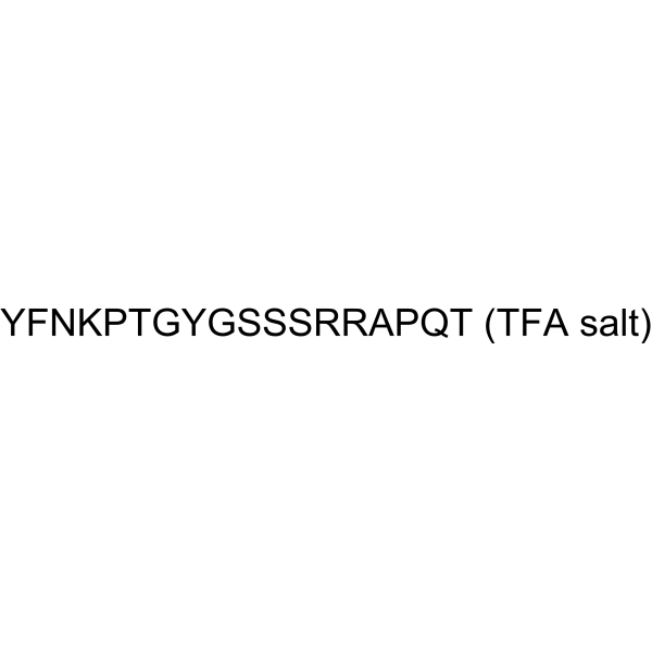
Tyr-Phe-Asn-Lys-Pro-Thr-Gly-Tyr-Gly-Ser-Ser-Ser-Arg-Arg-Ala-Pro-Gln-Thr
-
Sequence Shortening
YFNKPTGYGSSSRRAPQT
-
Shipping
Room temperature in continental US; may vary elsewhere.
-
Storage
Please store the product under the recommended conditions in the Certificate of Analysis.


Solvent & Solubility
In Vitro
H2O
Peptide Solubility and Storage Guidelines:
1. Calculate the length of the peptide.
2. Calculate the overall charge of the entire peptide according to the following table:
| Contents | Assign value | |
|---|---|---|
| Acidic amino acid | Asp (D), Glu (E), and the C-terminal -COOH. | -1 |
| Basic amino acid | Arg (R), Lys (K), His (H), and the N-terminal -NH2 | +1 |
| Neutral amino acid | Gly (G), Ala (A), Leu (L), Ile (I), Val (V), Cys (C), Met (M), Thr (T), Ser (S), Phe (F), Tyr (Y), Trp (W), Pro (P), Asn (N), Gln (Q) | 0 |
3. Recommended solution:
| Overall charge of peptide | Details |
|---|---|
| Negative (<0) |
1. Try to dissolve the peptide in water first. 2. If water fails, add NH4OH (<50 μL). 3. If the peptide still does not dissolve, add DMSO (50-100 μL) to solubilize the peptide. |
| Positive (>0) |
1. Try to dissolve the peptide in water first. 2. If water fails, try dissolving the peptide in a 10%-30% acetic acid solution. 3. If the peptide still does not dissolve, try dissolving the peptide in a small amount of DMSO. |
| Zero (=0) |
1. Try to dissolve the peptide in organic solvent (acetonitrile, methanol, etc.) first. 2. For very hydrophobic peptides, try dissolving the peptide in a small amount of DMSO, and then dilute the solution with water to the desired concentration. |

Protocols
-
Research Protocol for Endocrine Diseases
Endocrine diseases often arise from disrupted hormone production, hormone signaling, or target-tissue responsiveness; for diabetes-focused endocrine disease models, insulin signaling regulates glucose uptake, hepatic glucose output, lipid metabolism, and β-cell compensation. Type 2 diabetes develops through interacting defects in insulin resistance, β-cell dysfunction, adipose inflammation, hepatic glucose overproduction, altered incretin signaling, and ectopic lipid metabolism. A major unresolved question is whether endocrine dysfunction is driven primarily by target-tissue insulin resistance, intrinsic β-cell failure, immune/inflammatory stress, or combined multi-organ failure that differs by disease stage.
-
Research Protocol for Inflammation-related Diseases
The NLRP3 inflammasome is a cytosolic innate immune signaling platform that integrates priming signals and danger-signal activation to promote caspase-1 activation, maturation of IL-1β and IL-18, and gasdermin D-mediated pyroptotic cell death. The core experimental logic is to determine whether inflammatory disease phenotypes are driven by increased NLRP3 expression, ASC-containing inflammasome assembly, caspase-1 cleavage, GSDMD cleavage, and extracellular release of IL-1β/IL-18 rather than by nonspecific cell injury alone. The pathway is strongly linked to inflammation-related disease phenotypes because monosodium urate crystals activate NALP3/NLRP3 inflammasome signaling in gout-like crystal inflammation, cholesterol crystals activate NLRP3 inflammasomes in atherogenesis models, and DSS-induced intestinal inflammation has been reported to involve NLRP3 inflammasome activity. However, experimental colitis studies also show context-dependent protective effects of NLRP3 inflammasome co


Purity & Documentation
References
[1]. Puche JE, et al. Human conditions of insulin-like growth factor-I (IGF-I) deficiency. J Transl Med. 2012 Nov 14;10:224. [Content Brief]
[2]. D Santucci, et al. IGF-I and IGF-I24-41 but not IGF-I57-70 affect somatic and neurobehavioral development of newborn male mice. Brain Res Bull. 1994;35(4):367-71. [Content Brief]
[3]. Dan Meng, et al. Insulin-like growth factor-I (IGF-I) induces epidermal growth factor receptor transactivation and cell proliferation through reactive oxygen species. Free Radic Biol Med. 2007 Jun 1;42(11):1651-60. [Content Brief]

Calculators
Concentration (start) × Volume (start) = Concentration (final) × Volume (final)


IGF-I (24-41) TFA Related Classifications
HY-P1777A Related Classifications
 Powered by Bioz