


Lapatinib ditosylate monohydrate
Based on 95 publication(s) in Google Scholar
Lapatinib ditosylate monohydrate (GW572016 ditosylate monohydrate) is a potent, orally active inhibitor of the ErbB-2 and EGFR tyrosine kinase domains with IC50 values against purified EGFR and ErbB-2 of 10.2 and 9.8 nM, respectively.
For research use only. We do not sell to patients.
- Purity: 99.99%
- CAS No.: 388082-78-8
- Formula: C43H44ClFN4O11S3
- Molecular Weight:943.48
-
Storage:
4°C, sealed storage, away from moisture
* In solvent : -80°C, 6 months; -20°C, 1 month (sealed storage, away from moisture)

To place orders, for customer services and technical support, please contact: MedChemExpress USA
Tel: 609-228-6898 E-mail: [email protected] [email protected]


-
 Biological Activity
-
Chemical Information
-
Solvent & Solubility
- Purity & Documentation
- References
-
 Help & FAQs
-


Anti-Infection Compound Library
HY-L002
-
Immunology/Inflammation Compound Library
HY-L007
-
JAK/STAT Compound Library
HY-L008
-
Kinase Inhibitor Library
HY-L009
-
Protein Tyrosine Kinase Compound Library
HY-L016
-
FDA-Approved Drug Library
HY-L022
-
Anti-Cancer Compound Library
HY-L025
-
Autophagy Compound Library
HY-L029
-
Drug Repurposing Compound Library
HY-L035
-
Differentiation Inducing Compound Library
HY-L038
-
Ferroptosis Compound Library
HY-L051
-
Orally Active Compound Library
HY-L061
-
FDA Approved & Pharmacopeial Drug Library
HY-L066
-
Anti-Hepatitis C Virus Compound Library
HY-L073
-
Anti-Breast Cancer Compound Library
HY-L074
-
Anti-Lung Cancer Compound Library
HY-L075
-
Anti-Pancreatic Cancer Compound Library
HY-L077
-
Anti-Obesity Compound Library
HY-L087
-
Angiogenesis-Related Compound Library
HY-L088
-
Anti-Liver Cancer Compound Library
HY-L101
-
Rare Diseases Drug Library
HY-L102
-
Anti-Colorectal Cancer Compound Library
HY-L103
-
Chemotherapy Drug Library
HY-L112
-
EMA-Approved Drug Library
HY-L116
-
FDA-Approved Anticancer Drug Library
HY-L122
-
Human Metabolite Library
HY-L123
-
Anti-Prostate Cancer Compound Library
HY-L124
-
Cancer Stem Cells Compound Library
HY-L135
-
Heterocyclic Compound Library
HY-L138
-
Off-patent Drug Library
HY-L141
-
Membrane Protein-targeted Compound Library
HY-L149
-
Membrane Receptor-targeted Compound Library
HY-L150
-
Cytokine Inhibitors Library
HY-L161
-
Cell Death Library
HY-L162
-
Anti-Ovarian Cancer Compound Library
HY-L173
-
Multi-Target Compound Library
HY-L176
-
Bioactive Compound Library Max
HY-L181
-
MCE Bioactive Compound Library
HY-L001V
-
Drug Repurposing Compound Library Plus
HY-L035P
-
FDA-Approved Drug Library Plus
HY-L022P
-
FDA-Approved Drug Library Mini
HY-L022M
-
Bioactive Compound Library
HY-L001
-
Anti-Gastric Cancer Compound Library
HY-L184
-
Anti-Brain Cancer Compound Library
HY-L188
-
Protein Kinase Compound Library
HY-L196
-
Cell Proliferation Compound Library
HY-L201
-
High-Throughput Bioactive Compound Library
HY-L205
-
Mass Spectrometry Human Metabolite Library
HY-L215
-
Posttranslational Modification Library
HY-L226
-
FDA Kinase Inhibitor Library
HY-L230
-
RNA Binding Bioactive Compound Library
HY-L248


Publications Citing Use of MedChemExpress (MCE) Lapatinib ditosylate monohydrate
More- Nat Med. 2016 Jul;22(7):723-6. [Abstract]
- Nature. 2017 Aug 24;548(7668):471-475. [Abstract]
- Cancer Cell. 2025 Jul 25:S1535-6108(25)00310-1. [Abstract]
- Nat Cancer. 2026 Jan;7(1):116-130. [Abstract]
- Nat Immunol. 2018 Mar;19(3):233-245. [Abstract]
- Nat Commun. 2023 Jun 15;14(1):3560. [Abstract]
- Nat Commun. 2023 Apr 13;14(1):2110. [Abstract]
- Sci Transl Med. 2018 Jul 18;10(450):eaaq1093. [Abstract]
- Adv Sci (Weinh). 2024 Nov 20:e2407662. [Abstract]
- Adv Sci (Weinh). 2023 Feb;10(5):e2203884. [Abstract]
- Nat Chem Biol. 2025 Aug;21(8):1226-1237. [Abstract]
- J Exp Clin Cancer Res. 2024 Nov 20;43(1):308. [Abstract]
- J Exp Clin Cancer Res. 2018 Jun 25;37(1):123. [Abstract]
- Redox Biol. 2024 Nov 5:78:103419. [Abstract]
- Cell Rep Med. 2025 Apr 2:102053. [Abstract]
- Pharmacol Res. 2025 Nov:221:107993. [Abstract]
- Cancer Lett. 2024 Jun 5:217008. [Abstract]
- Cancer Lett. 2021 Oct 10:518:82-93. [Abstract]
- Cancer Lett. 2021 Apr 28;504:125-136. [Abstract]
- Cancer Lett. 2020 Apr 10;475:53-64. [Abstract]
- Int J Biol Sci. 2025 Jun 12;21(9):4081-4097. [Abstract]
- Cell Death Dis. 2025 Mar 12;16(1):170. [Abstract]
- Acta Pharmacol Sin. 2021 Jan;42(1):108-114. [Abstract]
- NPJ Precis Oncol. 2026 Apr 8;10(1):211. [Abstract]
- Cell Syst. 2020 Nov 18;11(5):478-494.e9. [Abstract]
- Oncogene. 2022 May;41(22):3064-3078. [Abstract]
- Oncogene. 2016 Jun 9;35(23):2961-70. [Abstract]
- Cell Death Discov. 2024 Nov 1;10(1):462. [Abstract]
- Cell Rep. 2025 Mar 25;44(4):115466. [Abstract]
- Br J Cancer. 2022 Mar;126(5):778-790. [Abstract]
- Clin Transl Med. 2020 Aug;10(4):e148. [Abstract]
- Cell Mol Life Sci. 2023 Mar 18;80(4):100. [Abstract]
- Front Immunol. 2023 Mar 17:14:1153423. [Abstract]
- Eur J Med Chem. 2019 Aug 15:176:393-409. [Abstract]
- Mol Cancer Ther. 2018 Mar;17(3):603-613. [Abstract]
- Cells. 2026 Feb 28;15(5):430. [Abstract]
- Commun Biol. 2024 Dec 31;7(1):1719. [Abstract]
- Life Sci. 2021 May 15:273:119239. [Abstract]
- Drug Des Devel Ther. 2020 Feb 25;14:783-793. [Abstract]
- Stem Cell Reports. 2017 Dec 12;9(6):1948-1960. [Abstract]
- Pharmaceuticals (Basel). 2026 Feb 27;19(3):381. [Abstract]
- Front Pharmacol. 2021 Mar 8;12:644342. [Abstract]
- RSC Adv. 2019 Jun 20;9(34):19325-19332. [Abstract]
- Cell Rep Methods. 2026 Jun 15;6(6):101339. [Abstract]
- Cell Rep Methods. 2023 Oct 23;3(10):100599. [Abstract]
- Mol Pharm. 2022 Nov 7;19(11):4320-4332. [Abstract]
- Cancers (Basel). 2024 Apr 25;16(9):1651. [Abstract]
- Cancers (Basel). 2022 Nov 28;14(23):5854. [Abstract]
- Biol Proced Online. 2023 Jun 27;25(1):19. [Abstract]
- ACS Omega. 2022 Mar 3;7(10):9004-9014. [Abstract]
- Biochim Biophys Acta Mol Basis Dis. 2024 Aug 9:167458. [Abstract]
- iScience. 2023 Oct 6;26(11):108152. [Abstract]
- Transl Oncol. 2022 Jul; 21: 101444. [Abstract]
- Sci Rep. 2026 Apr 20;16(1):12890. [Abstract]
- J Biol Chem. 2022 Sep;298(9):102310. [Abstract]
- ACS Chem Biol. 2016 Apr 15;11(4):992-1000. [Abstract]
- J Proteome Res. 2022 Apr 1;21(4):953-964. [Abstract]
- Biotechnol Bioeng. 2021 Dec;118(12):4687-4698. [Abstract]
- Exp Cell Res. 2020 Aug 1;393(1):112054. [Abstract]
- Front Oncol. 2021 Mar 26:11:608201. [Abstract]
- Analyst. 2017 Jan 26;142(3):525-536. [Abstract]
- Cancer Med. 2025 Sep;14(18):e71227. [Abstract]
- Carcinogenesis. 2022 Dec 25;43(11):1071-1082. [Abstract]
- Clin Chim Acta. 2018 Oct:485:298-304. [Abstract]
- J Chromatogr B Analyt Technol Biomed Life Sci. 2024 Apr 15:1237:124100. [Abstract]
- J Chromatogr B Analyt Technol Biomed Life Sci. 2017 Apr 1:1049-1050:30-40. [Abstract]
- Separations. 2023 May 26, 10(6), 330.
- PLoS One. 2024 Nov 1;19(11):e0308647. [Abstract]
- J Nanopart Res. 2022; 24:261
- PLoS One. 2019 Apr 4;14(4):e0214598. [Abstract]
- Fundam Clin Pharmacol. 2021 Oct;35(5):919-929. [Abstract]
- Gene. 2022 Mar 30:816:146171. [Abstract]
- Eur J Drug Metab Pharmacokinet. 2021 Sep;46(5):625-635. [Abstract]
- Cancer Chemother Pharmacol. 2018 Sep;82(3):383-394. [Abstract]
- Biochem Biophys Res Commun. 2026 Feb 12:800:153165.
- Head Neck. 2023 Jul;45(7):1801-1811. [Abstract]
- Cell Physiol Biochem. 2015;37(6):2275-87. [Abstract]
- Open Life Sci. 2023 Jan 10;18(1):20220535. [Abstract]
- chemRxiv. 2026 Apr 6.
- bioRxiv. 2025 Nov 18.
- SSRN. 2025 Oct 13.
- bioRxiv. 2025 Sep 21.
- bioRxiv. 2025 May 09.
- bioRxiv. 2024 Nov 6:2024.11.04.621884. [Abstract]
- Patent. US20240293405A1.
- University of Gothenburg. 2023 Jun 27.
- Patent. US20220305013A1.
- Biomedical Engineering Advances. June 2022, 100040.
- bioRxiv. 2021 Jan 9.
- Oncotarget. 2020 Nov 3;11(44):3921-3932. [Abstract]
- Patent. US20200108066A1
- bioRxiv. 2019 Sep.
- Personalized Medicine Universe. 2019 May.
- Oncotarget. 2017 Aug 24;8(62):104894-104912. [Abstract]
- Tumour Biol. 2016 Nov;37(11):14831-14839. [Abstract]
 Customer Validation & Images

-
WB
-
WB
-
WB
-
WB
-
WB
All EGFR Isoforms
More

Biological Activity
|
EGFR 10.8 nM (IC50) |
ErbB2 9.2 nM (IC50) |
|
Cell Line
|
Type | Value | Description | References |
|---|---|---|---|---|
| A-431 | IC50 |
0.14 μM
Compound: Tykerb
|
Inhibition of EGFR in human A431 cells by HTRF assay
Inhibition of EGFR in human A431 cells by HTRF assay
|
[PMID: 19815412] |
| BT-474 | EC50 |
20 nM
Compound: Tykerb
|
Antiproliferative activity against human BT474 cells overexpressing ERBb2 after 3 days by methylene blue staining
Antiproliferative activity against human BT474 cells overexpressing ERBb2 after 3 days by methylene blue staining
|
[PMID: 19028424] |
| BT-474 | IC50 |
0.1 μM
Compound: 1, Tykerb
|
Antiproliferative activity against human BT474 cells by methylene blue staining method
Antiproliferative activity against human BT474 cells by methylene blue staining method
|
[PMID: 21334203] |
| BT-474 | IC50 |
100 nM
Compound: 3, Tykerb
|
Antiproliferative activity against human BT474 cells expressing ErbB2 by MEB assay
Antiproliferative activity against human BT474 cells expressing ErbB2 by MEB assay
|
[PMID: 19111461] |
| BT-474 | IC50 |
37.4 nM
Compound: GW572016, tykerb
|
Cytotoxicity against human BT474 cells after 72 hrs by SRB assay
Cytotoxicity against human BT474 cells after 72 hrs by SRB assay
|
[PMID: 24121234] |
| HN5 | EC50 |
15 nM
Compound: Tykerb
|
Antiproliferative activity against human HN5 cells overexpressing EGFR after 3 days by methylene blue staining
Antiproliferative activity against human HN5 cells overexpressing EGFR after 3 days by methylene blue staining
|
[PMID: 19028424] |
| HN5 | IC50 |
120 nM
Compound: 3, Tykerb
|
Antiproliferative activity against human HN5 cells expressing EGFR by MEB assay
Antiproliferative activity against human HN5 cells expressing EGFR by MEB assay
|
[PMID: 19111461] |
| NCI-N87 | IC50 |
46.2 nM
Compound: GW572016, tykerb
|
Cytotoxicity against human NCI-N87 cells after 72 hrs by SRB assay
Cytotoxicity against human NCI-N87 cells after 72 hrs by SRB assay
|
[PMID: 24121234] |
| SK-BR-3 | IC50 |
0.124 μM
Compound: Tykerb
|
Inhibition of HER2 in human SKBR3 cells by HTRF assay
Inhibition of HER2 in human SKBR3 cells by HTRF assay
|
[PMID: 19815412] |
Lapatinib (GW2016; 0.03-10 μM; 6 hours; BT474 and HN5 cells) treatment inhibits receptor autophosphorylation of EGFR and ErbB-2 in a dose-responsive manner. Phosphorylation of serine 473 of AKT was inhibited by GW2016 in a dose-dependent manner[1].
Lapatinib (GW2016; 72 hours; HN5, A-43, BT474, N87, and CaLu-3 cells) treatment has a selective inhibition of the proliferation of human tumor cell lines[1].
Lapatinib (GW2016; 1-10 μM; 72 hours; HN5 cells) treatment results in induces G1 arrest[1].
MedChemExpress (MCE) has not independently confirmed the accuracy of these methods. They are for reference only.
-
Cell Line:BT474 and HN5 cells
-
Concentration:0.03 µM, 0.1 µM, 0.3 µM, 1 µM, 3 µM, or 10 µM
-
Incubation Time:6 hours
-
Result:Inhibited receptor autophosphorylation of EGFR and ErbB-2 in a dose-responsive manner. Phosphorylation of serine 473 of AKT was also inhibited in a dose-dependent manner.
-
Cell Line:HN5, A-43, BT474, N87, and CaLu-3 cells
-
Concentration:
-
Incubation Time:72 hours
-
Result:Inhibited the growth of tumor cells overexpressing EGFR or ErbB-2.
-
Cell Line:HN5 cells
-
Concentration:1 µM, or 10 µM
-
Incubation Time:72 hours
-
Result:Resulted in induction of G1 arrest.
MedChemExpress (MCE) has not independently confirmed the accuracy of these methods. They are for reference only.
-
Animal Model:CD-1 nude female mice (4-6 weeks old) with HN5 cells[1]
-
Dosage:30 mg/kg, 100 mg/kg
-
Administration:Oral administration; twice daily; for 21 days
-
Result:Inhibited tumor xenograft growth of the HN5 cells in a dose-responsive manner.


Chemical Information
-
CAS No. 388082-78-8
-
Appearance Solid
-
Molecular Weight 943.48
-
Formula C43H44ClFN4O11S3
-
Color Light yellow to yellow
-
SMILES
O=S(CCNCC1=CC=C(C2=CC3=C(NC4=CC=C(OCC5=CC=CC(F)=C5)C(Cl)=C4)N=CN=C3C=C2)O1)(C)=O.O=S(C6=CC=C(C)C=C6)(O)=O.O=S(C7=CC=C(C)C=C7)(O)=O.O
-
Synonyms
GW572016 ditosylate monohydrate; GW2016 ditosylate monohydrate
-
Shipping
Room temperature in continental US; may vary elsewhere.
-
Storage
4°C, sealed storage, away from moisture
* In solvent : -80°C, 6 months; -20°C, 1 month (sealed storage, away from moisture)


Publications (95)
-
Journal Impact Factor
-
Most Recent
-
Nat Med
Combination inhibition of PI3K and mTORC1 yields durable remissions in mice bearing orthotopic patient-derived xenografts of HER2-positive breast cancer brain metastases. [Abstract]2016 Jul;22(7):723-6. PMID: 27270588 -
Nature
2017 Aug 24;548(7668):471-475. PMID: 28813415
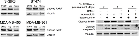
Lapatinib ditosylate monohydrate purchased from MedChemExpress. Usage Cited in: Nature. 2017 Aug 24;548(7668):471-475. [Abstract]
Western blot of SKBR3, BT474, MDA-MB-453, and MDA-MB-361 cells treated with DMSO, Lapatinib, or Abemaciclib for 48 h. Western blot of MDA-MB-453 cells pretreated with DMSO or Abemaciclib (500 nM) for 0, 1, or 7 days before exposure to Staurosporine (500 nM) for 4 h.
-
Cancer Cell
A pan-KRAS inhibitor and its derived degrader elicit multifaceted anti-tumor efficacy in KRAS-driven cancers. [Abstract]2025 Jul 25:S1535-6108(25)00310-1. PMID: 40780213 -
Nat Cancer
The MEK-RAF molecular glue IK-595 has potent antitumor activity across RAS/MAPK pathway-altered cancers. [Abstract]2026 Jan;7(1):116-130. PMID: 41482524 -
Nat Immunol
2018 Mar;19(3):233-245. PMID: 29358709 -
Nat Commun
CNK2 promotes cancer cell motility by mediating ARF6 activation downstream of AXL signalling. [Abstract]2023 Jun 15;14(1):3560. PMID: 37322019 -
Nat Commun
Nucleocytoplasmic transport of active HER2 causes fractional escape from the DCIS-like state. [Abstract]2023 Apr 13;14(1):2110. PMID: 37055441 -
Sci Transl Med
PP2A inhibition is a druggable MEK inhibitor resistance mechanism in KRAS-mutant lung cancer cells. [Abstract]2018 Jul 18;10(450):eaaq1093. PMID: 30021885 -
Adv Sci (Weinh)
Osteoblast-Derived ECM1 Promotes Anti-Androgen Resistance in Bone Metastatic Prostate Cancer. [Abstract]2024 Nov 20:e2407662. PMID: 39563492 -
Adv Sci (Weinh)
RNF126-Mediated MRE11 Ubiquitination Activates the DNA Damage Response and Confers Resistance of Triple-Negative Breast Cancer to Radiotherapy. [Abstract]2023 Feb;10(5):e2203884. PMID: 36563124 -
Nat Chem Biol
2025 Aug;21(8):1226-1237. PMID: 39870764 -
J Exp Clin Cancer Res
Dual inhibition of HERs and PD-1 counteract resistance in KRASG12C-mutant head and neck cancer. [Abstract]2024 Nov 20;43(1):308. PMID: 39567998 -
J Exp Clin Cancer Res
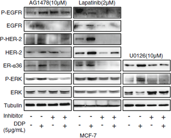
ER-α36 mediates cisplatin resistance in breast cancer cells through EGFR/HER-2/ERK signaling pathway. [Abstract]2018 Jun 25;37(1):123. PMID: 29940998
Lapatinib ditosylate monohydrate purchased from MedChemExpress. Usage Cited in: J Exp Clin Cancer Res. 2018 Jun 25;37(1):123. [Abstract]
MCF-7 cells are treated with or without 5 μg/mL cisplatin (DDP) for 48 h after preincubated with or without Lapatinib at the indicated concentrations for 6 h, respectively. Then the levels of ER-α36, total EGFR and P-EGFR, total HER-2 and P-HER-2, total ERK and P-ERK are evaluated using western blot.
-
Redox Biol
Reactivation of MAPK-SOX2 pathway confers ferroptosis sensitivity in KRASG12C inhibitor resistant tumors. [Abstract]2024 Nov 5:78:103419. PMID: 39527862 -
Cell Rep Med
CAN-Scan: A multi-omic phenotype-driven precision oncology platform identifies prognostic biomarkers of therapy response for colorectal cancer. [Abstract]2025 Apr 2:102053. PMID: 40187357 -
Pharmacol Res
Ceritinib inhibits growth and ACTH production of PitNETs: Insights from patient-derived organoids. [Abstract]2025 Nov:221:107993. PMID: 41083089 -
Cancer Lett
Sideroflexin-1 promotes progression and sensitivity to lapatinib in triple-negative breast cancer by inhibiting TOLLIP-mediated autophagic degradation of CIP2A. [Abstract]2024 Jun 5:217008. PMID: 38849012 -
Cancer Lett
Induction of EnR stress by Melatonin enhances the cytotoxic effect of Lapatinib in HER2-positive breast cancer. [Abstract]2021 Oct 10:518:82-93. PMID: 34153400 -
Cancer Lett
Targeting ubiquitin conjugating enzyme UbcH5b by a triterpenoid PC3-15 from Schisandra plants sensitizes triple-negative breast cancer cells to lapatinib. [Abstract]2021 Apr 28;504:125-136. PMID: 33607208 -
Cancer Lett
Targeting the EphB4 receptor tyrosine kinase sensitizes HER2-positive breast cancer cells to Lapatinib. [Abstract]2020 Apr 10;475:53-64. PMID: 32006616 -
Int J Biol Sci
Promoter Hyper-methylation of ZNF662 Restrains its Tumor Suppressing Function in Triple-Negative Breast Cancer Through Regulating NGF Signaling Axis. [Abstract]2025 Jun 12;21(9):4081-4097. PMID: 40612670 -
Cell Death Dis
Both direct and indirect suppression of MCL1 synergizes with BCLXL inhibition in preclinical models of gastric cancer. [Abstract]2025 Mar 12;16(1):170. PMID: 40075071 -
Acta Pharmacol Sin
Osimertinib successfully combats EGFR-negative glioblastoma cells by inhibiting the MAPK pathway. [Abstract]2021 Jan;42(1):108-114. PMID: 32398685 -
NPJ Precis Oncol
High-throughput screening identifies NT-1 that synergizes with MRTX1133 against acquired resistant KRASG12D colorectal cancer. [Abstract]2026 Apr 8;10(1):211. PMID: 41951768 -
Cell Syst
Receptor-Driven ERK Pulses Reconfigure MAPK Signaling and Enable Persistence of Drug-Adapted BRAF-Mutant Melanoma Cells. [Abstract]2020 Nov 18;11(5):478-494.e9. PMID: 33113355 -
Oncogene
2022 May;41(22):3064-3078. PMID: 35461328 -
Oncogene
PIK3CA(H1047R)- and Her2-initiated mammary tumors escape PI3K dependency by compensatory activation of MEK-ERK signaling. [Abstract]2016 Jun 9;35(23):2961-70. PMID: 26640141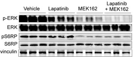
Lapatinib ditosylate monohydrate purchased from MedChemExpress. Usage Cited in: Oncogene. 2016 Jun 9;35(23):2961-70. [Abstract]
The combined use of MEK162 with HER kinase inhibitor Lapatinib, almost completely abolishes MAPK signaling as evidenced by diminished phospho-Erk levels. Western blot analyses of ERK signaling in tumor transplants from mice treated as indicated. Three hours after their dose on day four of treatment, the mice are sacrificed for analysis. Vinculin is used as a loading control.
Lapatinib ditosylate monohydrate purchased from MedChemExpress. Usage Cited in: Oncogene. 2016 Jun 9;35(23):2961-70. [Abstract]
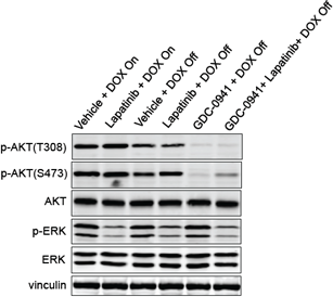
Western blot analysis of p-AKT(T308), p-AKT(S473) and p-ERK in transplanted NIC+PIK3CAH1047R tumors treated as indicated. Transplants of NIC+PIK3CAH1047R primary mammary tumors are first established in immunodeficient nude mice maintained on Doxycycline. Treatment starts when tumor transplants reach 500 mm3. DOX On, on Doxycycline; DOX Off, Doxycycline withdrawal. Lapatinib, 100mg/kg/day, p.o; GDC-0941, 120mg/kg/ day, p.o. Tumo
-
Cell Death Discov
Targeting the HECTD3-p62 axis increases the radiosensitivity of triple negative breast cancer cells. [Abstract]2024 Nov 1;10(1):462. PMID: 39487119 -
Cell Rep
Structural basis for the reversal of human MRP4-mediated multidrug resistance by lapatinib. [Abstract]2025 Mar 25;44(4):115466. PMID: 40138312 -
Br J Cancer
PCK1 regulates neuroendocrine differentiation in a positive feedback loop of LIF/ZBTB46 signalling in castration-resistant prostate cancer. [Abstract]2022 Mar;126(5):778-790. PMID: 34815524 -
Clin Transl Med
Pyrotinib combined with CDK4/6 inhibitor in HER2-positive metastatic gastric cancer: A promising strategy from AVATAR mouse to patients. [Abstract]2020 Aug;10(4):e148. PMID: 32898333 -
Cell Mol Life Sci
Loss of Kmt2c in vivo leads to EMT, mitochondrial dysfunction and improved response to lapatinib in breast cancer. [Abstract]2023 Mar 18;80(4):100. PMID: 36933062 -
Front Immunol
Prognostic signatures of sphingolipids: Understanding the immune landscape and predictive role in immunotherapy response and outcomes of hepatocellular carcinoma. [Abstract]2023 Mar 17:14:1153423. PMID: 37006285 -
Eur J Med Chem
Design, synthesis and biological evaluation of novel substituted purine isosters as EGFR kinase inhibitors, with promising pharmacokinetic profile and in vivo efficacy. [Abstract]2019 Aug 15:176:393-409. PMID: 31125894 -
Mol Cancer Ther
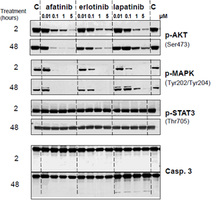
Afatinib Is a New Therapeutic Approach in Chordoma with a Unique Ability to Target EGFR and Brachyury. [Abstract]2018 Mar;17(3):603-613. PMID: 29237806
Lapatinib ditosylate monohydrate purchased from MedChemExpress. Usage Cited in: Mol Cancer Ther. 2018 Mar;17(3):603-613. [Abstract]
Immunoblot analysis of U-CH1 cells treated with the indicated doses of inhibitors (Afatinib, Erlotinib and Lapatinib) for 2 h (upper panel) or 48 h (lower panel). Protein cell extracts are resolved on SDS-PAGE gel and membranes probed with the indicated antibodies. IC50s of the different inhibitors are reported.
-
Cells
Machine Learning-Driven Multi-Omics Analysis Identifies CHP2 as a Key PANoptosis-Related Dual-Function Biomarker in Colorectal Cancer. [Abstract]2026 Feb 28;15(5):430. PMID: 41827864 -
Commun Biol
Proteolysis targeting chimera (PROTAC)-driven antibody internalization of oncogenic cell surface receptors. [Abstract]2024 Dec 31;7(1):1719. PMID: 39741170 -
Life Sci
Qiliqiangxin alleviates Ang II-induced CMECs apoptosis by downregulating autophagy via the ErbB2-AKT-FoxO3a axis. [Abstract]2021 May 15:273:119239. PMID: 33652033 -
Drug Des Devel Ther
Metabolic Stability Assessment of New PARP Inhibitor Talazoparib Using Validated LC-MS/MS Methodology: In silico Metabolic Vulnerability and Toxicity Studies. [Abstract]2020 Feb 25;14:783-793. PMID: 32158196 -
Stem Cell Reports
Inhibition of Farnesyltransferase Potentiates NOTCH-Targeted Therapy against Glioblastoma Stem Cells. [Abstract]2017 Dec 12;9(6):1948-1960. PMID: 29198824 -
Pharmaceuticals (Basel)
Synthesis and Development of 3-((2,4-Difluorophenyl)Amino)Propanoic Acid Derivatives as an Antiproliferative Medicinal Chemistry Scaffold Targeting Growth Factor Receptors. [Abstract]2026 Feb 27;19(3):381. PMID: 41901229 -
Front Pharmacol
2021 Mar 8;12:644342. PMID: 33790797 -
RSC Adv
Validated liquid chromatography tandem mass spectrometry for simultaneous quantification of foretinib and lapatinib, and application to metabolic stability investigation. [Abstract]2019 Jun 20;9(34):19325-19332. PMID: 35519400 -
Cell Rep Methods
Tumor immune microenvironment reconstitution in patient-derived organoids enables therapy modeling for NSCLC. [Abstract]2026 Jun 15;6(6):101339. PMID: 42134319 -
Cell Rep Methods
RECOVER identifies synergistic drug combinations in vitro through sequential model optimization. [Abstract]2023 Oct 23;3(10):100599. PMID: 37797618 -
Mol Pharm
2022 Nov 7;19(11):4320-4332. PMID: 36269563 -
Cancers (Basel)
Liver X Receptor Ligand GAC0001E5 Downregulates Antioxidant Capacity and ERBB2/HER2 Expression in HER2-Positive Breast Cancer Cells. [Abstract]2024 Apr 25;16(9):1651. PMID: 38730603 -
Cancers (Basel)
Drug Repurposing Applications to Overcome Male Predominance via Targeting G2/M Checkpoint in Human Esophageal Squamous Cell Carcinoma. [Abstract]2022 Nov 28;14(23):5854. PMID: 36497337 -
Biol Proced Online
Trastuzumab-resistant breast cancer cells-derived tumor xenograft models exhibit distinct sensitivity to lapatinib treatment in vivo. [Abstract]2023 Jun 27;25(1):19. PMID: 37370010 -
ACS Omega
Lapatinib Acts against Biofilm Formation and the Hemolytic Activity of Staphylococcus aureus. [Abstract]2022 Mar 3;7(10):9004-9014. PMID: 35309438 -
Biochim Biophys Acta Mol Basis Dis
Additional statin treatment enhances the efficacy of HER2 blockade and improves prognosis in Rac1-high/HER2-positive breast cancer. [Abstract]2024 Aug 9:167458. PMID: 39128642 -
iScience
FUS-dependent microRNA deregulations identify TRIB2 as a druggable target for ALS motor neurons. [Abstract]2023 Oct 6;26(11):108152. PMID: 37920668 -
Transl Oncol
A novel treatment strategy of HER2-targeted therapy in combination with Everolimus for HR+/HER2- advanced breast cancer patients with HER2 mutations. [Abstract]2022 Jul; 21: 101444. PMID: 35523006 -
Sci Rep
A degrader of HER2 and EGFR abolishes p95HER2 and shows robust antitumor efficacy in HER2-positive breast cancer. [Abstract]2026 Apr 20;16(1):12890. PMID: 42009845 -
J Biol Chem
MAP3K4 promotes fetal and placental growth by controlling the receptor tyrosine kinases IGF1R/IR and Akt signaling pathway. [Abstract]2022 Sep;298(9):102310. PMID: 35921893 -
ACS Chem Biol
2016 Apr 15;11(4):992-1000. PMID: 26741163 -
J Proteome Res
An Integrative Proteome-Based Pharmacologic Characterization and Therapeutic Strategy Exploration of SAHA in Solid Malignancies. [Abstract]2022 Apr 1;21(4):953-964. PMID: 35172096 -
Biotechnol Bioeng
An integrated biomimetic array chip for establishment of collagen-based 3D primary human hepatocyte model for prediction of clinical drug-induced liver injury. [Abstract]2021 Dec;118(12):4687-4698. PMID: 34478150 -
Exp Cell Res
Network-based analysis with primary cells reveals drug response landscape of acute myeloid leukemia. [Abstract]2020 Aug 1;393(1):112054. PMID: 32376287 -
Front Oncol
Discovery and Pharmacological Evaluation of STEAP4 as a Novel Target for HER2 Overexpressing Breast Cancer. [Abstract]2021 Mar 26:11:608201. PMID: 33842315 -
Analyst
Assessment of coulometric array electrochemical detection coupled with HPLC-UV for the absolute quantitation of pharmaceuticals. [Abstract]2017 Jan 26;142(3):525-536. PMID: 28098264 -
Cancer Med
Focal Adhesion Kinase Intersects With the BRD4-MYC Axis and YAP1 to Drive Tumor Cell Growth, Phenotypic Plasticity, Stemness, and Metastatic Potential in Colorectal Cancer. [Abstract]2025 Sep;14(18):e71227. PMID: 40959971 -
Carcinogenesis
SNCA inhibits epithelial-mesenchymal transition and correlates to favorable prognosis of breast cancer. [Abstract]2022 Dec 25;43(11):1071-1082. PMID: 36179220 -
Clin Chim Acta
2018 Oct:485:298-304. PMID: 30006284 -
J Chromatogr B Analyt Technol Biomed Life Sci
Simultaneous determination of 11 oral targeted antineoplastic drugs and 2 active metabolites by LC-MS/MS in human plasma and its application to therapeutic drug monitoring in cancer patients. [Abstract]2024 Apr 15:1237:124100. PMID: 38547701 -
J Chromatogr B Analyt Technol Biomed Life Sci
UPLC-ESI-MS/MS study of the effect of green tea extract on the oral bioavailability of erlotinib and lapatinib in rats: Potential risk of pharmacokinetic interaction. [Abstract]2017 Apr 1:1049-1050:30-40. PMID: 28260629 -
-
PLoS One
A novel small molecule screening assay using normal human chondrocytes toward osteoarthritis drug discovery. [Abstract]2024 Nov 1;19(11):e0308647. PMID: 39485774 -
-
PLoS One
Validated LC-MS/MS assay for quantification of the newly approved tyrosine kinase inhibitor, dacomitinib, and application to investigating its metabolic stability. [Abstract]2019 Apr 4;14(4):e0214598. PMID: 30947315 -
Fundam Clin Pharmacol
2021 Oct;35(5):919-929. PMID: 33523504 -
Gene
Pharmacotranscriptomic profiling of resistant triple-negative breast cancer cells treated with lapatinib and berberine shows upregulation of PI3K/Akt signaling under cytotoxic stress. [Abstract]2022 Mar 30:816:146171. PMID: 35026293 -
Eur J Drug Metab Pharmacokinet
Differential Inhibition of Equilibrative Nucleoside Transporter 1 (ENT1) Activity by Tyrosine Kinase Inhibitors. [Abstract]2021 Sep;46(5):625-635. PMID: 34275128 -
Cancer Chemother Pharmacol
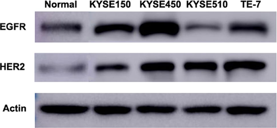
Lapatinib in combination with paclitaxel plays synergistic antitumor effects on esophageal squamous cancer. [Abstract]2018 Sep;82(3):383-394. PMID: 29909520
Lapatinib ditosylate monohydrate purchased from MedChemExpress. Usage Cited in: Cancer Chemother Pharmacol. 2018 Sep;82(3):383-394. [Abstract]
The expression level of EGFR and HER2 on normal esophageal epithelium cell and four esophageal squamous cancer cell lines analyzed by western blot.
-
-
Head Neck
Effect of HER2-targeted therapy on PDX and PDX-derived organoids generated from HER2-positive salivary duct carcinoma. [Abstract]2023 Jul;45(7):1801-1811. PMID: 37184432 -
Cell Physiol Biochem
2015;37(6):2275-87. PMID: 26624928 -
Open Life Sci
Systemic investigation of inetetamab in combination with small molecules to treat HER2-overexpressing breast and gastric cancers. [Abstract]2023 Jan 10;18(1):20220535. PMID: 36694697 -
-
-
-
-
-
bioRxiv
PAIRWISE: Deep Learning-based Prediction of Effective Personalized Drug Combinations in Cancer. [Abstract]2024 Nov 6:2024.11.04.621884. PMID: 39574568 -
-
-
-
-
-
Oncotarget
2020 Nov 3;11(44):3921-3932. PMID: 33216841 -
-
-
-
Oncotarget
A c-Jun N-terminal kinase inhibitor, JNK-IN-8, sensitizes triple negative breast cancer cells to lapatinib. [Abstract]2017 Aug 24;8(62):104894-104912. PMID: 29285221
Lapatinib ditosylate monohydrate purchased from MedChemExpress. Usage Cited in: Oncotarget. 2017 Aug 24;8(62):104894-104912. [Abstract]
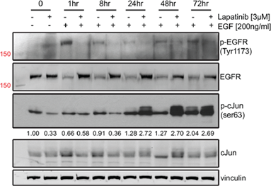
MDA-MB-231 cells are serum-starved overnight and pre-treated with 3 μM Lapatinib or DMSO for 3 hours. After that time, cells are lysed (0hr) or stimulated with EGF for 1, 8, 24, 48, and 72 hours. Western blots of p-EGFR (Tyr1173), EGFR, p-cJun (ser63), cJun, and vinculin are shown with band densitometries beneath.
-
Tumour Biol
Addition of the p110α inhibitor BYL719 overcomes targeted therapy resistance in cells from Her2-positive-PTEN-loss breast cancer. [Abstract]2016 Nov;37(11):14831-14839. PMID: 27639383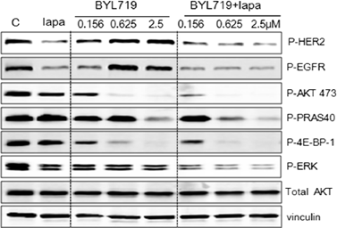
Lapatinib ditosylate monohydrate purchased from MedChemExpress. Usage Cited in: Tumour Biol. 2016 Nov;37(11):14831-14839. [Abstract]
Western blot analysis of PI3K signaling in cell lysates treated with Lapatinib (1 μM) with or without BYL719.


Solvent & Solubility
DMSO : 50 mg/mL (53.00 mM; ultrasonic and warming and heat to 60°C; Hygroscopic DMSO has a significant impact on the solubility of product, please use newly opened DMSO)
Please refer to the solubility information to select the appropriate solvent. Once prepared, please aliquot and store the solution to prevent product inactivation from repeated freeze-thaw cycles.
Storage method and period of stock solution: -80°C, 6 months; -20°C, 1 month (sealed storage, away from moisture). When stored at -80°C, please use it within 6 months. When stored at -20°C, please use it within 1 month.
Concentration (start) × Volume (start) = Concentration (final) × Volume (final)
Select the appropriate dissolution method based on your experimental animal and administration route.
- For the following dissolution methods, please ensure to first prepare a clear stock solution using an In Vitro approach and then sequentially add co-solvents:
- To ensure reliable experimental results, the clarified stock solution can be appropriately stored based on storage conditions. As for the working solution for In Vivo experiments, it is recommended to prepare freshly and use it on the same day.
- The percentages shown for the solvents indicate their volumetric ratio in the final prepared solution. If precipitation or phase separation occurs during preparation, heat and/or sonication can be used to aid dissolution.
Add each solvent one by one: 10% DMSO 90% (20% SBE-β-CD in Saline)
Solubility: 2.5 mg/mL (2.65 mM); Suspended solution; Need ultrasonic
This protocol yields a suspended solution of 2.5 mg/mL. Suspended solution can be used for oral and intraperitoneal injection.
Taking 1 mL working solution as an example, add 100 μL DMSO stock solution (25.0 mg/mL) to 900 μL 20% SBE-β-CD in Saline, and mix evenly.
Preparation of 20% SBE-β-CD in Saline (4°C, storage for one week): 2 g SBE-β-CD powder is dissolved in 10 mL Saline, completely dissolve until clear.
Please enter the basic information of animal experiments:
-
-
-
-
Recommended: Prepare an additional quantity of animals to account for potential losses during experiments.
Please enter your animal formula composition:
-
%DMSO +
Recommended: Keep the proportion of DMSO in working solution below 2% if your animal is weak.
-
%+
-
+%Tween-80 + +
-
%Saline +
The co-solvents required include: DMSO, . All of co-solvents are available by MedChemExpress (MCE). , Tween 80. All of co-solvents are available by MedChemExpress (MCE).
Working solution concentration: 0.22 mg/mL
Method for preparing stock solution: mg drug dissolved in μL DMSO. Stock solution concentration: mg/mL. * In solvent : -80°C, 6 months; -20°C, 1 month (sealed storage, away from moisture)
1. Take μL DMSO stock solution;
2. Add μL .
μL , mix evenly;
3. Then add μL Tween 80, mix evenly;
4. Then add μL
Please ensure that the stock solution in the first step is dissolved to a clear state, and add co-solvents in sequence. You can use ultrasonic heating (ultrasonic cleaner, recommended frequency 20-40 kHz), vortexing, etc. to assist dissolution.


Purity & Documentation
-
Data Sheet (280 KB)
-
SDS (393 KB)
- English - EN (393 KB)
- Français - FR (393 KB)
- Deutsch - DE (393 KB)
- Norwegian - NO (393 KB)
- Español - ES (393 KB)
- Swedish - SV (393 KB)
- Italian - IT (393 KB)
- Korean - KR (393 KB)
- Portuguese - PT (393 KB)
-
Handling Instructions (2659 KB)
References


Complete Stock Solution Preparation Table
Please refer to the solubility information to select the appropriate solvent. Once prepared, please aliquot and store the solution to prevent product inactivation from repeated freeze-thaw cycles.
Storage method and period of stock solution: -80°C, 6 months; -20°C, 1 month (sealed storage, away from moisture). When stored at -80°C, please use it within 6 months. When stored at -20°C, please use it within 1 month.
| Optional Solvent | Concentration Solvent Mass | 1 mg | 5 mg | 10 mg | 25 mg |
|---|---|---|---|---|---|
| DMSO | 1 mM | 1.0599 mL | 5.2995 mL | 10.5991 mL | 26.4976 mL |
| 5 mM | 0.2120 mL | 1.0599 mL | 2.1198 mL | 5.2995 mL | |
| 10 mM | 0.1060 mL | 0.5300 mL | 1.0599 mL | 2.6498 mL | |
| 15 mM | 0.0707 mL | 0.3533 mL | 0.7066 mL | 1.7665 mL | |
| 20 mM | 0.0530 mL | 0.2650 mL | 0.5300 mL | 1.3249 mL | |
| 25 mM | 0.0424 mL | 0.2120 mL | 0.4240 mL | 1.0599 mL | |
| 30 mM | 0.0353 mL | 0.1767 mL | 0.3533 mL | 0.8833 mL | |
| 40 mM | 0.0265 mL | 0.1325 mL | 0.2650 mL | 0.6624 mL | |
| 50 mM | 0.0212 mL | 0.1060 mL | 0.2120 mL | 0.5300 mL |


 Powered by Bioz