



Melevodopa hydrochloride

Based on 1 Customer Validation
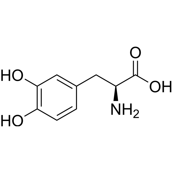
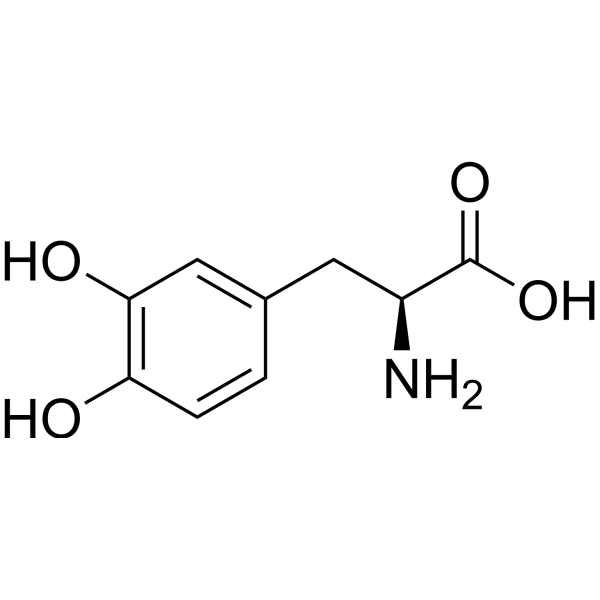
Melevodopa (Levodopa methylester) hydrochloride is an orally active methylated prodrug of Levodopa (HY-N0304), a dopamine precursor. When used in combination with Carbidopa (HY-B0311) in an effervescent tablet formulation, Melevodopa hydrochloride inhibits motor fluctuations in Parkinson's disease. Melevodopa hydrochloride suppresses subcutaneous tumor growth, pulmonary metastasis proliferation, and primary mass invasiveness of melanoma. Melevodopa hydrochloride induces lipid peroxidation by increasing malondialdehyde levels in melanoma and improves the survival rate of melanoma-bearing mice. Melevodopa hydrochloride can be used in research related to Parkinson's disease (with motor fluctuations) and melanoma.
For research use only. We do not sell to patients.
- Purity : 99.76%
- CAS No.: 1421-65-4
- Formula: C10H14ClNO4
- Molecular Weight:247.68
-
Storage:
4°C, stored under nitrogen
* In solvent : -80°C, 6 months; -20°C, 1 month (stored under nitrogen)


To place orders, for customer services and technical support, please contact: MedChemExpress USA
Tel: 609-228-6898 E-mail: [email protected] [email protected]


-
 Biological Activity
-
Chemical Information
-
Solvent & Solubility
- Protocols
- Purity & Documentation
- References
-
 Help & FAQs


All Dopamine Receptor Isoforms
More

Biological Activity
Description
In Vivo
MedChemExpress (MCE) has not independently confirmed the accuracy of these methods. They are for reference only.
-
Animal Model:B6D2F1 (female); C57BL/6 (male, female; inoculated subcutaneously with 106 viable B16 melanoma cells)[2]
-
Dosage:1000 mg/kg; preceded by carbidopa 100 mg/kg (90-minute pre-injection)
-
Administration:i.p.; daily; 15 days
-
Result:Inhibited tumor growth during 15-day treatment period and for 8 days after treatment cessation, with mean tumor volume never reaching that of untreated mice.
Increased median survival from 22 days to 27 days (23% increase) in basal diet-fed mice, and from 28 days to 38 days (36% increase) in restricted diet-fed mice.
Reduced tumor invasion into the pulmonary cavity in mice fed either diet.
Decreased food intake by 0.3 g in basal diet-fed mice and 1.3 g in restricted diet-fed mice during treatment period.
Increased tumor malondialdehyde levels 1.4-fold in basal diet-fed mice and 2.4-fold in restricted diet-fed mice compared to intradietary untreated controls.
-
Animal Model:B6D2F1 (female; inoculated intravenously with 2×10⁴ viable B16-BL6 melanoma cells to induce experimental lung metastases)[2]
-
Dosage:1000 mg/kg; preceded by benserazide 100 mg/kg (90-minute pre-injection)
-
Administration:i.p.; daily; 15 days
-
Result:Shifted lung tumor colony size distribution to a higher proportion of small (17.9% vs 8.9% in untreated) and medium (38.3% vs 25.2% in untreated) colonies, and a lower proportion of large colonies (43.8% vs 65.9% in untreated) in basal diet-fed mice.
Shifted colony size distribution to a higher proportion of small colonies (51.4% vs 27.8% in untreated) and a lower proportion of medium (43.3% vs 48.3% in untreated) and large (5.3% vs 23.9% in untreated) colonies in restricted diet-fed mice.
Did not significantly alter the number of lung colonies in either diet group.
- How to Select the Route of Administration for Mammals
- Human pluripotent stem cell midbrain dopaminergic neuron differentiation
- Patient-Derived Xenograft (PDX)
- Patient-Derived Orthotopic Xenograft (PDOX)
- Ferroptosis Solutions
- Subcutaneous Cell-Line-Derived Xenograft
- Orthotopic Cell-Line Xenograft
- Cell Viability Determination by MTT Colorimetric Assay


Chemical Information
-
CAS No. 1421-65-4
-
Appearance Solid
-
Molecular Weight 247.68
-
Formula C10H14ClNO4
-
Color White to off-white
-
SMILES
N[C@@H](CC1=CC=C(O)C(O)=C1)C(OC)=O.[H]Cl
-
Synonyms
Levodopa methylester hydrochloride
-
Shipping
Room temperature in continental US; may vary elsewhere.
-
Storage
4°C, stored under nitrogen
* In solvent : -80°C, 6 months; -20°C, 1 month (stored under nitrogen)


Solvent & Solubility
In Vitro:
H2O : ≥ 100 mg/mL (403.75 mM)
DMSO : 100 mg/mL (403.75 mM; Need ultrasonic; Hygroscopic DMSO has a significant impact on the solubility of product, please use newly opened DMSO)
* "≥" means soluble, but saturation unknown.
Please refer to the solubility information to select the appropriate solvent. Once prepared, please aliquot and store the solution to prevent product inactivation from repeated freeze-thaw cycles.
Storage method and period of stock solution: -80°C, 6 months; -20°C, 1 month (stored under nitrogen). When stored at -80°C, please use it within 6 months. When stored at -20°C, please use it within 1 month.
* Note: If you choose water as the stock solution, please dilute it to the working solution, then filter and sterilize it with a 0.22 μm filter before use.
Please refer to the solubility information to select the appropriate solvent. Once prepared, please aliquot and store the solution to prevent product inactivation from repeated freeze-thaw cycles.
Storage method and period of stock solution: -80°C, 6 months; -20°C, 1 month (stored under nitrogen). When stored at -80°C, please use it within 6 months. When stored at -20°C, please use it within 1 month.
* Note: If you choose water as the stock solution, please dilute it to the working solution, then filter and sterilize it with a 0.22 μm filter before use.
Concentration (start) × Volume (start) = Concentration (final) × Volume (final)
In Vivo:
Select the appropriate dissolution method based on your experimental animal and administration route.
- For the following dissolution methods, please ensure to first prepare a clear stock solution using an In Vitro approach and then sequentially add co-solvents:
- To ensure reliable experimental results, the clarified stock solution can be appropriately stored based on storage conditions. As for the working solution for In Vivo experiments, it is recommended to prepare freshly and use it on the same day.
- The percentages shown for the solvents indicate their volumetric ratio in the final prepared solution. If precipitation or phase separation occurs during preparation, heat and/or sonication can be used to aid dissolution.
Add each solvent one by one: 10% DMSO 40% PEG300 5% Tween-80 45% Saline
Solubility: ≥ 2.5 mg/mL (10.09 mM); Clear solution
This protocol yields a clear solution of ≥ 2.5 mg/mL (saturation unknown).
Taking 1 mL working solution as an example, add 100 μL DMSO stock solution (25.0 mg/mL) to 400 μL PEG300, and mix evenly; then add 50 μL Tween-80 and mix evenly; then add 450 μL Saline to adjust the volume to 1 mL.
Preparation of Saline: Dissolve 0.9 g sodium chloride in ddH₂O and dilute to 100 mL to obtain a clear Saline solution.
Add each solvent one by one: 10% DMSO 90% (20% SBE-β-CD in Saline)
Solubility: ≥ 2.5 mg/mL (10.09 mM); Clear solution
This protocol yields a clear solution of ≥ 2.5 mg/mL (saturation unknown).
Taking 1 mL working solution as an example, add 100 μL DMSO stock solution (25.0 mg/mL) to 900 μL 20% SBE-β-CD in Saline, and mix evenly.
Preparation of 20% SBE-β-CD in Saline (4°C, storage for one week): 2 g SBE-β-CD powder is dissolved in 10 mL Saline, completely dissolve until clear.
In Vivo Dissolution Calculator
Please enter the basic information of animal experiments:
-
-
-
-
Recommended: Prepare an additional quantity of animals to account for potential losses during experiments.
Working solution concentration: 0.22 mg/mL
This product has good water solubility, please refer to the measured solubility data in water/PBS/Saline for details.


Purity & Documentation
-
Data Sheet (279 KB)
-
SDS (394 KB)
- English - EN (394 KB)
- Français - FR (394 KB)
- Deutsch - DE (394 KB)
- Norwegian - NO (394 KB)
- Español - ES (394 KB)
- Swedish - SV (394 KB)
- Italian - IT (394 KB)
- Korean - KR (394 KB)
- Portuguese - PT (394 KB)
-
Handling Instructions (2659 KB)
References
[1]. Stocchi F, et al. Melevodopa/carbidopa effervescent formulation in the treatment of motor fluctuations in advanced Parkinson's disease. Movement disorders : official journal of the Movement Disorder Society. 2010 Sep 15;25(12):1881-7. [Content Brief]
[2]. Meadows GG, et al. Ascorbate in the treatment of experimental transplanted melanoma. The American journal of clinical nutrition. 1991 Dec;54(6 Suppl):1284S-1291S. [Content Brief]


Complete Stock Solution Preparation Table
Please refer to the solubility information to select the appropriate solvent. Once prepared, please aliquot and store the solution to prevent product inactivation from repeated freeze-thaw cycles.
Storage method and period of stock solution: -80°C, 6 months; -20°C, 1 month (stored under nitrogen). When stored at -80°C, please use it within 6 months. When stored at -20°C, please use it within 1 month.
| Optional Solvent | Concentration Solvent Mass | 1 mg | 5 mg | 10 mg | 25 mg |
|---|---|---|---|---|---|
| H2O / DMSO | 1 mM | 4.0375 mL | 20.1873 mL | 40.3747 mL | 100.9367 mL |
| 5 mM | 0.8075 mL | 4.0375 mL | 8.0749 mL | 20.1873 mL | |
| 10 mM | 0.4037 mL | 2.0187 mL | 4.0375 mL | 10.0937 mL | |
| 15 mM | 0.2692 mL | 1.3458 mL | 2.6916 mL | 6.7291 mL | |
| 20 mM | 0.2019 mL | 1.0094 mL | 2.0187 mL | 5.0468 mL | |
| 25 mM | 0.1615 mL | 0.8075 mL | 1.6150 mL | 4.0375 mL | |
| 30 mM | 0.1346 mL | 0.6729 mL | 1.3458 mL | 3.3646 mL | |
| 40 mM | 0.1009 mL | 0.5047 mL | 1.0094 mL | 2.5234 mL | |
| 50 mM | 0.0807 mL | 0.4037 mL | 0.8075 mL | 2.0187 mL | |
| 60 mM | 0.0673 mL | 0.3365 mL | 0.6729 mL | 1.6823 mL | |
| 80 mM | 0.0505 mL | 0.2523 mL | 0.5047 mL | 1.2617 mL | |
| 100 mM | 0.0404 mL | 0.2019 mL | 0.4037 mL | 1.0094 mL |
* Note: If you choose water as the stock solution, please dilute it to the working solution, then filter and sterilize it with a 0.22 μm filter before use.


Melevodopa hydrochloride Related Classifications
HY-W011963 Related Classifications
 Powered by Bioz