



Nur77 modulator 3

Based on 1 Customer Validation
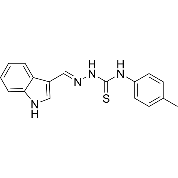
Nur77 modulator 3 is a Nur77 modulator. Nur77 modulator 3 induces Nur77 expression, inhibits hepatic stellate cells (HSCs) activation, and reduces extracellular matrix (ECM) deposition. Nur77 modulator 3 enhances Nur77-denpendent autophagic flux and significantly inhibits the mTORC1 signaling pathway. Nur77 modulator 3 ameliorates HSCs activation, inflammation and hepatic fibrosis in vivo.
For research use only. We do not sell to patients.
- Purity : 99.01%
- CAS No.: 2097787-75-0
- Formula: C17H16N4S
- Molecular Weight:308.40
-
Storage:Powder -20°C, 3 years ; In solvent -80°C, 6 months , -20°C, 1 month

To place orders, for customer services and technical support, please contact: MedChemExpress USA
Tel: 609-228-6898 E-mail: [email protected] [email protected]


-
 Biological Activity
-
Chemical Information
-
Solvent & Solubility
- Protocols
- Purity & Documentation
- References
-
 Help & FAQs


All Nuclear Hormone Receptor 4A/NR4A Isoforms
More

Biological Activity
Description
IC50 & Target
[1]|
Nur77/NR4A1 |
mTORC1 |
Cellular Effect
|
Cell Line

|
Type | Value | Description | References |
|---|---|---|---|---|
| 786-0 | GI50 |
3.99 μM
Compound: 2c
|
Growth inhibition of human 786-0 cells after 48 hrs by sulforhodamine B dye-based spectrophotometric assay
Growth inhibition of human 786-0 cells after 48 hrs by sulforhodamine B dye-based spectrophotometric assay
|
[PMID: 28505535] |
| HaCaT | GI50 |
0.99 μM
Compound: 2c
|
Growth inhibition of human HaCaT cells after 48 hrs by sulforhodamine B dye-based spectrophotometric assay
Growth inhibition of human HaCaT cells after 48 hrs by sulforhodamine B dye-based spectrophotometric assay
|
[PMID: 28505535] |
| HT-29 | GI50 |
1.45 μM
Compound: 2c
|
Growth inhibition of human HT-29 cells after 48 hrs by sulforhodamine B dye-based spectrophotometric assay
Growth inhibition of human HT-29 cells after 48 hrs by sulforhodamine B dye-based spectrophotometric assay
|
[PMID: 28505535] |
| K562 | GI50 |
0.16 μM
Compound: 2c
|
Growth inhibition of human K562 cells after 48 hrs by sulforhodamine B dye-based spectrophotometric assay
Growth inhibition of human K562 cells after 48 hrs by sulforhodamine B dye-based spectrophotometric assay
|
[PMID: 28505535] |
| MCF7 | GI50 |
2.52 μM
Compound: 2c
|
Growth inhibition of human MCF7 cells after 48 hrs by sulforhodamine B dye-based spectrophotometric assay
Growth inhibition of human MCF7 cells after 48 hrs by sulforhodamine B dye-based spectrophotometric assay
|
[PMID: 28505535] |
| NCI/ADR-RES | GI50 |
1.46 μM
Compound: 2c
|
Growth inhibition of human NCI-ADR-RES cells after 48 hrs by sulforhodamine B dye-based spectrophotometric assay
Growth inhibition of human NCI-ADR-RES cells after 48 hrs by sulforhodamine B dye-based spectrophotometric assay
|
[PMID: 28505535] |
| NCI-H460 | GI50 |
11.72 μM
Compound: 2c
|
Growth inhibition of human NCI-H460 cells after 48 hrs by sulforhodamine B dye-based spectrophotometric assay
Growth inhibition of human NCI-H460 cells after 48 hrs by sulforhodamine B dye-based spectrophotometric assay
|
[PMID: 28505535] |
| OVCAR-3 | GI50 |
3.88 μM
Compound: 2c
|
Growth inhibition of human OVCAR3 cells after 48 hrs by sulforhodamine B dye-based spectrophotometric assay
Growth inhibition of human OVCAR3 cells after 48 hrs by sulforhodamine B dye-based spectrophotometric assay
|
[PMID: 28505535] |
| U-251 | GI50 |
8.11 μM
Compound: 2c
|
Growth inhibition of human U251 cells after 48 hrs by sulforhodamine B dye-based spectrophotometric assay
Growth inhibition of human U251 cells after 48 hrs by sulforhodamine B dye-based spectrophotometric assay
|
[PMID: 28505535] |
In Vitro
Nur77 modulator 3 (Compound 9e) (5 and 10 μM, 24 h) inhibits TGF-β1-induced HSCs (LX-2, HSC-T6) activation in a Nur77-dependent manner[1].
Nur77 modulator 3 (5 and 10 μM, 36 h) inhibits TGF-β1-mediated HSCs (LX-2) activation by promoting autophagic flux[1].
Nur77 modulator 3 (5 and 10 μM, 24 h) enhances autophagic flux by inhibiting TGF-β1-induced mTOR phosphorylation in LX-2 cells[1].
MedChemExpress (MCE) has not independently confirmed the accuracy of these methods. They are for reference only.
-
Cell Line:HSCs (LX-2 and HSC-T6 cell)
-
Concentration:5, 10 μM
-
Incubation Time:24 h
-
Result:Dose-dependently upregulated Nur77 expression, which was downregulated by TGF-β1.
Suppressed TGF-β1-mediated α-SMA protein expression.
-
Cell Line:HSCs (LX-2 cell)
-
Concentration:5, 10 μM
-
Incubation Time:36 h
-
Result:Increased LC3B/LC3A ratio but inhibited TGF-β1-induced p62 expression.
Induced colocalization of EGFP and mRFP, suggesting that Nur77 modulator 3 promoted autophagic flux.
Induced more autophagic vesicles (AVs) and autolysosome-containing engulfed organelles than control cells (TEM assay).
Inhibited TGF-β1 induced HSCs activation and ECM deposition, and these effects were greatly reversed by Chloroquine (HY-17589A).
In Vivo
MedChemExpress (MCE) has not independently confirmed the accuracy of these methods. They are for reference only.
-
Animal Model:CCl4 (2.0 mL/kg, i.p., three weekly for 8 weeks) induced male C57BL/6 mice (6-8 weeks, 20-22 g)[1]
-
Dosage:20 mg/kg
-
Administration:i.p., daily, starting from the 4th week until the eighth week.
-
Result:Ameliorated tissue disorders in the liver, lung, spleen, and kidney induced by CCl4.
Inhibited the mRNA expression levels of α-SMA, IL-1β and IL-6 up-regulated by CCl4.
Suppressed CCl4 induced α-SMA and COL1A1 protein production in mouse liver tissue samples.
- Fibrosis/Collagen Morphometry
- RNA extraction experimental
- Collagen: Sirius Red Staining
- ECM-Embedded Organoid (Matrigel/Dome) Culture
- Connective Tissue: Masson's Trichrome/Collagen Trichrome Staining
- Autophagy
- Matrigel/ECM Transwell Invasion Assay
- Research Protocol for Inflammation-related Diseases
- Macroautophagy Solutions
- Matrigel Transwell/Boyden Chamber Invasion Assay


Chemical Information
-
CAS No. 2097787-75-0
-
Appearance Solid
-
Molecular Weight 308.40
-
Formula C17H16N4S
-
Color White to off-white
-
SMILES
S=C(N/N=C/C1=CNC2=CC=CC=C12)NC3=CC=C(C)C=C3
-
Shipping
Room temperature in continental US; may vary elsewhere.
-
Storage
Powder -20°C 3 years In solvent -80°C 6 months -20°C 1 month


Solvent & Solubility
In Vitro:
DMSO : 250 mg/mL (810.64 mM; Need ultrasonic; Hygroscopic DMSO has a significant impact on the solubility of product, please use newly opened DMSO)
Please refer to the solubility information to select the appropriate solvent. Once prepared, please aliquot and store the solution to prevent product inactivation from repeated freeze-thaw cycles.
Storage method and period of stock solution: -80°C, 6 months; -20°C, 1 month. When stored at -80°C, please use it within 6 months. When stored at -20°C, please use it within 1 month.
Please refer to the solubility information to select the appropriate solvent. Once prepared, please aliquot and store the solution to prevent product inactivation from repeated freeze-thaw cycles.
Storage method and period of stock solution: -80°C, 6 months; -20°C, 1 month. When stored at -80°C, please use it within 6 months. When stored at -20°C, please use it within 1 month.
Concentration (start) × Volume (start) = Concentration (final) × Volume (final)


Purity & Documentation
-
Data Sheet (274 KB)
-
SDS (251 KB)
- English - EN (251 KB)
- Français - FR (251 KB)
- Deutsch - DE (251 KB)
- Norwegian - NO (251 KB)
- Español - ES (251 KB)
- Swedish - SV (251 KB)
- Italian - IT (251 KB)
- Korean - KR (251 KB)
- Portuguese - PT (251 KB)
-
Handling Instructions (2659 KB)
References


Complete Stock Solution Preparation Table
Please refer to the solubility information to select the appropriate solvent. Once prepared, please aliquot and store the solution to prevent product inactivation from repeated freeze-thaw cycles.
Storage method and period of stock solution: -80°C, 6 months; -20°C, 1 month. When stored at -80°C, please use it within 6 months. When stored at -20°C, please use it within 1 month.
| Optional Solvent | Concentration Solvent Mass | 1 mg | 5 mg | 10 mg | 25 mg |
|---|---|---|---|---|---|
| DMSO | 1 mM | 3.2425 mL | 16.2127 mL | 32.4254 mL | 81.0636 mL |
| 5 mM | 0.6485 mL | 3.2425 mL | 6.4851 mL | 16.2127 mL | |
| 10 mM | 0.3243 mL | 1.6213 mL | 3.2425 mL | 8.1064 mL | |
| 15 mM | 0.2162 mL | 1.0808 mL | 2.1617 mL | 5.4042 mL | |
| 20 mM | 0.1621 mL | 0.8106 mL | 1.6213 mL | 4.0532 mL | |
| 25 mM | 0.1297 mL | 0.6485 mL | 1.2970 mL | 3.2425 mL | |
| 30 mM | 0.1081 mL | 0.5404 mL | 1.0808 mL | 2.7021 mL | |
| 40 mM | 0.0811 mL | 0.4053 mL | 0.8106 mL | 2.0266 mL | |
| 50 mM | 0.0649 mL | 0.3243 mL | 0.6485 mL | 1.6213 mL | |
| 60 mM | 0.0540 mL | 0.2702 mL | 0.5404 mL | 1.3511 mL | |
| 80 mM | 0.0405 mL | 0.2027 mL | 0.4053 mL | 1.0133 mL | |
| 100 mM | 0.0324 mL | 0.1621 mL | 0.3243 mL | 0.8106 mL |


Nur77 modulator 3 Related Classifications
HY-162147 Related Classifications
 Powered by Bioz