



OTUB2-IN-1
Based on 1 publication(s) in Google Scholar
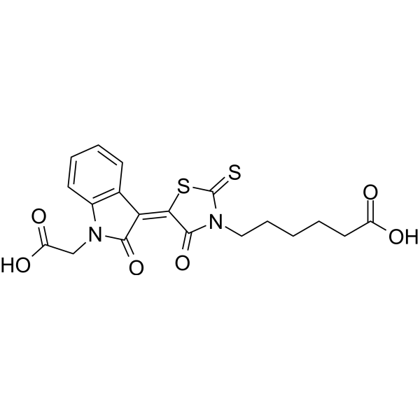
OTUB2-IN-1, a specific inhibitor of OTUB2 (KD: ~12 μM), reduces PD-L1 protein expression in tumor cells and inhibits tumor growth by promoting robust intra-tumor infiltration of cytotoxic T lymphocytes (CTL) .
연구목적의 판매만을 진행합니다. 환자를 대상으로 한 판매는 하지 않습니다.
- Purity : 98.79%
- 화학식: C19H18N2O6S2
- 분자량:434.49
-
보관:
4°C, protect from light
* In solvent : -80°C, 6 months; -20°C, 1 month (protect from light)

To place orders, for customer services and technical support, please contact: MedChemExpress USA
Tel: 609-228-6898 E-mail: [email protected] [email protected]


-
 Biological Activity
-
Chemical Information
-
용액&용해도
- Protocol
- 순도&문서
- References
-
 Help & FAQs


Publications Citing Use of MedChemExpress (MCE) OTUB2-IN-1
More

Biological Activity
제품 설명
In Vitro
OTUB2-IN-1 (0-40 μM) reduces PD-L1 levels in tumor cells (NCI-H358, SK-MES-1, and NCI-H226) in a dose-dependent manner, but it is unable to affect OTUB2 stability[1].
OTUB2-IN-1 (0-50 μM; 1 h) does not interfere with protein interactions between OTUB2 and PD-L1[1].
OTUB2-IN-1 (10 μM; 0-4 d) can’t inhibit the viability of B16-F10 tumor cells[1].
MedChemExpress (MCE) has not independently confirmed the accuracy of these methods. They are for reference only.
-
Cell Line:B16-F10 tumor cells
-
Concentration:10 μM
-
Incubation Time:0-4 d
-
Result:OTUB2-IN-1 did not show significant inhibitory effects on the viability of B16-F10 tumor cells over a period of up to four days.
-
Cell Line:NCI-H358,SK-MES-1, NCI-H226
-
Concentration:0-40 μM
-
Incubation Time:
-
Result:Reduced PD-L1 levels in a dose-dependent manner across these cell lines, indicating effective inhibition of OTUB2's functional activity regarding PD-L1 stabilization.
However, it did not affect the stability of OTUB2 itself, suggesting that the inhibitor specifically disrupts the PD-L1 regulatory function of OTUB2 without altering OTUB2 protein stability.
In Vivo
OTUB2-IN-1 (20 mg/kg; i.p.; daily for five days)reduces the expression of YAP and phosphorylated p65 in mice implanted with LL/2 cells; reduces phosphorylated Akt expression in mice implanted with B16-F10 cells and it reduces phosphorylated p65 expression in mice implanted with KLN205 cells[1].
MedChemExpress (MCE) has not independently confirmed the accuracy of these methods. They are for reference only.
-
Animal Model:C57BL/6 mice implanted with B16-F10 cells or LL/2 cells [1]
-
Dosage:20 mg/kg; daily for five days
-
Administration:i.p
-
Result:Reduced the expression of PD-L1 on tumor cells, suggesting its potential in enhancing immune recognition and response against tumor cells.
Did not significantly impact tumor cell viability directly, indicating its action might be more about modulating immune evasion mechanisms rather than cytotoxic effects.
Increased the infiltration of cytotoxic T cells within the tumors, suggesting that reducing PD-L1 levels can indeed make the tumor more susceptible to immune attack.within the tumors, suggesting that reducing PD-L1 levels can indeed make the tumor more susceptible to immune attack.


Chemical Information
-
Appearance Solid
-
분자량 434.49
-
화학식 C19H18N2O6S2
-
Color Orange to red
-
SMILES
OC(CCCCCN1C(S/C(C1=O)=C2C(C=CC=C3)=C3N(CC(O)=O)C\2=O)=S)=O
-
선적
Room temperature in continental US; may vary elsewhere.
-
보관
4°C, protect from light
* In solvent : -80°C, 6 months; -20°C, 1 month (protect from light)


Publications (1)
-
Journal Impact Factor
-
Most Recent
-
Virol Sin
2026 Jul 27:S1995-820X(26)00121-5. PMID: 42508733


용액&용해도
In Vitro:
DMSO : 50 mg/mL (115.08 mM; Need ultrasonic; Hygroscopic DMSO has a significant impact on the solubility of product, please use newly opened DMSO)
Please refer to the solubility information to select the appropriate solvent. Once prepared, please aliquot and store the solution to prevent product inactivation from repeated freeze-thaw cycles.
Storage method and period of stock solution: -80°C, 6 months; -20°C, 1 month (protect from light). When stored at -80°C, please use it within 6 months. When stored at -20°C, please use it within 1 month.
Please refer to the solubility information to select the appropriate solvent. Once prepared, please aliquot and store the solution to prevent product inactivation from repeated freeze-thaw cycles.
Storage method and period of stock solution: -80°C, 6 months; -20°C, 1 month (protect from light). When stored at -80°C, please use it within 6 months. When stored at -20°C, please use it within 1 month.
Concentration (start) × Volume (start) = Concentration (final) × Volume (final)
In Vivo:
Select the appropriate dissolution method based on your experimental animal and administration route.
- For the following dissolution methods, please ensure to first prepare a clear stock solution using an In Vitro approach and then sequentially add co-solvents:
- To ensure reliable experimental results, the clarified stock solution can be appropriately stored based on storage conditions. As for the working solution for In Vivo experiments, it is recommended to prepare freshly and use it on the same day.
- The percentages shown for the solvents indicate their volumetric ratio in the final prepared solution. If precipitation or phase separation occurs during preparation, heat and/or sonication can be used to aid dissolution.
For the following dissolution methods, please prepare the working solution directly:
It is recommended to prepare fresh solutions and use them promptly within a short period of time.
The percentages shown for the solvents indicate their volumetric ratio in the final prepared solution. If precipitation or phase separation occurs during preparation, heat and/or sonication can be used to aid dissolution.
Add each solvent one by one: 50% PEG300 50% Saline
Solubility: 20 mg/mL (46.03 mM); Suspended solution; Need ultrasonic
Add each solvent one by one: 15% Cremophor EL 85% Saline
Solubility: 10 mg/mL (23.02 mM); Suspended solution; Need ultrasonic
In Vivo Dissolution Calculator
Please enter the basic information of animal experiments:
-
-
-
-
Recommended: Prepare an additional quantity of animals to account for potential losses during experiments.
Please enter your animal formula composition:
-
%DMSO +
Recommended: Keep the proportion of DMSO in working solution below 2% if your animal is weak.
-
%+
-
+%Tween-80 + +
-
%Saline +
The co-solvents required include: DMSO, . All of co-solvents are available by MedChemExpress (MCE). , Tween 80. All of co-solvents are available by MedChemExpress (MCE).
Working solution concentration: 0.22 mg/mL
Method for preparing stock solution: mg drug dissolved in μL DMSO. Stock solution concentration: mg/mL. * In solvent : -80°C, 6 months; -20°C, 1 month (protect from light)
1. Take μL DMSO stock solution;
2. Add μL .
μL , mix evenly;
3. Then add μL Tween 80, mix evenly;
4. Then add μL
Please ensure that the stock solution in the first step is dissolved to a clear state, and add co-solvents in sequence. You can use ultrasonic heating (ultrasonic cleaner, recommended frequency 20-40 kHz), vortexing, etc. to assist dissolution.


순도&문서
-
Data Sheet (276 KB)
-
SDS (251 KB)
- English - EN (251 KB)
- Français - FR (251 KB)
- Deutsch - DE (251 KB)
- Norwegian - NO (251 KB)
- Español - ES (251 KB)
- Swedish - SV (251 KB)
- Italian - IT (251 KB)
- Korean - KR (251 KB)
- Portuguese - PT (251 KB)
-
Handling Instructions (2659 KB)
References


Complete Stock Solution Preparation Table
Please refer to the solubility information to select the appropriate solvent. Once prepared, please aliquot and store the solution to prevent product inactivation from repeated freeze-thaw cycles.
Storage method and period of stock solution: -80°C, 6 months; -20°C, 1 month (protect from light). When stored at -80°C, please use it within 6 months. When stored at -20°C, please use it within 1 month.
| Optional Solvent | Concentration Solvent Mass | 1 mg | 5 mg | 10 mg | 25 mg |
|---|---|---|---|---|---|
| DMSO | 1 mM | 2.3015 mL | 11.5077 mL | 23.0155 mL | 57.5387 mL |
| 5 mM | 0.4603 mL | 2.3015 mL | 4.6031 mL | 11.5077 mL | |
| 10 mM | 0.2302 mL | 1.1508 mL | 2.3015 mL | 5.7539 mL | |
| 15 mM | 0.1534 mL | 0.7672 mL | 1.5344 mL | 3.8359 mL | |
| 20 mM | 0.1151 mL | 0.5754 mL | 1.1508 mL | 2.8769 mL | |
| 25 mM | 0.0921 mL | 0.4603 mL | 0.9206 mL | 2.3015 mL | |
| 30 mM | 0.0767 mL | 0.3836 mL | 0.7672 mL | 1.9180 mL | |
| 40 mM | 0.0575 mL | 0.2877 mL | 0.5754 mL | 1.4385 mL | |
| 50 mM | 0.0460 mL | 0.2302 mL | 0.4603 mL | 1.1508 mL | |
| 60 mM | 0.0384 mL | 0.1918 mL | 0.3836 mL | 0.9590 mL | |
| 80 mM | 0.0288 mL | 0.1438 mL | 0.2877 mL | 0.7192 mL | |
| 100 mM | 0.0230 mL | 0.1151 mL | 0.2302 mL | 0.5754 mL |


OTUB2-IN-1 Related Classifications
HY-160768 Related Classifications
 Powered by Bioz