



Perfluorobutanesulfonic acid
Based on 1 publication(s) in Google Scholar
Perfluorobutanesulfonic acid (PFBS) is a short-chain perfluoroalkyl substance and the main replacement for perfluorooctanesulfonic acid. Perfluorobutanesulfonic acid induces fat accumulation in human HepG2 hepatoma cells. Perfluorobutanesulfonic acid promotes lipid accumulation by activating PPARγ pathway and triggering oxidative stress, endoplasmic reticulum stress and calcium dyshomeostasis. Perfluorobutanesulfonic acid impairs reproduction and causes developmental disorders in offspring of Caenorhabditis elegans. Perfluorobutanesulfonic acid disrupts pancreatic organogenesis and lipid homeostasis in zebrafish embryos. Perfluorobutanesulfonic acid can be used in environmental toxicology, lipid metabolism and developmental toxicity studies.
For research use only. We do not sell to patients.
- Purity : 98.0%
- CAS No.: 375-73-5
- Formula: C4HF9O3S
- Molecular Weight:300.10
-
Storage:
4°C, stored under nitrogen
* In solvent : -80°C, 6 months; -20°C, 1 month (stored under nitrogen)


To place orders, for customer services and technical support, please contact: MedChemExpress USA
Tel: 609-228-6898 E-mail: [email protected] [email protected]


-
 Biological Activity
-
Chemical Information
-
Solvent & Solubility
- Protocols
- Purity & Documentation
- References
-
 Help & FAQs


-

HY-W275295
Perfluorododecanoic acidToxic Compound -

HY-W004100
2-(Trifluoromethyl)benzenesulfonyl chlorideBiochemical Assay Reagent -

HY-21194
Perfluorohexanoic acid -
HY-A0067
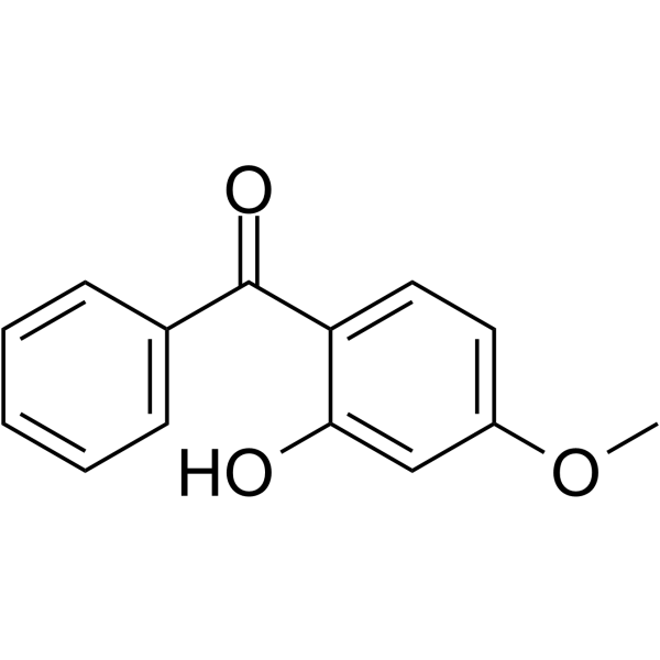
OxybenzoneUltraviolet Light Filter -
HY-B0346
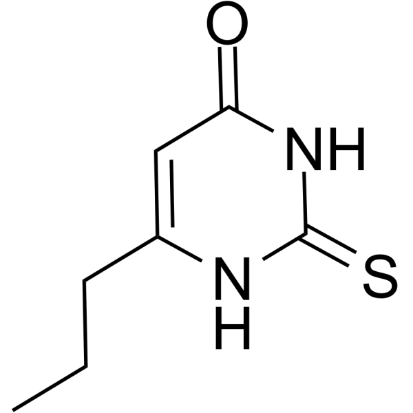
PropylthiouracilAntithyroid Agent -
HY-108298
PerfluorodecalinActive Compound -
HY-A0067R
Oxybenzone (Standard)Ultraviolet Light Filter -
HY-Y1115
2-Fluorobenzoic acidBiochemical Reagent -
HY-W008974
Sulfobetaine-18Biochemical Assay Reagent -
HY-W007331
5-FluorosalicylaldehydeBiochemical Reagent -
HY-W002068
2,4-Difluorobenzoic acidDifluorinated Aromatic Carboxylic Acid -

HY-W002172
2-Amino-5-chloro-2'-fluorobenzophenoneMidazolam Precursor
Publications Citing Use of MedChemExpress (MCE) Perfluorobutanesulfonic acid
More

Biological Activity
Description
IC50 & Target
[4]|
PPARγ |
In Vitro
Perfluorobutanesulfonic acid binds with high affinity to estrogen receptor isoforms from zebrafish, largemouth bass, fathead minnow, and Atlantic salmon, with binding affinities ranging from -7.4 to -9.1 kCal/mol, indicating strong potential for estrogen receptor-mediated endocrine disruption in these fish species[3].
Perfluorobutanesulfonic acid (PFBS) (50-200 μmol/L; 48 h) does not reduce viability of HepG2 human hepatoma cells, with or without concurrent fatty acid exposure[4].
Perfluorobutanesulfonic acid (200 μmol/L; 48 h) significantly increases triglyceride accumulation in HepG2 human hepatoma cells after 48 h of treatment when combined with 300 μmol/L fatty acid mixture, but not in the absence of fatty acid[4].
Perfluorobutanesulfonic acid (200 μmol/L; 48 h) significantly upregulates the mRNA expression of lipogenesis-related genes (ACC, FAS, SREBP1, DGAT2), fatty acid uptake-related genes (CD36, PPARγ), fatty acid β‑oxidation-related genes (CPT1a, PPARα), and the endoplasmic reticulum stress marker CHOP in HepG2 human hepatoma cells[4].
Perfluorobutanesulfonic acid (200 μmol/L; 1-12 h) significantly increases intracellular ROS production in HepG2 human hepatoma cells[4].
Perfluorobutanesulfonic acid (200 μmol/L; 1 h) significantly increases cytosolic calcium levels in HepG2 human hepatoma cells after 1 h of treatment[4].
Perfluorobutanesulfonic acid (200 μmol/L; 48 h) increases triglyceride accumulation in HepG2 human hepatoma cells after 48 h of treatment with 300 μmol/L fatty acid mixture via a PPARγ-dependent pathway[4].
MedChemExpress (MCE) has not independently confirmed the accuracy of these methods. They are for reference only. Further protocols information, click here.
-
Cell Line:HepG2 human hepatoma cells
-
Concentration:50 μmol/L; 100 μmol/L; 200 μmol/L
-
Incubation Time:48 h
-
Result:Exerted no cytotoxic effect on HepG2 cells, either with or without fatty acid treatment.
Maintained cell viability at ~85-105% of control levels across all tested concentrations.
-
Cell Line:HepG2 human hepatoma cells
-
Concentration:200 μmol/L
-
Incubation Time:48 h
-
Result:Upregulated mRNA levels of ACC by 75%, FAS by 98%, SREBP1 by 74%, and DGAT2 by 39%; elevated CD36 by 26% and PPARγ by 47%; increased CPT1a by 149% and PPARα by 67%; and enhanced CHOP expression by 82% versus controls, regardless of fatty acid cotreatment.
In Vivo
Perfluorobutanesulfonic acid (8.25-8250 µM; daily exposure in 0.3x Danieau’s medium; daily; 3 hours post fertilization to 5 dpf) causes dose-dependent decreases in zebrafish survival, hatching, and swim bladder inflation[1].
Perfluorobutanesulfonic acid (PFBS) (1000-2000 μM; in S-complete liquid media with heat-killed E. coli; 1 day exposure) significantly impairs reproductive capacity in wild-type C. elegans without altering hatchability[2].
Perfluorobutanesulfonic acid (1000-2000 μM; in vitro in S-complete liquid media with heat-killed E. coli; 2 day exposure) at 1000 μM reduces embryonic triglyceride and protein levels by ~24% each in C. elegans, with internal embryonic concentrations reaching 1.8 ng/mg DNA at 1000 μM and 3.3 ng/mg DNA at 2000 μM, while 2000 μM does not produce significant embryonic nutrient reductions[2].
Perfluorobutanesulfonic acid (1000-2000 μM; in vitro in S-complete liquid media with heat-killed E. coli; 1 day exposure) significantly reduces VIT-2::GFP intensity (a marker of oocyte yolk uptake) in transgenic C. elegans by 25% and 23%, respectively, with internal parent worm concentrations reaching 2.8 ng/mg DNA and 4.6 ng/mg DNA[2].
Perfluorobutanesulfonic acid (1000-2000 μM; in vitro in S-complete liquid media with heat-killed E. coli; 2 day exposure to F0) significantly retards growth rate in C. elegans F1 offspring[2].
Perfluorobutanesulfonic acid (PFBS) (16-32 μM; waterborne; continuous; fertilization to 120 hpf) causes developmental malformations, growth stunting, and metabolic pathway dysregulation in zebrafish embryos with high survival rates[3].
Perfluorobutanesulfonic acid (waterborne; continuous; fertilization to 6 days post-fertilization; 3000 mg/L) induces hypoactivity and alters locomotor behavior in zebrafish larvae[3].
Perfluorobutanesulfonic acid (waterborne; continuous; fertilization to 120 hpf, fertilization to 4 days post-fertilization) induces concentration-dependent hyperactivity, hypoactivity, and seizure-like locomotor effects in Danio rerio larvae[3].
Perfluorobutanesulfonic acid (10-100 μg/L; waterborne; continuous; 28 days) disrupts intestinal integrity, alters gut microbiota composition, and induces sex-specific metabolic dysregulation in adult zebrafish[3].
Perfluorobutanesulfonic acid (0.2-200 μM; waterborne; continuous; fertilization to 120 hpf) alters PPAR and insulin signaling pathways in developing zebrafish[3].
Perfluorobutanesulfonic acid (10 mg/L; waterborne; continuous; fertilization to 4 days post-fertilization) increases cortisol levels, indicating stress axis disruption, in Danio rerio larvae[3].
Perfluorobutanesulfonic acid (3000 mg/L; waterborne; continuous; fertilization to 5 days post-fertilization) induces developmental malformations and reduced heart rate in zebrafish embryos with low mortality, with an EC50 of 1529 mg/L for malformations[3].
MedChemExpress (MCE) has not independently confirmed the accuracy of these methods. They are for reference only.
-
Animal Model:transgenic Tg(insulin:GFP); Tg(ptf1a:GFP) (homozygous populations, embryonic stage)[1]
-
Dosage:16 µM; 32 µM
-
Administration:daily exposure in 0.3x Danieau’s medium; daily; 1 dpf to 7 dpf
-
Result:Showed high survival rates at 4 dpf without significant differences from controls.
Increased growth retardation, tail deformities, craniofacial defects, and swim bladder and yolk utilization disorders at 4 dpf.
Reduced exocrine pancreas length and elevated abnormal islet morphology at 4 dpf.
Altered multiple gene expressions and upregulated exocrine pancreatic genes by 11-53.7% at 4 dpf.
Activated lipid and xenobiotic metabolism pathways while suppressing p53, glycolysis/gluconeogenesis, and pentose phosphate pathways at 4 dpf.
-
Animal Model:Danio rerio (embryonic stage)[1]
-
Dosage:8.25 µM; 82.5 µM; 825 µM; 8250 µM
-
Administration:daily exposure in 0.3x Danieau’s medium; daily; 3 hours post fertilization to 5 dpf
-
Result:Reduced survival, hatching, and swim bladder inflation rates in a concentration‑dependent manner, with an LC50 of 1310 μM for PFBS.
-
Animal Model:DH1033 (sqt-1(sc103) II; bIs1 X)[2]
-
Dosage:1000 μM; 2000 μM
-
Administration:in S-complete liquid media with heat-killed E. coli; 1 day exposure
-
Result:Reduced VIT-2::GFP intensity by 25% at 1000 μM compared to control.
Reduced VIT-2::GFP intensity by 23% at 2000 μM compared to control.
Reached internal parent worm concentration of 2.8 ng/mg DNA at 1000 μM and 4.6 ng/mg DNA at 2000 μM.
-
Animal Model:Danio rerio (early life stage embryos/larvae; exposed from fertilization to 120 hpf)[3]
-
Dosage:16 μM; 32 μM
-
Administration:waterborne; continuous; fertilization to 120 hpf
-
Result:Achieved 96% survival at 16 μM and 95% at 32 μM.
Caused shorter body length, stunted growth, and increased incidence of blunt and curly tails at both doses.
Induced craniofacial malformations and decreased swim bladder inflation at 16 μM.
Caused impaired yolk utilization and decreased pancreas length at 32 μM.
Downregulated energy metabolism pathways (gluconeogenesis/glycolysis) and p53 signaling at both doses.
Upregulated lipid and xenobiotics metabolism pathways at both doses.
-
Animal Model:Oryzias melastigma (embryos and adult fish; embryos exposed from fertilization to 15 dpf; adult fish exposed for multi-generational assessment)[3]
-
Dosage:1 mg/L; 3.3 mg/L; 10 mg/L (embryos); 9.5 μg/L (adult fish)
-
Administration:waterborne; continuous; fertilization to 15 dpf (embryos); waterborne; continuous; 6 months (adult fish)
-
Result:Showed no impact on survival, heart rate, or malformation rate in embryos, but increased hatching rate in a concentration-dependent manner.
Decreased 17β-estradiol (E2) levels at all embryo doses.
Decreased testosterone (T) levels only at 1 mg/L in embryos, and decreased E2/T ratio in a concentration-dependent manner.
Upregulated transcript levels for gnrh isoforms and their receptors, while downregulated steroidogenesis, reproduction, estrogen receptor, choriogenin, and vtg-1 genes at 10 mg/L in embryos.
Reduced plasma 3,5,3′-triiodothyronine (T3) and brain thyroxine (T4) levels in F0 females; increased plasma T4 levels and downregulated hypothalamus-pituitary-thyroid (HPT) signaling in both F0 males and females.
Increased T3 and thyroxine-binding globulin (TBG) levels and downregulated HPT signaling in F1 larvae; resolved most thyroid disturbances but had significantly higher T4 levels in F2 larvae from F0 fish exposed to 9.5 μg/L.
Blocked oocyte development, reduced ovary size, altered follicle-stimulating hormone (FSH) and T levels, and caused predominantly male sex ratio in F0 fish.
Detected 10 ng/g dw PFBS in F0 whole fish, up to 2.9 ng/g dw PFBS in F1 unexposed larvae, and no detectable PFBS in F2 larvae.
-
Animal Model:Danio rerio (early life stage larvae; exposed from fertilization to 6 days post-fertilization)[3]
-
Dosage:3000 mg/L
-
Administration:waterborne; continuous; fertilization to 6 days post-fertilization
-
Result:Decreased overall larval activity (hypoactivity).
Showed positive correlation with active swimming speed.
Showed negative correlation with swimming distance, swimming time, and average swimming speed.
-
Animal Model:Danio rerio (early life stage embryos/larvae; exposed from fertilization to 120 hpf or 4 days post-fertilization)[3]
-
Dosage:7 μM; 43 μM; 57 μM; 700 μM; 3500 μM; 7000 μM; 10 mg/L
-
Administration:waterborne; continuous; fertilization to 120 hpf; waterborne; continuous; fertilization to 4 days post-fertilization
-
Result:Caused hypoactivity at 43 μM and hyperactivity at 57 μM.
Increased total movement at 3500 and 7000 μM during light phases.
Increased total movement at 7-700 μM and decreased total movement at 3500 and 7000 μM during dark phases.
Caused lethargic phototaxis behavior and reduced locomotor activity at 10 mg/L.
-
Animal Model:Danio rerio (adult fish; exposed for 28 days)[3]
-
Dosage:10 μg/L; 100 μg/L
-
Administration:waterborne; continuous; 28 days
-
Result:Decreased tight junction protein 2 (TJP2) in both male and female intestines at 100 μg/L; increased interleukin 1β levels (intestinal inflammation) in females at 100 μg/L.
Increased relative abundances of Acidobacteria and Chloroflexi phyla in females at 10 μg/L; decreased Bifidobacterium abundance in females at 100 μg/L.
Increased abundances of short chain fatty acid-producing bacteria (Bifidobacterium, Blautia, Faecalibacterium) in males at 10 μg/L.
Increased total bile acids in females but decreased total bile acids in males; decreased L-carnitine levels and downregulated apolipoprotein hepatic synthesis in females.
Disrupted bile acid secretion and fatty acid oxidation, increased blood glucose (2.5-fold), and caused hepatic vacuolization in males at 10 μg/L; decreased blood glucagon (2.9-fold) and increased glycogen (3.2-fold) in females at 10 μg/L.
-
Animal Model:Oryzias melastigma (adult fish; exposed for 7 or 21 days under normoxia)[3]
-
Dosage:10 μg/L
-
Administration:waterborne; continuous; 7 or 21 days
-
Result:Increased bile acid levels in males after 7 days.
Increased glycerol levels and free fatty acid content after 21 days.
-
Animal Model:Danio rerio (early life stage embryos/larvae; exposed from fertilization to 120 hpf)[3]
-
Dosage:0.2 μM; 2 μM; 20 μM; 200 μM
-
Administration:waterborne; continuous; fertilization to 120 hpf
-
Result:Enriched pathways related to PPAR and insulin signaling via KEGG analysis.
-
Animal Model:Danio rerio (early life stage embryos/larvae; exposed from fertilization to 5 days post-fertilization)[3]
-
Dosage:10 mg/L
-
Administration:waterborne; continuous; fertilization to 4 days post-fertilization
-
Result:Significantly increased cortisol levels in larvae.
-
Animal Model:Danio rerio (early life stage embryos; exposed from fertilization to 5 days post-fertilization)[3]
-
Dosage:3000 mg/L
-
Administration:waterborne; continuous; fertilization to 5 days post-fertilization
-
Result:Caused low mortality.
Significantly decreased heart rate after 72 hpf.
Induced malformations including uninflated swim bladders and tail deformities.
Achieved a median effective concentration (EC50) of 1529 mg/L for malformations, and a lethal concentration 50 (LC50) of > 3000 mg/L.


Chemical Information
-
CAS No. 375-73-5
-
Appearance Liquid (Density: 1.849±0.06 g/cm3)
-
Molecular Weight 300.10
-
Formula C4HF9O3S
-
Color Colorless to light yellow
-
SMILES
O=S(C(F)(F)C(F)(F)C(F)(F)C(F)(F)F)(O)=O
-
Synonyms
PFBS
-
Shipping
Room temperature in continental US; may vary elsewhere.
-
Storage
4°C, stored under nitrogen
* In solvent : -80°C, 6 months; -20°C, 1 month (stored under nitrogen)


Publications (1)
-
Journal Impact Factor
-
Most Recent


Solvent & Solubility
In Vitro:
DMSO : 50 mg/mL (166.61 mM; Need ultrasonic; Hygroscopic DMSO has a significant impact on the solubility of product, please use newly opened DMSO)
Please refer to the solubility information to select the appropriate solvent. Once prepared, please aliquot and store the solution to prevent product inactivation from repeated freeze-thaw cycles.
Storage method and period of stock solution: -80°C, 6 months; -20°C, 1 month (stored under nitrogen). When stored at -80°C, please use it within 6 months. When stored at -20°C, please use it within 1 month.
Please refer to the solubility information to select the appropriate solvent. Once prepared, please aliquot and store the solution to prevent product inactivation from repeated freeze-thaw cycles.
Storage method and period of stock solution: -80°C, 6 months; -20°C, 1 month (stored under nitrogen). When stored at -80°C, please use it within 6 months. When stored at -20°C, please use it within 1 month.
Concentration (start) × Volume (start) = Concentration (final) × Volume (final)
In Vivo:
Select the appropriate dissolution method based on your experimental animal and administration route.
- For the following dissolution methods, please ensure to first prepare a clear stock solution using an In Vitro approach and then sequentially add co-solvents:
- To ensure reliable experimental results, the clarified stock solution can be appropriately stored based on storage conditions. As for the working solution for In Vivo experiments, it is recommended to prepare freshly and use it on the same day.
- The percentages shown for the solvents indicate their volumetric ratio in the final prepared solution. If precipitation or phase separation occurs during preparation, heat and/or sonication can be used to aid dissolution.
Add each solvent one by one: 10% DMSO 40% PEG300 5% Tween-80 45% Saline
Solubility: ≥ 5 mg/mL (16.66 mM); Clear solution
This protocol yields a clear solution of ≥ 5 mg/mL (saturation unknown).
Taking 1 mL working solution as an example, add 100 μL DMSO stock solution (50.0 mg/mL) to 400 μL PEG300, and mix evenly; then add 50 μL Tween-80 and mix evenly; then add 450 μL Saline to adjust the volume to 1 mL.
Preparation of Saline: Dissolve 0.9 g sodium chloride in ddH₂O and dilute to 100 mL to obtain a clear Saline solution.
Add each solvent one by one: 10% DMSO 90% (20% SBE-β-CD in Saline)
Solubility: ≥ 5 mg/mL (16.66 mM); Clear solution
This protocol yields a clear solution of ≥ 5 mg/mL (saturation unknown).
Taking 1 mL working solution as an example, add 100 μL DMSO stock solution (50.0 mg/mL) to 900 μL 20% SBE-β-CD in Saline, and mix evenly.
Preparation of 20% SBE-β-CD in Saline (4°C, storage for one week): 2 g SBE-β-CD powder is dissolved in 10 mL Saline, completely dissolve until clear.
In Vivo Dissolution Calculator
Please enter the basic information of animal experiments:
-
-
-
-
Recommended: Prepare an additional quantity of animals to account for potential losses during experiments.
Please enter your animal formula composition:
-
%DMSO +
Recommended: Keep the proportion of DMSO in working solution below 2% if your animal is weak.
-
%+
-
+%Tween-80 + +
-
%Saline +
The co-solvents required include: DMSO, . All of co-solvents are available by MedChemExpress (MCE). , Tween 80. All of co-solvents are available by MedChemExpress (MCE).
Working solution concentration: 0.22 mg/mL
Method for preparing stock solution: mg drug dissolved in μL DMSO. Stock solution concentration: mg/mL. * In solvent : -80°C, 6 months; -20°C, 1 month (stored under nitrogen)
1. Take μL DMSO stock solution;
2. Add μL .
μL , mix evenly;
3. Then add μL Tween 80, mix evenly;
4. Then add μL
Please ensure that the stock solution in the first step is dissolved to a clear state, and add co-solvents in sequence. You can use ultrasonic heating (ultrasonic cleaner, recommended frequency 20-40 kHz), vortexing, etc. to assist dissolution.

Protocols
-
Reproductive and Developmental Toxicity Study
Reproductive and developmental toxicity studies detect adverse effects of prenatal or peri/postnatal exposure on maternal condition, pregnancy maintenance, embryo-fetal survival, fetal growth, structural development, and offspring reproductive or developmental endpoints; classic rat protocols generate readouts by comparing treated groups with vehicle, pair-fed, or untreated controls for implantation, resorption, fetal weight, crown-rump length, external morphology, visceral morphology, skeletal ossification, anogenital distance, nipple/areola retention, and postnatal cohort outcomes.
-
3T3-L1 preadipocyte-to-adipocyte differentiation
3T3-L1 preadipocytes are induced to differentiate after growth arrest using adipogenic media containing insulin, dexamethasone, and IBMX; differentiation is assessed by lipid-droplet accumulation, triglyceride increase, Oil Red O staining, and adipocyte-marker induction such as PPARγ and C/EBPα.
-
ROS/oxidative-stress fluorescent staining
ROS/oxidative-stress fluorescent staining uses cell-permeant fluorogenic probes that become fluorescent after oxidation inside cells or tissues; commonly used examples include DCFH-DA/DCFDA for broad cellular oxidant detection, DHE for superoxide-related signal detection, MitoSOX for mitochondrial superoxide-related signal detection, and CellROX probes for oxidative-stress-associated fluorescence readouts. The assay detects probe oxidation rather than a single ROS species unless the probe and analysis method have been chemically validated for that species. DCFH-DA enters cells, is deacetylated by intracellular esterases to DCFH, and produces fluorescent DCF after oxidation, so the readout is used as an operational measure of total cellular oxidative stress rather than a species-specific ROS measurement. DHE and MitoSOX can report superoxide-related oxidation, but red fluorescence alone can include non-specific ethidium-like oxidation products; HPLC or optimized spectral approaches are
-
Subchronic/Chronic Toxicity Study
A subchronic/chronic oral toxicity study detects systemic adverse effects caused by repeated administration of a test article, using mortality, clinical signs, body weight, food/water intake, ophthalmology, urinalysis, hematology, serum biochemistry, organ weights, gross necropsy, and histopathology as integrated readouts. The readout reflects dose-related physiological injury, target-organ pathology, reversibility after recovery, and derivation of NOAEL, LOAEL, or related point-of-departure values when the dataset supports them.
-
Liver Cancer Modeling
Liver cancer can be classified into primary liver cancer and secondary liver cancer. Secondary liver cancer is the metastatic liver cancer. Primary liver cancer includes hepatocellular carcinoma (HCC), intrahepatic cholangiocarcinoma (ICC) and fibrolamellar HCC, of which HCC is the most common form, accounting for approximately 90% of primary liver cancers[1]. HCC mouse models include chemical agent-induced models, transplanted tumor models, and genetic engineered models.
-
Acute Systemic Toxicity Study
Acute systemic toxicity studies evaluate adverse effects occurring after a single exposure, or repeated exposure within a short acute window, and the main in vivo readouts are mortality, moribund condition, clinical signs, body-weight change, and gross pathological findings; acute oral toxicity methods were developed to replace classical LD50 testing with reduced-animal designs such as fixed-dose procedure, acute toxic class method, and up-and-down procedure. The fixed-dose procedure classifies acute toxicity by administering predefined dose levels and observing evident toxicity rather than using death as the primary endpoint, whereas the acute toxic class method uses sequential groups of three animals per step and the up-and-down procedure doses animals sequentially to estimate an LD50 with fewer animals than conventional LD50 testing.
-
Lipid Droplets: Oil Red O/Sudan Dye Lipid Staining
Lipid droplets are intracellular organelles with a neutral-lipid core that stores triacylglycerols and sterol esters, and Oil Red O or Sudan dyes detect these hydrophobic lipid deposits by partitioning into retained lipids in fresh or frozen specimens. Oil Red O stains neutral triglycerides and lipids in frozen tissue sections or air-dried cytologic preparations, while Sudan Black B has also been used as a histochemical fat stain for lipid-rich tissue structures.


Purity & Documentation
-
Data Sheet (298 KB)
-
SDS (615 KB)
- English - EN (615 KB)
- Français - FR (615 KB)
- Deutsch - DE (615 KB)
- Norwegian - NO (615 KB)
- Español - ES (615 KB)
- Swedish - SV (615 KB)
- Italian - IT (615 KB)
- Korean - KR (615 KB)
- Portuguese - PT (615 KB)
-
Handling Instructions (2659 KB)
References
[1]. Sant KE, et al. Perfluorobutanesulfonic Acid Disrupts Pancreatic Organogenesis and Regulation of Lipid Metabolism in the Zebrafish, Danio rerio. Toxicol Sci. 2019;167(1):258-268. [Content Brief]
[2]. Yue Y, et al. Perfluorooctanesulfonic acid (PFOS) and perfluorobutanesulfonic acid (PFBS) impaired reproduction and altered offspring physiological functions in Caenorhabditis elegans. Food Chem Toxicol. 2020;145:111695. [Content Brief]
[3]. Ivantsova E, et al. Occurrence and toxicity mechanisms of perfluorobutanoic acid (PFBA) and perfluorobutane sulfonic acid (PFBS) in fish. Chemosphere. 2024;349:140815. [Content Brief]
[4]. Qi W, et al. Perfluorobutanesulfonic Acid (PFBS) Induces Fat Accumulation in HepG2 Human Hepatoma. Toxicol Environ Chem. 2020;102(10):585-606. [Content Brief]


Complete Stock Solution Preparation Table
Please refer to the solubility information to select the appropriate solvent. Once prepared, please aliquot and store the solution to prevent product inactivation from repeated freeze-thaw cycles.
Storage method and period of stock solution: -80°C, 6 months; -20°C, 1 month (stored under nitrogen). When stored at -80°C, please use it within 6 months. When stored at -20°C, please use it within 1 month.
| Optional Solvent | Concentration Solvent Mass | 1 mg | 5 mg | 10 mg | 25 mg |
|---|---|---|---|---|---|
| DMSO | 1 mM | 3.3322 mL | 16.6611 mL | 33.3222 mL | 83.3056 mL |
| 5 mM | 0.6664 mL | 3.3322 mL | 6.6644 mL | 16.6611 mL | |
| 10 mM | 0.3332 mL | 1.6661 mL | 3.3322 mL | 8.3306 mL | |
| 15 mM | 0.2221 mL | 1.1107 mL | 2.2215 mL | 5.5537 mL | |
| 20 mM | 0.1666 mL | 0.8331 mL | 1.6661 mL | 4.1653 mL | |
| 25 mM | 0.1333 mL | 0.6664 mL | 1.3329 mL | 3.3322 mL | |
| 30 mM | 0.1111 mL | 0.5554 mL | 1.1107 mL | 2.7769 mL | |
| 40 mM | 0.0833 mL | 0.4165 mL | 0.8331 mL | 2.0826 mL | |
| 50 mM | 0.0666 mL | 0.3332 mL | 0.6664 mL | 1.6661 mL | |
| 60 mM | 0.0555 mL | 0.2777 mL | 0.5554 mL | 1.3884 mL | |
| 80 mM | 0.0417 mL | 0.2083 mL | 0.4165 mL | 1.0413 mL | |
| 100 mM | 0.0333 mL | 0.1666 mL | 0.3332 mL | 0.8331 mL |


Perfluorobutanesulfonic acid Related Classifications
HY-21191 Related Classifications
 Powered by Bioz
Keywords
- Perfluorobutanesulfonic acid
- 375-73-5
- PFBS
- Biochemical Assay Reagents
- PPAR
- Reactive Oxygen Species (ROS)
- Caenorhabditis elegans
- estrogen receptors
- Esr2a
- PPARγ
- nonalcoholic fatty liver disease
- peroxisome proliferator-activated receptor gamma
- Esr2b
- zebrafish embryos
- HepG2 human hepatoma cells
- Esr1
- Inhibitor
- inhibitor
- inhibit