



Trimethoprim sulfate
Based on 13 publication(s) in Google Scholar
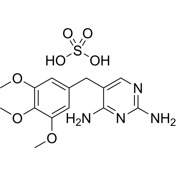
Trimethoprim sulfate is a bacteriostatic antibiotic and an orally active dihydrofolate reductase inhibitor. Trimethoprim sulfate is active against a wide range of Gram-positive and Gram-negative aerobic bacteria. Trimethoprim sulfate has the potential for the research of urinary tract infections, Shigellosis and Pneumocystis pneumonia. Trimethoprim sulfate can inhibit infection of Influenza A virus in chick embryo when combinated with zinc.
For research use only. We do not sell to patients.
- CAS No.: 56585-33-2
- Formula: C14H20N4O7S
- Molecular Weight:388.40
-
Storage:
Please store the product under the recommended conditions in the Certificate of Analysis.

To place orders, for customer services and technical support, please contact: MedChemExpress USA
Tel: 609-228-6898 E-mail: [email protected] [email protected]


-
 Biological Activity
-
Chemical Information
- Protocols
- Purity & Documentation
- References
-
 Help & FAQs


Publications Citing Use of MedChemExpress (MCE) Trimethoprim sulfate
More- Autophagy. 2023 Oct;19(10):2702-2718. [Abstract]
- Water Res. 2023 Jul 15:240:120110. [Abstract]
- J Exp Med. 2026 Mar 2;223(3):e20241287. [Abstract]
- Food Chem X. 2026 Feb 8:34:103646. [Abstract]
- Virulence. 2026 Dec 31;17(1):2646808. [Abstract]
- J Mol Med (Berl). 2019 Aug;97(8):1183-1193. [Abstract]
- Microorganisms. 2025 Apr 18;13(4):938. [Abstract]
- Infect Drug Resist. 2026 May 8;19.
- bioRxiv. 2026 Apr 29.
- Res Sq. 2026 Mar 9.
- bioRxiv. 2025 January 15.
- bioRxiv. 2024 May 10.
- Chemosphere. 2019 Jun:225:378-387. [Abstract]
 Customer Validation & Images

-
Bio/Physico-chemical Assay
All Antibiotic Isoforms
More

Biological Activity
Description
IC50 & Target
In Vitro
Trimethoprim interrupts folate metabolism by inhibition of the activity of dihydrofolase reductase (DHFR), which reduces dihydrofolate to tetrahydrofolate (THF)[1].
Trimethoprim (3 μg/mL; 1 h) induces protein aggregation and main heat shock proteins (Hsps) in E. coli cells, which indicates that Trimethoprim sulfate presence leads to protein misfolding[1].
Trimethoprim (1.5-3 μg/mL; 1 h) causes induction of DnaK, DnaJ, GroEL, ClpB, and IbpA/B Hsps in E. coli cells exposed to folate and heat stress[1].
MedChemExpress (MCE) has not independently confirmed the accuracy of these methods. They are for reference only. Further protocols information, click here.
In Vivo
Trimethoprim can be connected with the thiomaltose (TM-TMP) and shows stability with a half-life of about 1 hour in complete serum, and has an MIC value around 1 μM against E. coli[2].
Trimethoprim (10 mg/mL; 0.5 mL; inject with Trimethoprim-Zn combined suspension) decreases the virus titer and increases the survival rate of chicken embryo[4].
MedChemExpress (MCE) has not independently confirmed the accuracy of these methods. They are for reference only.
-
Animal Model:Female C3H/HeOuJ mice (transurethrally infected with a 50 μL suspension containing 1-2×10⁷ CFU of E. coli under 3% isoflurane)[2]
-
Dosage:10 mg/kg
-
Administration:i.v.; once every 12 h; for 3 d
-
Result:Showed antibacterial activity against H. influenzae, S. pneumoniae, E. coli and N. meningitidis with CD50s of 150 mg/kg, 335 mg/kg, 27.5 mg/kg and 8.4 mg/kg, respectively in infected mice.
-
Animal Model:Fertilized eggs (injected H3N2 virus into amniotic and allantoic space at day 8)[4]
-
Dosage:10 mg/mL; 0.5 mL
-
Administration:The Trimethoprim-Zn combined suspension was injected into the air sac; single dosage
-
Result:Decreased the virus titer and increased the survival rate of chicken embryo.
The survival rate peaked at ratio about 0.18 (Zn/Trimethoprim).


Chemical Information
-
CAS No. 56585-33-2
-
Molecular Weight 388.40
-
Formula C14H20N4O7S
-
SMILES
NC1=NC(N)=NC=C1CC2=CC(OC)=C(OC)C(OC)=C2.O=S(O)(O)=O
-
Shipping
Room temperature in continental US; may vary elsewhere.
-
Storage
Please store the product under the recommended conditions in the Certificate of Analysis.


Publications (13)
-
Journal Impact Factor
-
Most Recent
-
Autophagy
2023 Oct;19(10):2702-2718. PMID: 37312409 -
Water Res
Quantifying community-wide antibiotic usage via urban water fingerprinting: Focus on contrasting resource settings in South Africa. [Abstract]2023 Jul 15:240:120110. PMID: 37247434 -
J Exp Med
2026 Mar 2;223(3):e20241287. PMID: 41400657 -
Food Chem X
Machine learning prioritization of antibiotic residues in aquatic foods reveals exposure-driven genotoxic risk mediated by BCL2. [Abstract]2026 Feb 8:34:103646. PMID: 41756597 -
Virulence
Antibacterial efficacy and mechanism of the novel antimicrobial peptide lachnospirin-1 against Acinetobacter baumannii. [Abstract]2026 Dec 31;17(1):2646808. PMID: 41838520 -
J Mol Med (Berl)
2019 Aug;97(8):1183-1193. PMID: 31201471 -
Microorganisms
Isolation, Antimicrobial Susceptibility, and Genotypes of Three Pasteurellaeae Species Prevalent on Pig Farms in China Between 2021 and 2023. [Abstract]2025 Apr 18;13(4):938. PMID: 40284774 -
-
-
-
Trimethoprim sulfate purchased from MedChemExpress. Usage Cited in: bioRxiv. 2025 January 15.
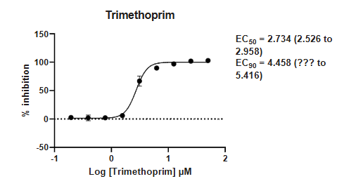
Inhibitory effect of Trimethoprim on Babesia.
-
-
Chemosphere
Mass-balance-model-based evaluation of sewage treatment plant contribution to residual pharmaceuticals in environmental waters. [Abstract]2019 Jun:225:378-387. PMID: 30884299

Protocols
-
Research Protocol for Infectious Diseases
Infectious-disease experiments test how pathogens interact with host barriers, innate immune receptors, inflammatory signaling, pathogen replication, and tissue injury; pattern-recognition receptors such as TLRs, RIG-I-like receptors, NOD-like receptors, and inflammasomes detect microbial molecules and activate NF-κB, interferon, and cytokine responses. The central hypothesis is that infection severity reflects the balance between pathogen burden and host response: protective inflammation restricts pathogen growth, whereas excessive or mislocalized inflammation contributes to tissue damage and disease phenotype. Unresolved questions include which host pathways are protective versus pathogenic, why some infection models fail to translate to human disease, and which combined readouts best predict clinically relevant infection outcomes.
-
Aerobic Bacterial Batch Culture on Broth/Agar
Aerobic bacterial batch culture grows a closed inoculated population in broth or on agar without continuous medium replacement; growth readouts include turbidity/OD for total suspended biomass and colony-forming units for viable cells able to form colonies on agar. OD-based growth curves reflect light scattering by cells, but OD is instrument-, pathlength-, species-, cell-size-, and density-dependent, so OD should be calibrated or interpreted alongside viable counts when quantitative cell density is required.
-
Gram Staining of Tissue Sections
Gram staining of tissue sections is a histochemical technique used to differentiate Gram-positive and Gram-negative bacteria within histological specimens based on differences in bacterial cell wall structure and dye retention, adapted from classical bacteriological Gram staining into tissue-compatible “histological Gram stain” variants. In tissue applications, modifications of the Brown-Hopps and Brown-Brenn methods are commonly used to improve differentiation of microorganisms embedded within host connective tissue and to reduce overstaining or loss of Gram-negative signal, which are known limitations of earlier approaches. The principle relies on crystal violet-iodine complex retention in Gram-positive organisms and subsequent decolorization and counterstaining steps that allow contrast visualization of Gram-negative organisms against tissue background.


Purity & Documentation
References
[1]. Laskowska, E., et al., Trimethoprim sulfate induces heat shock proteins and protein aggregation in E. coli cells. Curr Microbiol, 2003. 47(4): p. 286-9. [Content Brief]
[2]. Xiaojian Wang, et al. A Trimethoprim sulfate Conjugate of Thiomaltose Has Enhanced Antibacterial Efficacy In Vivo. Bioconjug Chem. 2018 May 16;29(5):1729-1735. [Content Brief]
[3]. Brogden, R.N., et al., Trimethoprim sulfate: a review of its antibacterial activity, pharmacokinetics and therapeutic use in urinary tract infections. Drugs, 1982. 23(6): p. 405-30. [Content Brief]
[4]. El Habbal MH. Combination therapy of zinc and trimethoprim inhibits infection of influenza A virus in chick embryo. Virol J. 2021 Jun 3;18(1):113. [Content Brief]

Calculators
Concentration (start) × Volume (start) = Concentration (final) × Volume (final)


Trimethoprim sulfate Related Classifications
HY-B0510A Related Classifications
 Powered by Bioz