


Hyaluronidase, Bovine testes
Based on 19 publication(s) in Google Scholar
Hyaluronidase, Bovine testes (Hyaluronate 4-glycanohydrolase; Hyaluronoglucosaminidase) is an endoglycosidase that depolymerizes Hyaluronic acid (HA) (HY-B0633A) by cleavage of glycosidic bonds. Hyaluronidase degrades HA and activates membrane receptors that trigger pathways converging in NF-κB activation. Hyaluronidase is employed in the research of granulomatous foreign body reactions, soft-tissue necrosis caused by vascular compromise and uncomplicated nodules, overcorrection, inflamed nodules or tissue ischemia associated with HA filler injection.
For research use only. We do not sell to patients.
- CAS No.: 37326-33-3
- Molecular Weight:55000 (average)
-
Storage:
Please store the product under the recommended conditions in the Certificate of Analysis.

To place orders, for customer services and technical support, please contact: MedChemExpress USA
Tel: 609-228-6898 E-mail: [email protected] [email protected]


-
 Biological Activity
-
Chemical Information
-
Solvent & Solubility
- Purity & Documentation
- References
-
 Help & FAQs


Publications Citing Use of MedChemExpress (MCE) Hyaluronidase, Bovine testes
More- Mol Cancer. 2024 Jan 10;23(1):12. [Abstract]
- Adv Mater. 2025 Nov 28:e18378. [Abstract]
- Cancer Res. 2025 Aug 1;85(15):2805-2819. [Abstract]
- Nat Commun. 2023 May 26;14(1):3050. [Abstract]
- Theranostics. 2023 Mar 27;13(6):2040-2056. [Abstract]
- Adv Sci (Weinh). 2025 Apr 3:e2412356. [Abstract]
- J Control Release. 2026 Jun 10:394:114858. [Abstract]
- Cell Death Dis. 2025 Mar 20;16(1):190. [Abstract]
- Mater Today Bio. 2026 Mar 31:38:103086. [Abstract]
- Cell Biosci. 2023 Aug 10;13(1):147. [Abstract]
- J Mol Cell Biol. 2026 Jun 25:mjag019. [Abstract]
- Int J Mol Sci. 2025 Nov 27;26(23):11475. [Abstract]
- Cell Oncol (Dordr). 2024 Dec;47(6):2439-2460. [Abstract]
- Reproduction. 2026 Jan 15;171(1):xaaf013. [Abstract]
- Animals (Basel). 2025 Nov 16;15(22):3304. [Abstract]
- Oral Dis. 2026 Apr 15. [Abstract]
- Drug Dev Ind Pharm. 2023 Feb;49(2):189-206. [Abstract]
- Vet Med Int. 2026 May 21:2026:6119294. [Abstract]
- Preprints. 2025 Nov 26.
 Customer Validation & Images

-
IHC
-
In Vivo Efficacy Study
-
In Vivo Efficacy Study


Biological Activity
1. Hyaluronidase inhibition assay[3]
(1) Mix 10 μL of 8 mg/mL Hyaluronidase with the sample in a buffer solution (0.1 M acetate buffer, pH 3.6) in a test tube, and incubate at 37 °C for 20 minutes.
(2) Add 20 μL of 12.5 mM calcium chloride to the mixture and incubate at 37 °C for another 20 minutes.
(3) Treat the activated Ca2+ mixture with 50 μL of 2.4 μg/mL hyaluronic acid in the buffer, and incubate further at 37 °C for 40 minutes.
(4) Add 2 μL of 0.4 N sodium hydroxide and 20 μL of 0.4 N potassium tetraborate tetrahydrate, then incubate in a water bath at 100 °C for 3 minutes.
(5) After cooling to room temperature, add 600 μL of DMAB solution to the mixture and incubate at 37 °C for 20 minutes.
(6) Measure the absorbance at 585 nm using an enzyme-linked immunosorbent assay (ELISA) reader.
MedChemExpress (MCE) has not independently confirmed the accuracy of these methods. They are for reference only.
(1) Anesthetize 10 rabbits weighing 3-4 kg using intramuscular injections of ketamine (50 mg/kg) and xylazine (10 mg/kg).
(2) Shave the hair off the ear, then use electrocoagulation to seal the blood vessels behind the ear. Inject 1% lidocaine (HY-B0185) to numb the skin near the ear, and make an incision to expose the anterior branch of the posterior auricular artery.
(3) Under the surgical microscope, a 30-gauge needle was used to puncture the artery, and 0.25 mL of HA filler was injected. The small initial amount of HA filler injected into the artery can expand the arterial lumen, helping to position the needle within the artery.
(4) Inject HA filler into both ears, with one ear receiving a subcutaneous injection of 750 IU of Hyaluronidase. Five rabbits (4-hour intervention group) were injected with Hyaluronidase 4 hours after HA injection, while another five rabbits (24-hour intervention group) received Hyaluronidase 24 hours after the HA injection.
(5) Capture the progression of skin changes and measure the final necrotic areas. Use the Statistical Package for Social Sciences (SPSS) software for statistical analysis.
2. The effect of hyaluronidase on the anti-tumor activity based on nanoparticles [8]
(1) This experiment used female Balb/c mice. 4T1 cells were suspended in PBS at a density of 1×10⁶/50μL and were injected subcutaneously into the back of the mice either unilaterally or bilaterally.
(2) When the tumor volume reaches 110-150 mm3, in vivo experiments are conducted. Mice are randomly divided into 4 groups (n=5): saline injection group, Hyaluronidase injection group, NM-Ce6 induced PDT group, and Hyaluronidase + NM-Ce6 induced PDT group. In the Hyaluronidase injection group, 1500 U of Hyaluronidase is dissolved in 50 µL PBS and injected into each tumor. One hour after the Hyaluronidase injection, each mouse receives an intravenous injection of 200 µL NM-Ce6, with a Ce6 concentration of 1 mg/mL (Ce6 dose = 10 mg/kg). Twenty-four hours after the NM-Ce6 injection, the tumors are exposed to 660 nm light for 60 minutes at a power density of 2 mW/cm2 to perform PDT.
(3) On the 6th day after the initial treatment, repeat the same procedure. Record tumor volume and mouse weight for a total of 12 days. After the treatment, remove 4 tumors from each group and weigh them, and remove the main organs from each group for H&E staining.
MedChemExpress (MCE) has not independently confirmed the accuracy of these methods. They are for reference only.


Chemical Information
-
CAS No. 37326-33-3
-
Appearance Solid
-
Molecular Weight 55000 (average)
-
Color White to light yellow
-
SMILES
[Hyaluronidase, Bovine testes]
-
Synonyms
Hyaluronate 4-glycanohydrolase, Bovine testes; Hyaluronoglucosaminidase, Bovine testes
-
Shipping
Room temperature in continental US; may vary elsewhere.
-
Storage
Please store the product under the recommended conditions in the Certificate of Analysis.


Publications (19)
-
Journal Impact Factor
-
Most Recent
-
Mol Cancer
Organoids derived from patients provide a new opportunity for research and individualized treatment of malignant peritoneal mesothelioma. [Abstract]2024 Jan 10;23(1):12. PMID: 38200517 -
Adv Mater
Disrupting Complement-Inflammation Positive Feedback Circuit via Oligonucleotide Hydrogel Microspheres for Reversing Joint Inflammation. [Abstract]2025 Nov 28:e18378. PMID: 41312643 -
Cancer Res
Pancreatic Neuroendocrine Tumors Secrete Apolipoprotein E to Induce Tip Endothelial Cells That Remodel the Tumor-Stroma Ratio and Promote Cancer Progression. [Abstract]2025 Aug 1;85(15):2805-2819. PMID: 40623046
Hyaluronidase, Bovine testes purchased from MedChemExpress. Usage Cited in: Cancer Res. 2025 Aug 1;85(15):2805-2819. [Abstract]
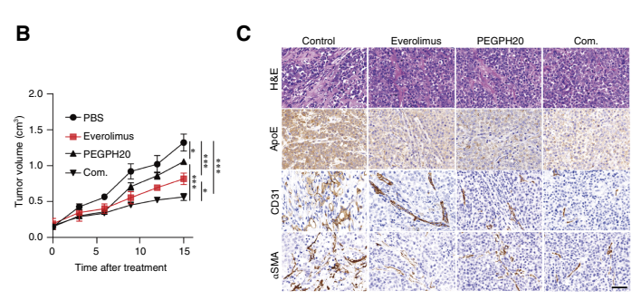
B, Growth curves of PDX mice treated with PBS, everolimus, PEGPH20 (Hyaluronidase), and everolimus combined with PEGPH20 (n = 5 per group). C, H&E staining and IHC were used to demonstrate the effects of different drug administration strategies on tumor stroma and CD31+ cell and αSMA+ cell number
Hyaluronidase, Bovine testes purchased from MedChemExpress. Usage Cited in: Cancer Res. 2025 Aug 1;85(15):2805-2819. [Abstract]
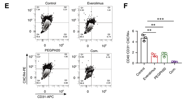
E and F, Flow cytometry analysis was used to demonstrate the effects of different drug administration strategies on the proportion of TipECs. Hyaluronidase (PEGPH20)
Hyaluronidase, Bovine testes purchased from MedChemExpress. Usage Cited in: Cancer Res. 2025 Aug 1;85(15):2805-2819. [Abstract]
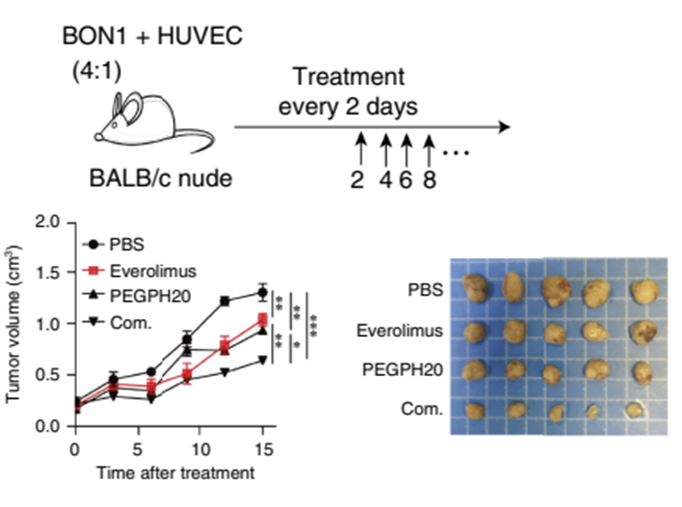
The effects of different drug administration strategies in the BON1+ HUVEC (4:1) model. Hyaluronidase (PEGPH20)
-
Nat Commun
TRABID inhibition activates cGAS/STING-mediated anti-tumor immunity through mitosis and autophagy dysregulation. [Abstract]2023 May 26;14(1):3050. PMID: 37237031 -
Theranostics
Platelets promote CRC by activating the C5a/C5aR1 axis via PSGL-1/JNK/STAT1 signaling in tumor-associated macrophages. [Abstract]2023 Mar 27;13(6):2040-2056. PMID: 37064877 -
Adv Sci (Weinh)
Screening and Identification of Novel DNA Aptamer for Targeted Delivery to Injured Podocytes in Glomerular Diseases. [Abstract]2025 Apr 3:e2412356. PMID: 40178289 -
J Control Release
Hydrogel-immobilized coacervate droplets enable microenvironment-responsive sustained drug release to accelerate diabetic foot ulcers healing. [Abstract]2026 Jun 10:394:114858. PMID: 41881075 -
Cell Death Dis
The role of vitamin K and its antagonist in the process of ferroptosis-damaged RPE-mediated CNV. [Abstract]2025 Mar 20;16(1):190. PMID: 40108164 -
Mater Today Bio
Mannosylated graphene oxide nanotherapeutics co-delivering docetaxel and a STING agonist reprogram myeloid cells and potentiate antitumor immunity. [Abstract]2026 Mar 31:38:103086. PMID: 42006712 -
Cell Biosci
Tumor-derived interleukin-1 receptor antagonist exhibits immunosuppressive functions and promotes pancreatic cancer. [Abstract]2023 Aug 10;13(1):147. PMID: 37563620 -
J Mol Cell Biol
Microtubule-associated CCDC112 is essential for spermiogenesis and male fertility in mice. [Abstract]2026 Jun 25:mjag019. PMID: 42348320 -
Int J Mol Sci
Melatonin Rescues Heat Stress-Induced Suppression of TCA Cycle and Mitochondrial Damage in Goat Sertoli Cells. [Abstract]2025 Nov 27;26(23):11475. PMID: 41373630 -
Cell Oncol (Dordr)
GALNT6 drives lenvatinib resistance in hepatocellular carcinoma through autophagy and cancer-associated fibroblast activation. [Abstract]2024 Dec;47(6):2439-2460. PMID: 39718738 -
Reproduction
2026 Jan 15;171(1):xaaf013. PMID: 41575147 -
Animals (Basel)
Optimization of Hydrogen Peroxide Concentrations for Inducing Oxidative Stress in Bovine Oocytes Prior to In Vitro Maturation. [Abstract]2025 Nov 16;15(22):3304. PMID: 41302012 -
Oral Dis
2026 Apr 15. PMID: 41986910 -
Drug Dev Ind Pharm
Multiple stimulus-response berberine plus baicalin micelles with particle size-charge-release triple variable properties for breast cancer therapy. [Abstract]2023 Feb;49(2):189-206. PMID: 36971392 -
Vet Med Int
Sericin Supplementation Prior to and During In Vitro Maturation Mitigates H2O2-Induced Oxidative Stress in Bovine Oocytes. [Abstract]2026 May 21:2026:6119294. PMID: 42180161 -


Solvent & Solubility
H2O : 100 mg/mL (Need ultrasonic)


Purity & Documentation
-
Data Sheet (275 KB)
-
SDS (419 KB)
- English - EN (419 KB)
- Français - FR (419 KB)
- Deutsch - DE (419 KB)
- Norwegian - NO (419 KB)
- Español - ES (419 KB)
- Swedish - SV (419 KB)
- Italian - IT (419 KB)
- Korean - KR (419 KB)
- Portuguese - PT (419 KB)
-
Handling Instructions (2659 KB)
References
[1]. Vartanian AJ, et al. Injected hyaluronidase reduces restylane-mediated cutaneous augmentation. Arch Facial Plast Surg. 2005 Jul-Aug;7(4):231-7. [Content Brief]
[3]. Apaza Ticona L, et al. Rubus urticifolius Compounds with Antioxidant Activity, and Inhibition Potential against Tyrosinase, Melanin, Hyaluronidase, Elastase, and Collagenase[J]. Pharmaceuticals (Basel). 2024 Jul 13;17(7):937. [Content Brief]
[4]. Jung H. Hyaluronidase: An overview of its properties, applications, and side effects[J]. Arch Plast Surg. 2020 Jul;47(4):297-300. [Content Brief]
[5]. Lee W, et al. Comparative Effectiveness of Different Interventions of Perivascular Hyaluronidase[J]. Plast Reconstr Surg. 2020 Apr;145(4):957-964. [Content Brief]
[6]. Wang M, et al. Comparison of Intra-arterial and Subcutaneous Testicular Hyaluronidase Injection Treatments and the Vascular Complications of Hyaluronic Acid Filler[J]. Dermatol Surg. 2017 Feb;43(2):246-254. [Content Brief]
[7]. Kim DW, et al. Vascular complications of hyaluronic acid fillers and the role of hyaluronidase in management. J Plast Reconstr Aesthet Surg. 2011 Dec;64(12):1590-5. [Content Brief]
[8]. Gong H, et al. Hyaluronidase To Enhance Nanoparticle-Based Photodynamic Tumor Therapy. Nano Lett. 2016 Apr 13;16(4):2512-21. [Content Brief]

Calculators
Concentration (start) × Volume (start) = Concentration (final) × Volume (final)


 Powered by Bioz