



KY386

Based on 1 Customer Validation
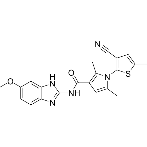
KY386 is a DHX33 helicase inhibitor with an IC50 of 0.019 μM. KY386 inhibits the cell viability of various cancer cells. KY386 induces ferroptosis in cancer cells, and induces apoptosis in some cancer cell lines. KY386 increases the intracellular levels of ROS, LPO and Fe2+, and decreases the level of GSH in cancer cells. KY386 inhibits the growth of gastric cancer and colon cancer xenografts in nude mice. KY386 is applicable to the related research on liver cancer, lung cancer, pancreatic cancer, colorectal cancer, gastric cancer, breast cancer, leukemia, renal cancer, prostate cancer, esophageal cancer, cervical cancer, brain cancer (glioblastoma) and melanoma.
For research use only. We do not sell to patients.
- Purity : 99.86%
- CAS No.: 2787598-01-8
- Formula: C21H19N5O2S
- Molecular Weight:405.47
-
Storage:Powder -20°C, 3 years ; In solvent -80°C, 6 months , -20°C, 1 month

To place orders, for customer services and technical support, please contact: MedChemExpress USA
Tel: 609-228-6898 E-mail: [email protected] [email protected]


-
 Biological Activity
-
Chemical Information
-
Solvent & Solubility
- Protocols
- Purity & Documentation
- References
-
 Help & FAQs


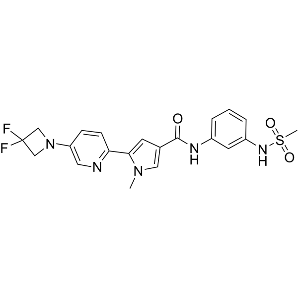
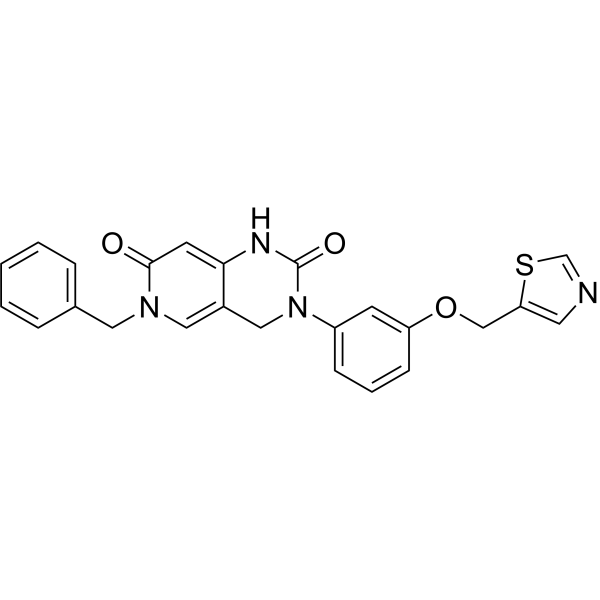
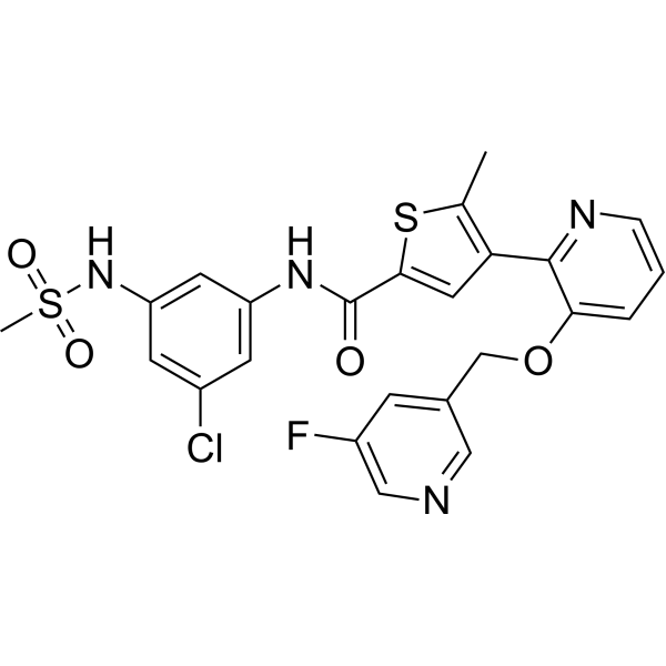
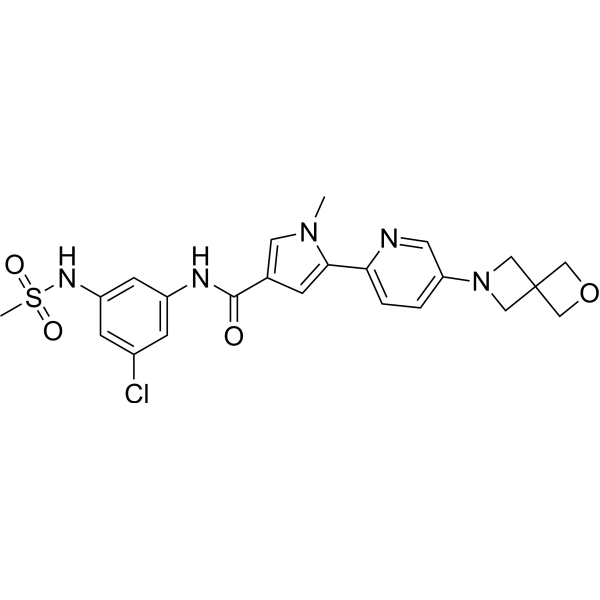
All DNA/RNA Synthesis Isoforms
More

Biological Activity
Description
IC50 & Target
|
Helicase |
Cellular Effect
|
Cell Line

|
Type | Value | Description | References |
|---|---|---|---|---|
| U-251 | IC50 |
0.02 μM
Compound: IV-a; KY386
|
Antiproliferative activity against human U-251MG cells overexpressing DHX33 assessed as inhibition of cell growth measured after 48 hrs by CCK-8 assay
Antiproliferative activity against human U-251MG cells overexpressing DHX33 assessed as inhibition of cell growth measured after 48 hrs by CCK-8 assay
|
[PMID: 37838340] |
In Vitro
KY386 (0-640 nM; 60 min) potently and selectively inhibits the helicase activity of recombinant wild-type DHX33 protein, with an IC50 of 0.019 μM[1].
KY386 (5 nM-20 μM; 48 h) potently inhibits the viability of U251-MG cancer cells overexpressing DHX33, with an IC50 of 0.02 μM[1].
KY386 (72 h) exhibits broad nanomolar anticancer activity against most of the DU145, HCT-116, HepG2, K-562, LoVo, MDA-MB-231, MIA PaCa-2, PC-3, SK-BR-3, NCI-H1299, NCI-H1975,
HCC1806, SNU-1, BT549, HCT-15, U-937, A-498, KYSE-150, A549, Daudi, Jurkat T, SNU-387, SW480, Caki-1, SNU-423, CALU-1, COLO 829, T84, U-87MG, NCI-N87, MeWo, HEEC, HGC27 and SGC7901 cells[2].
KY386 (72 h) inhibits the cell viability of human cancer cell lines A875, A375, T24, 5637, SGC7901, HGC27 and SNU668, with IC50 values ranging from 13 nM (A375) to 35 nM (A875)[2].
KY386 (0-500 nM; 24-72 h) potently induces dose- and time-dependent apoptosis in HuH-7 hepatocellular carcinoma cells and HCT116 colorectal cancer cells[2].
KY386 (0-50 nM; 4-24 h) induces dose- and time-dependent increases in intracellular ROS levels in a variety of human cancer cell lines, including HGC27, 5637, T24, A375, A875, DU145, PC3 and SNU668[2].
KY386 (0-40 nM; 8-16 h) induces a dose- and time-dependent decrease in intracellular GSH levels in HGC27 gastric cancer cells[2].
KY386 (0-60 nM; 8-16 h) induces dose- and time-dependent increases in intracellular LPO levels in HGC27, 5637, DU145 and A875 human cancer cells[2].
KY386 (0-60 nM; 16 h) induces a dose-dependent increase in intracellular Fe2+ levels in 5637, HGC27, DU145, PC3 and SNU668 human cancer cells[2].
KY386 (0-50 nM; 24 h) reduces the protein expression of lipid metabolism-related factors FADS1, FADS2 and SCD1 in human cancer cell lines HGC27, 5637, A875, PC-3 and SNU668, while exerting minimal effects on ferroptosis-related factors GPX4, SLC3A2 and SLC7A11[2].
KY386 (0-40 nM; 0-8 h) induces dose-dependent downregulation of FADS1, FADS2 and SCD1 mRNA levels in human cancer cell lines A875, A375, HGC27, T24 and DU145[2].
MedChemExpress (MCE) has not independently confirmed the accuracy of these methods. They are for reference only.
-
Cell Line:U251-MG DHX33-overexpressing glioblastoma cancer cells
-
Concentration:5 nM, 10 nM, 25 nM, 50 nM, 100 nM, 250
nM, 500 nM, 1000 nM, 2000 nM, 5000 nM, 10 μM or 20 μM -
Incubation Time:48 h
-
Result:Exhibited potent cytotoxicity against U251-MG cells, with an IC50 of 0.02 μM (20 nM).
Demonstrated moderate metabolic stability with an intrinsic clearance (CLint) of 22.4 mL/min/mg in human liver microsomes.
-
Cell Line:HuH-7, HCT116, HGC27, and DU145 human cancer cell lines
-
Concentration:20 nM, 100 nM, 500 nM (HuH-7, HCT116); 50 nM, 100 nM, 500 nM (HGC27, DU145, 24 h); 100 nM (HGC27, DU145, 48 h)
-
Incubation Time:48 h, 72 h (HuH-7, HCT116); 24 h, 48 h (HGC27, DU145)
-
Result:Induced apoptosis in a dose- and time-dependent manner in HuH-7 and HCT116 cells.
Did not effectively induce apoptosis in HGC27 and DU145 cells, even with increased dose or treatment time.
-
Cell Line:HGC27, 5637, A875, PC-3, and SNU668 human cancer cell lines
-
Concentration:20 nM, 40 nM (HGC27, SNU668); 10 nM, 20 nM, 40 nM (5637); 30 nM, 40 nM (A875); 30 nM, 50 nM (PC-3)
-
Incubation Time:24 h
-
Result:Reduced protein expression of FADS1, FADS2, and SCD1 in all tested cancer cell lines.
Had limited effect on GPX4, SLC3A2, and SLC7A11 expression.
-
Cell Line:A875, A375, HGC27, T24, and DU145 human cancer cell lines
-
Concentration:40 nM (A875, A375, HGC27, DU145); 20 nM, 40 nM (T24, DU145)
-
Incubation Time:4 h, 6 h, 8 h (A875, A375, HGC27, DU145); 6 h (T24, DU145)
-
Result:Significantly reduced mRNA levels of FADS1, FADS2, and SCD1 in a time- and dose-dependent manner in all tested cancer cell lines, with statistically significant decreases observed at all specified time points and doses.
In Vivo
KY386 (15-35 mg/kg on days 0-8, 25-45 mg/kg on days 9-20; i.p.; 2 times daily for 2 consecutive days + 3 times daily for 1 consecutive day; total duration of 21 days) significantly inhibits the growth of RASG12D-mutated colon cancer patient-derived xenografts in nude mice[2].
MedChemExpress (MCE) has not independently confirmed the accuracy of these methods. They are for reference only.
-
Animal Model:Nude mice (female, 5-week old)[2]
-
Dosage:25 mg/kg; 35 mg/kg; 45 mg/kg
-
Administration:i.p.; BID×2 + TID×1; 7 days
-
Result:Inhibited tumor growth in a dose-dependent manner.
Reduced mean tumor volumes compared to vehicle controls at 21 days post-treatment, with the 45 mg/kg dose showing the greatest inhibition.
Caused no significant reduction in mouse body weight, indicating minimal toxicity.
-
Animal Model:Nude mice (female, 8-week old)[2]
-
Dosage:15 mg/kg (Days 0-8) then 25 mg/kg (Days 9-20); 25 mg/kg (Days 0-8) then 35 mg/kg (Days 9-20); 35 mg/kg (Days 0-8) then 45 mg/kg (Days 9-20)
-
Administration:i.p.; BID×2 + TID×1; 21 days
-
Result:Significantly inhibited tumor growth in the RAS G12D mutant colon cancer PDX model.
Reduced mean tumor volumes compared to vehicle controls at 21 days post-treatment.
Caused no significant reduction in mouse body weight, indicating minimal toxicity.
- Mammalian live/dead viability and cytotoxicity staining
- Annexin V plus membrane-impermeant dye apoptosis staining
- ROS/oxidative-stress fluorescent staining
- CCK-8/WST-8 Cell Proliferation Assay
- Liver Cancer Modeling
- Breast Cancer Modeling
- Cell Viability Determination by MTT Colorimetric Assay
- Detection of Apoptosis
- TUNEL staining for apoptotic DNA fragmentation
- Cell Cytotoxicity Assay
- Apoptosis Solutions
- Ferroptosis Solutions
- MTT Cell Proliferation Assay


Chemical Information
-
CAS No. 2787598-01-8
-
Appearance Solid
-
Molecular Weight 405.47
-
Formula C21H19N5O2S
-
Color White to off-white
-
SMILES
COC1=CC=C2N=C(NC2=C1)NC(C3=C(N(C(C)=C3)C4=C(C=C(S4)C)C#N)C)=O
-
Shipping
Room temperature in continental US; may vary elsewhere.
-
Storage
Powder -20°C 3 years In solvent -80°C 6 months -20°C 1 month


Solvent & Solubility
In Vitro:
DMSO : 100 mg/mL (246.63 mM; Need ultrasonic; Hygroscopic DMSO has a significant impact on the solubility of product, please use newly opened DMSO)
Please refer to the solubility information to select the appropriate solvent. Once prepared, please aliquot and store the solution to prevent product inactivation from repeated freeze-thaw cycles.
Storage method and period of stock solution: -80°C, 6 months; -20°C, 1 month. When stored at -80°C, please use it within 6 months. When stored at -20°C, please use it within 1 month.
Please refer to the solubility information to select the appropriate solvent. Once prepared, please aliquot and store the solution to prevent product inactivation from repeated freeze-thaw cycles.
Storage method and period of stock solution: -80°C, 6 months; -20°C, 1 month. When stored at -80°C, please use it within 6 months. When stored at -20°C, please use it within 1 month.
Concentration (start) × Volume (start) = Concentration (final) × Volume (final)
In Vivo:
Select the appropriate dissolution method based on your experimental animal and administration route.
- For the following dissolution methods, please ensure to first prepare a clear stock solution using an In Vitro approach and then sequentially add co-solvents:
- To ensure reliable experimental results, the clarified stock solution can be appropriately stored based on storage conditions. As for the working solution for In Vivo experiments, it is recommended to prepare freshly and use it on the same day.
- The percentages shown for the solvents indicate their volumetric ratio in the final prepared solution. If precipitation or phase separation occurs during preparation, heat and/or sonication can be used to aid dissolution.
In Vivo Dissolution Calculator
Please enter the basic information of animal experiments:
-
-
-
-
Recommended: Prepare an additional quantity of animals to account for potential losses during experiments.
Please enter your animal formula composition:
-
%DMSO +
Recommended: Keep the proportion of DMSO in working solution below 2% if your animal is weak.
-
%+
-
+%Tween-80 + +
-
%Saline +
The co-solvents required include: DMSO, . All of co-solvents are available by MedChemExpress (MCE). , Tween 80. All of co-solvents are available by MedChemExpress (MCE).
Working solution concentration: 0.22 mg/mL
Method for preparing stock solution: mg drug dissolved in μL DMSO. Stock solution concentration: mg/mL.
1. Take μL DMSO stock solution;
2. Add μL .
μL , mix evenly;
3. Then add μL Tween 80, mix evenly;
4. Then add μL
Please ensure that the stock solution in the first step is dissolved to a clear state, and add co-solvents in sequence. You can use ultrasonic heating (ultrasonic cleaner, recommended frequency 20-40 kHz), vortexing, etc. to assist dissolution.


Purity & Documentation
-
Data Sheet (283 KB)
-
SDS (394 KB)
- English - EN (394 KB)
- Français - FR (394 KB)
- Deutsch - DE (394 KB)
- Norwegian - NO (394 KB)
- Español - ES (394 KB)
- Swedish - SV (394 KB)
- Italian - IT (394 KB)
- Korean - KR (394 KB)
- Portuguese - PT (394 KB)
-
Handling Instructions (2659 KB)
References
[1]. Wang Y, et al. Development of small molecule inhibitors targeting RNA helicase DHX33 as anti-cancer agents. Bioorg Med Chem Lett. 2023;96:129505. [Content Brief]
[2]. Tang X, et al. An RNA Helicase DHX33 Inhibitor Shows Broad Anticancer Activity via Inducing Ferroptosis in Cancer Cells. ACS Omega. 2024;9(26):28372-28384. Published 2024 Jun 17. [Content Brief]


Complete Stock Solution Preparation Table
Please refer to the solubility information to select the appropriate solvent. Once prepared, please aliquot and store the solution to prevent product inactivation from repeated freeze-thaw cycles.
Storage method and period of stock solution: -80°C, 6 months; -20°C, 1 month. When stored at -80°C, please use it within 6 months. When stored at -20°C, please use it within 1 month.
| Optional Solvent | Concentration Solvent Mass | 1 mg | 5 mg | 10 mg | 25 mg |
|---|---|---|---|---|---|
| DMSO | 1 mM | 2.4663 mL | 12.3314 mL | 24.6627 mL | 61.6568 mL |
| 5 mM | 0.4933 mL | 2.4663 mL | 4.9325 mL | 12.3314 mL | |
| 10 mM | 0.2466 mL | 1.2331 mL | 2.4663 mL | 6.1657 mL | |
| 15 mM | 0.1644 mL | 0.8221 mL | 1.6442 mL | 4.1105 mL | |
| 20 mM | 0.1233 mL | 0.6166 mL | 1.2331 mL | 3.0828 mL | |
| 25 mM | 0.0987 mL | 0.4933 mL | 0.9865 mL | 2.4663 mL | |
| 30 mM | 0.0822 mL | 0.4110 mL | 0.8221 mL | 2.0552 mL | |
| 40 mM | 0.0617 mL | 0.3083 mL | 0.6166 mL | 1.5414 mL | |
| 50 mM | 0.0493 mL | 0.2466 mL | 0.4933 mL | 1.2331 mL | |
| 60 mM | 0.0411 mL | 0.2055 mL | 0.4110 mL | 1.0276 mL | |
| 80 mM | 0.0308 mL | 0.1541 mL | 0.3083 mL | 0.7707 mL | |
| 100 mM | 0.0247 mL | 0.1233 mL | 0.2466 mL | 0.6166 mL |


KY386 Related Classifications
HY-156418 Related Classifications
 Powered by Bioz
Keywords
- KY386
- 2787598-01-8
- KY 386
- KY-386
- DNA/RNA Synthesis
- Ferroptosis
- Apoptosis
- Reactive Oxygen Species (ROS)
- DHX33 helicase inhibitor
- ferroptosis
- apoptosis
- U251-MG cells
- A875 cells
- A375 cells
- T24 cells
- 5637 cells
- SGC7901 cells
- HGC27 cells
- SNU668 cells
- liver cancer
- lung cancer
- pancreatic cancer
- colorectal cancer
- gastric cancer
- breast cancer
- leukemia
- renal cancer
- prostate cancer
- esophageal cancer
- cervical cancer
- brain cancer (glioblastoma)
- melanoma
- Nude mice
- Inhibitor
- inhibitor
- inhibit