



PRLX-93936

Based on 1 Customer Validation
PRLX-93936 is a molecular Glues that binds to and reprograms the TRIM21 ubiquitin ligase to degrade nuclear pore complexes. PRLX-93936 binds to TRIM21, forms a ternary complex with TRIM21 and NUP98, and mediates the ubiquitination and proteasomal degradation of NUP98 and other nuclear pore complex proteins. PRLX-93936 induces the loss of short-lived cytoplasmic mRNA transcripts, triggers cancer cell apoptosis (Apoptosis), and inhibits the activated Ras pathway. PRLX-93936 inhibits HIF-1 under hypoxic conditions (IC50 = 0.09 μM in cell-based reporter gene assay). PRLX-93936 suppresses tumor growth in mouse models and improves survival rates in mouse models of multiple myeloma. PRLX-93936 is applicable to research related to pancreatic cancer and multiple myeloma.
연구목적의 판매만을 진행합니다. 환자를 대상으로 한 판매는 하지 않습니다.
- Purity : 98.31%
- CAS No.: 903499-49-0
- 화학식: C21H24N4O2
- 분자량:364.44
-
보관:
4°C, sealed storage, away from moisture
* In solvent : -80°C, 6 months; -20°C, 1 month (sealed storage, away from moisture)


To place orders, for customer services and technical support, please contact: MedChemExpress USA
Tel: 609-228-6898 E-mail: [email protected] [email protected]


-
 Biological Activity
-
Chemical Information
-
용액&용해도
- Protocol
- 순도&문서
- References
-
 Help & FAQs


-
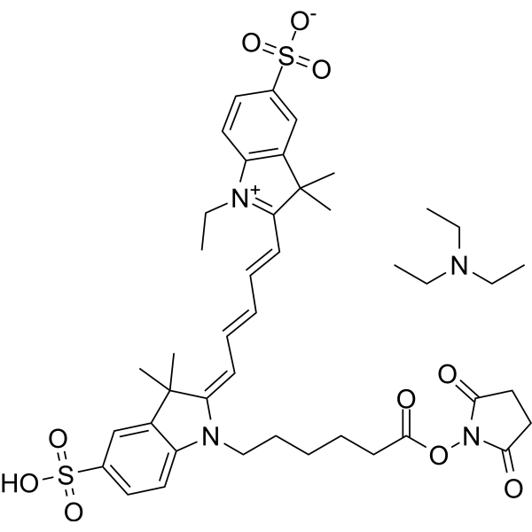
HY-D0819A
CY5-SE triethylamine saltFluorescent Probe -
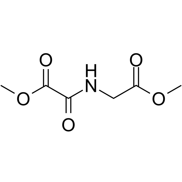
HY-15893
DMOGHIF-PH Inhibitor -
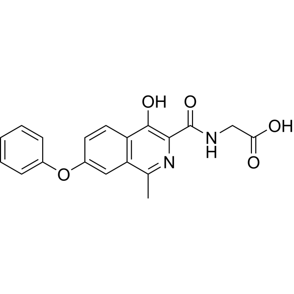
HY-13426
RoxadustatHIF-PH Inhibitor -
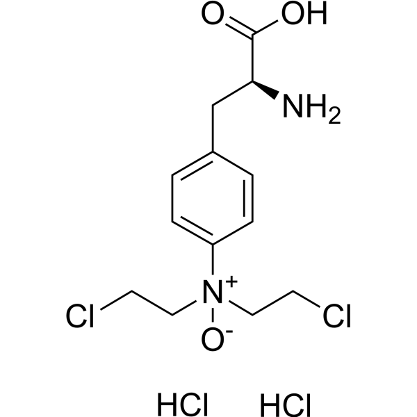
HY-10231
PX-478HIF-1α Inhibitor -
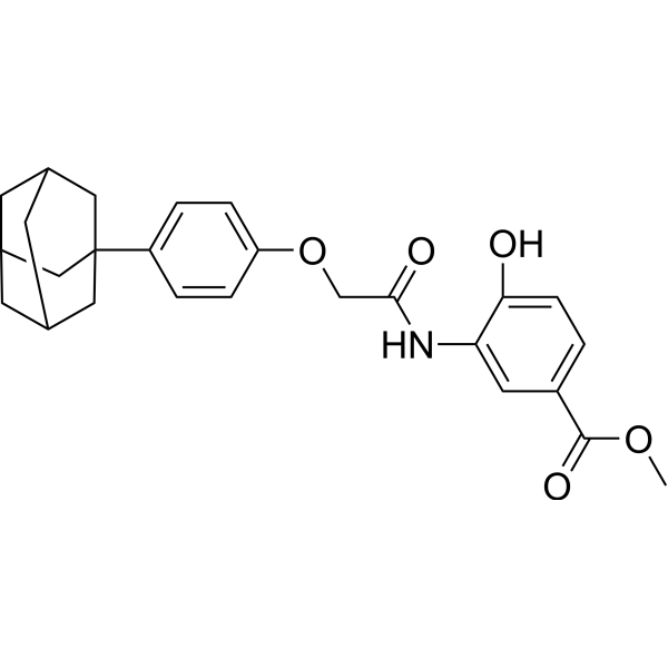
HY-13671
LW6Apoptosis Inducer -
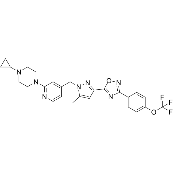
HY-15836
BAY 87-2243HIF-1 Inhibitor -

HY-15893R
DMOG (Standard)HIF-PH Inhibitor -
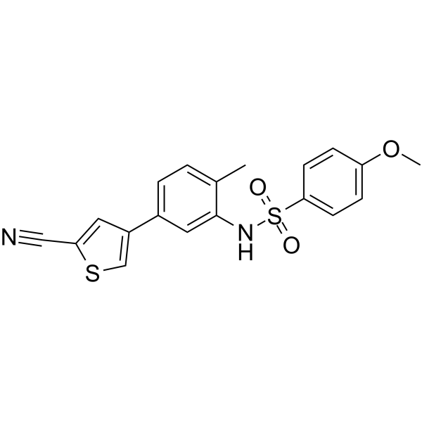
HY-16187
EL-102Hif1α Inhibitor -

HY-13426R
Roxadustat (Standard)HIF-PH Inhibitor -

HY-15893G
DMOG (GMP)HIF-1α Stabilizer -

HY-10231R
PX-478 (Standard)Reference Standard


Biological Activity
제품 설명
Cellular Effect
|
Cell Line

|
Type | Value | Description | References |
|---|---|---|---|---|
| ME-180 | IC50 |
0.09 μM
Compound: 16
|
Inhibition of hypoxia-induced HIF1alpha activation in human ME180 cells by HRE3-Bla-luciferase reporter gene assay
Inhibition of hypoxia-induced HIF1alpha activation in human ME180 cells by HRE3-Bla-luciferase reporter gene assay
|
[PMID: 21831635] |
In Vitro
PRLX-93936 (72 h) selectively inhibits the viability of human cancer cell lines, with the strongest activity observed in pancreatic and head and neck cancer cell lines, and reduces the survival rate of human cancer cell lines with high TRIM21 expression[1].
PRLX-93936 (500 nM; 30 min-10 h) induces TRIM21-dependent degradation of nuclear pore complex proteins and MCL1 in PANC-1 human pancreatic cancer cells[1].
PRLX-93936 (500 nM; 30 min) induces TRIM21-dependent ubiquitination of nuclear pore complex proteins in human pancreatic cancer cell line PANC-1 pretreated with MG132 (HY-13259)[1].
PRLX-93936 (cells are pretreated with 1 μM MG132 for 30 min, followed by treatment with 500 nM PRLX-93936 for 30 min or 2 h, and co-treated with 100 nM biotin for 30 min) induces the translocation of TRIM21 to the nuclear pore complex in human pancreatic cancer cell line PANC-1. Treatment with 500 nM PRLX-93936 for 30 min or 2 h results in close proximity between TRIM21 and nucleoporins such as NUP98[1].
PRLX-93936 (2 h) acts as a molecular glue that induces a direct, dose-dependent interaction between TRIM21 and NUP98 in HEK293T cells[1].
PRLX-93936 (for 5 days) potently reduces the viability of cholangiopancreatic organoids with high TRIM21 expression, and induces TRIM21-dependent degradation of nuclear pore complex proteins.
PRLX-93936 (1-10 μM) reduces HIF-1α protein accumulation under hypoxic conditions[3].
MedChemExpress (MCE) has not independently confirmed the accuracy of these methods. They are for reference only.
In Vivo
MedChemExpress (MCE) has not independently confirmed the accuracy of these methods. They are for reference only.
-
Animal Model:NOD.Cg-Prkdcscid Il2rgtm1Wjl/SzJ (female, 6 weeks old, subcutaneous xenograft with 5×10⁶ PANC-1 cells/HS766T cells/PANC-1 cells)[1]
-
Dosage:50, 100 mg/kg
-
Administration:i.p.; daily; 21 days/i.p.; two daily doses
-
Result:Reduced average tumor volume.
Depleted several nucleoporins (NUP98, NUP96, NUP35) in tumor tissue, confirming on-target degradation of nuclear pore complex components in vivo.
- RNA extraction experimental
- Dual Luciferin reporter gene assay
- Detection of Apoptosis
- TUNEL staining for apoptotic DNA fragmentation
- Annexin V plus membrane-impermeant dye apoptosis staining
- Patient-Derived Xenograft (PDX)
- Apoptosis Solutions
- Patient-Derived Orthotopic Xenograft (PDOX)
- Subcutaneous Cell-Line-Derived Xenograft
- Orthotopic Cell-Line Xenograft
- Directly Induced Neuron Culture
Clinical Trial
| NCT Number | Sponsor | Condition | Start Date |
Phase
|
|---|---|---|---|---|
| NCT01329991 | Plexxikon| | 2011-05 | PHASE1 |


Chemical Information
-
CAS No. 903499-49-0
-
Appearance Solid
-
분자량 364.44
-
화학식 C21H24N4O2
-
Color White to off-white
-
SMILES
O=C1N(C(CN2CCNCC2)=NC3=C1C=CC=C3)C4=CC=CC=C4OCC
-
선적
Room temperature in continental US; may vary elsewhere.
-
보관
4°C, sealed storage, away from moisture
* In solvent : -80°C, 6 months; -20°C, 1 month (sealed storage, away from moisture)


용액&용해도
In Vitro:
DMSO : ≥ 100 mg/mL (274.39 mM; Hygroscopic DMSO has a significant impact on the solubility of product, please use newly opened DMSO)
* "≥" means soluble, but saturation unknown.
Please refer to the solubility information to select the appropriate solvent. Once prepared, please aliquot and store the solution to prevent product inactivation from repeated freeze-thaw cycles.
Storage method and period of stock solution: -80°C, 6 months; -20°C, 1 month (sealed storage, away from moisture). When stored at -80°C, please use it within 6 months. When stored at -20°C, please use it within 1 month.
Please refer to the solubility information to select the appropriate solvent. Once prepared, please aliquot and store the solution to prevent product inactivation from repeated freeze-thaw cycles.
Storage method and period of stock solution: -80°C, 6 months; -20°C, 1 month (sealed storage, away from moisture). When stored at -80°C, please use it within 6 months. When stored at -20°C, please use it within 1 month.
Concentration (start) × Volume (start) = Concentration (final) × Volume (final)
In Vivo:
Select the appropriate dissolution method based on your experimental animal and administration route.
- For the following dissolution methods, please ensure to first prepare a clear stock solution using an In Vitro approach and then sequentially add co-solvents:
- To ensure reliable experimental results, the clarified stock solution can be appropriately stored based on storage conditions. As for the working solution for In Vivo experiments, it is recommended to prepare freshly and use it on the same day.
- The percentages shown for the solvents indicate their volumetric ratio in the final prepared solution. If precipitation or phase separation occurs during preparation, heat and/or sonication can be used to aid dissolution.
Add each solvent one by one: 10% DMSO 40% PEG300 5% Tween-80 45% Saline
Solubility: ≥ 2.5 mg/mL (6.86 mM); Clear solution
This protocol yields a clear solution of ≥ 2.5 mg/mL (saturation unknown).
Taking 1 mL working solution as an example, add 100 μL DMSO stock solution (25.0 mg/mL) to 400 μL PEG300, and mix evenly; then add 50 μL Tween-80 and mix evenly; then add 450 μL Saline to adjust the volume to 1 mL.
Preparation of Saline: Dissolve 0.9 g sodium chloride in ddH₂O and dilute to 100 mL to obtain a clear Saline solution.
Add each solvent one by one: 10% DMSO 90% (20% SBE-β-CD in Saline)
Solubility: ≥ 2.5 mg/mL (6.86 mM); Clear solution
This protocol yields a clear solution of ≥ 2.5 mg/mL (saturation unknown).
Taking 1 mL working solution as an example, add 100 μL DMSO stock solution (25.0 mg/mL) to 900 μL 20% SBE-β-CD in Saline, and mix evenly.
Preparation of 20% SBE-β-CD in Saline (4°C, storage for one week): 2 g SBE-β-CD powder is dissolved in 10 mL Saline, completely dissolve until clear.
In Vivo Dissolution Calculator
Please enter the basic information of animal experiments:
-
-
-
-
Recommended: Prepare an additional quantity of animals to account for potential losses during experiments.
Please enter your animal formula composition:
-
%DMSO +
Recommended: Keep the proportion of DMSO in working solution below 2% if your animal is weak.
-
%+
-
+%Tween-80 + +
-
%Saline +
The co-solvents required include: DMSO, . All of co-solvents are available by MedChemExpress (MCE). , Tween 80. All of co-solvents are available by MedChemExpress (MCE).
Working solution concentration: 0.22 mg/mL
Method for preparing stock solution: mg drug dissolved in μL DMSO. Stock solution concentration: mg/mL. * In solvent : -80°C, 6 months; -20°C, 1 month (sealed storage, away from moisture)
1. Take μL DMSO stock solution;
2. Add μL .
μL , mix evenly;
3. Then add μL Tween 80, mix evenly;
4. Then add μL
Please ensure that the stock solution in the first step is dissolved to a clear state, and add co-solvents in sequence. You can use ultrasonic heating (ultrasonic cleaner, recommended frequency 20-40 kHz), vortexing, etc. to assist dissolution.


순도&문서
-
Data Sheet (278 KB)
-
SDS (393 KB)
- English - EN (393 KB)
- Français - FR (393 KB)
- Deutsch - DE (393 KB)
- Norwegian - NO (393 KB)
- Español - ES (393 KB)
- Swedish - SV (393 KB)
- Italian - IT (393 KB)
- Korean - KR (393 KB)
- Portuguese - PT (393 KB)
-
Handling Instructions (2659 KB)
References
[1]. Yuan L, et al. Defining the Antitumor Mechanism of Action of a Clinical-stage Compound as a Selective Degrader of the Nuclear Pore Complex. Cancer Discov. 2025;15(12):2505-2529. [Content Brief]
[3]. Huang W, et al. Synthesis and evaluation of quinazolin-4-ones as hypoxia-inducible factor-1α inhibitors. Bioorg Med Chem Lett. 2011 Sep 15;21(18):5239-43. [Content Brief]


Complete Stock Solution Preparation Table
Please refer to the solubility information to select the appropriate solvent. Once prepared, please aliquot and store the solution to prevent product inactivation from repeated freeze-thaw cycles.
Storage method and period of stock solution: -80°C, 6 months; -20°C, 1 month (sealed storage, away from moisture). When stored at -80°C, please use it within 6 months. When stored at -20°C, please use it within 1 month.
| Optional Solvent | Concentration Solvent Mass | 1 mg | 5 mg | 10 mg | 25 mg |
|---|---|---|---|---|---|
| DMSO | 1 mM | 2.7439 mL | 13.7197 mL | 27.4394 mL | 68.5984 mL |
| 5 mM | 0.5488 mL | 2.7439 mL | 5.4879 mL | 13.7197 mL | |
| 10 mM | 0.2744 mL | 1.3720 mL | 2.7439 mL | 6.8598 mL | |
| 15 mM | 0.1829 mL | 0.9146 mL | 1.8293 mL | 4.5732 mL | |
| 20 mM | 0.1372 mL | 0.6860 mL | 1.3720 mL | 3.4299 mL | |
| 25 mM | 0.1098 mL | 0.5488 mL | 1.0976 mL | 2.7439 mL | |
| 30 mM | 0.0915 mL | 0.4573 mL | 0.9146 mL | 2.2866 mL | |
| 40 mM | 0.0686 mL | 0.3430 mL | 0.6860 mL | 1.7150 mL | |
| 50 mM | 0.0549 mL | 0.2744 mL | 0.5488 mL | 1.3720 mL | |
| 60 mM | 0.0457 mL | 0.2287 mL | 0.4573 mL | 1.1433 mL | |
| 80 mM | 0.0343 mL | 0.1715 mL | 0.3430 mL | 0.8575 mL | |
| 100 mM | 0.0274 mL | 0.1372 mL | 0.2744 mL | 0.6860 mL |


PRLX-93936 Related Classifications
HY-119264 Related Classifications
 Powered by Bioz