



Trastuzumab vedotin
Based on 3 publication(s) in Google Scholar
Trastuzumab vedotin (MRG002; Trastuzumab MMAE) is an antibody-drug conjugate and cytotoxin targeting HER2, with a Kd of 7.50E-11 M for human HER2. After binding to HER2, Trastuzumab vedotin undergoes internalization and lysosomal trafficking, delivering a cytotoxic payload to HER2-expressing cells and inducing tumor regression in in vivo xenograft models with HER2-expressing tumors. The anti-tumor activity of Trastuzumab vedotin is enhanced when used in combination with anti-PD-1 antibodies, and it exhibits preclinical anti-tumor activity in drug-resistant breast cancer, gastric cancer, and urothelial carcinoma PDX models. Trastuzumab vedotin has low antibody-dependent cellular cytotoxicity activity and can be used in studies related to HER2-positive breast cancer, HER2-positive gastric cancer, and unresectable locally advanced or metastatic HER2-positive urothelial carcinoma.
For research use only. We do not sell to patients.
- Purity : 98.05%
- Molecular Weight:150626 (average)
-
Storage:
-80°C, protect from light


To place orders, for customer services and technical support, please contact: MedChemExpress USA
Tel: 609-228-6898 E-mail: [email protected] [email protected]


-
 Biological Activity
-
Chemical Information
- Protocols
- Purity & Documentation
- References
-
 Help & FAQs


-

HY-P9907
TrastuzumabHER2 Blocking Antibody -

HY-138298A
Trastuzumab deruxtecanHER2 ADC -

HY-P9921
Trastuzumab emtansineAntibody-drug Conjugate -
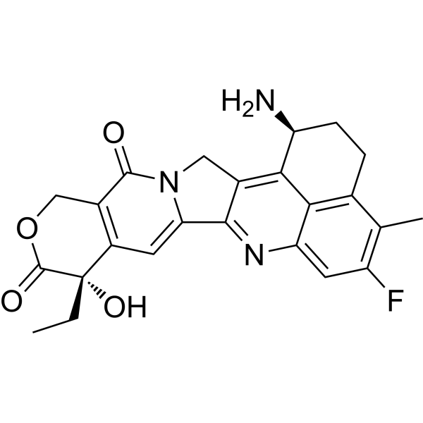
HY-13631
ExatecanTopoisomerase I Inhibitor -
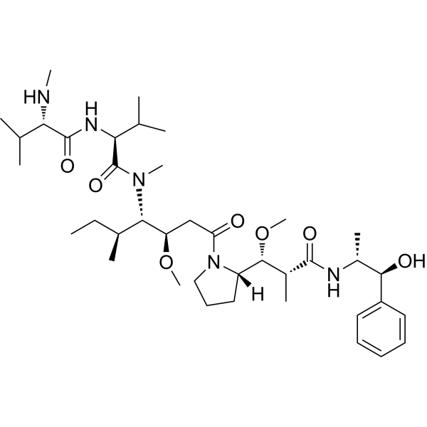
HY-15162
Monomethyl auristatin EAntimitotic Agent -

HY-132254
Sacituzumab govitecanTrop-2 ADC -

HY-10219
RapamycinmTOR Inhibitor -

HY-141601
Telisotuzumab vedotinAnti-c-MET ADC -

HY-P99001
Human IgG1 kappa, Isotype ControlIgG1κ Isotype Control -

HY-P9985
Disitamab vedotinHER2-Directed ADC -

HY-141598
Datopotamab deruxtecan (solution)TROP2-Targeted ADC -

HY-138298
Trastuzumab deruxtecan (solution)HER2 ADC
Publications Citing Use of MedChemExpress (MCE) Trastuzumab vedotin
MoreAll EGFR Isoforms
More

Biological Activity
Description
In Vitro
Trastuzumab vedotin (0.0128-1000 ng/mL; 60 min; RT) selectively binds to HER2 proteins of humans and monkeys, while no binding to HER2 proteins of rats or mice is observed[1].
Trastuzumab vedotin (4 μg/mL; 30 min, 37 °C) is efficiently internalized by human breast cancer SKBR3 cells at 37 °C, with an internalization rate comparable to that of its unconjugated antibody component MAB802[1].
Trastuzumab vedotin (4 μg; 4 h) exhibits no significant CDC activity against SKBR3 human breast cancer cells[1].
MedChemExpress (MCE) has not independently confirmed the accuracy of these methods. They are for reference only. Further protocols information, click here.
-
Cell Line:SKBR3 (high HER2), BT-474 (high HER2), MDA-MB-453 (medium HER2) human breast cancer cell lines; NCI-N87 (high HER2) human gastric cancer cell line
-
Concentration:0.02 ng/mL-200 μg/mL
-
Incubation Time:96 ± 2 h
-
Result:Displayed potent sub-nanomolar cytotoxicity across all tested cell lines, with IC50 values of 0.012 nM (SKBR3), 0.035 nM (BT-474), 0.398 nM (MDA-MB-453), and 0.151 nM (NCI-N87). Its cytotoxicity was stronger than that of Kadcyla® in all high HER2-expressing cell lines tested.
In Vivo
Trastuzumab vedotin (MRG002) (1-10 mg/kg; i.v.; dosing schedule matching qw ×3 or equivalent) exhibits potent antitumor activity in trastuzumab-resistant HER2-expressing breast cancer PDX models[1].
MedChemExpress (MCE) has not independently confirmed the accuracy of these methods. They are for reference only.
-
Animal Model:BALB/c naked mice (immunodeficient)[1]
-
Dosage:0.3 mg/kg; 1 mg/kg; 3 mg/kg
-
Administration:i.v.; once weekly; 3 doses
-
Result:Significantly inhibited tumor growth at all tested doses.\nMaintained suppressed tumor volume relative to vehicle controls at 0.3 mg/kg.\nFurther reduced tumor growth at 1 mg/kg, with a relative tumor growth (T/C) value of 45%.\nMaintained tumor volume near baseline levels at 3 mg/kg, with a relative tumor growth (T/C) value of 0%, corresponding to strong antitumor activity (++++).
-
Animal Model:BALB/c naked mice (immunodeficient)[1]
-
Dosage:0.3 mg/kg; 1 mg/kg; 3 mg/kg
-
Administration:i.v.; once weekly; 3 doses
-
Result:Significantly inhibited tumor growth relative to vehicle controls at all tested doses.\nReduced tumor volume compared to vehicle at 0.3 mg/kg.\nStrongly suppressed tumor growth at 1 mg/kg.\nMaintained tumor volume near baseline levels at 3 mg/kg.
-
Animal Model:BALB/c naked mice (immunodeficient)[1]
-
Dosage:1 mg/kg (BC#046); 3 mg/kg (BC#046, BC#197, BC#239); 10 mg/kg (BC#197, BC#239)
-
Administration:i.v.; dosing schedule matching qw ×3 or equivalent
-
Result:In BC#046 model, reached a relative tumor growth (T/C) of 45% at 1 mg/kg (++ activity) and 0% at 3 mg/kg (++++ activity).\nIn BC#197 model, reached a relative tumor growth (T/C) of 14% at 3 mg/kg and 2% at 10 mg/kg (++++ activity at both doses).\nIn BC#239 model, reached a relative tumor growth (T/C) of 10% at 3 mg/kg and 3% at 10 mg/kg (++++ activity at both doses).\nShowed stronger antitumor activity than the reference drug at equivalent doses.
-
Animal Model:Hu-HSC-NPG humanized mice (NPG mice transplanted with human CD34+ hematopoietic stem cells)[1]
-
Dosage:3 mg/kg
-
Administration:i.v.; once every 3 weeks; 2 doses
-
Result:Significantly inhibited tumor growth relative to vehicle controls as a single agent.\nEnhanced antitumor activity when combined with an anti-PD-1 antibody (HX008), with a statistically significant difference (p < 0.05) observed on Day 21 compared to the target reagent alone.


Chemical Information
-
Appearance Liquid
-
Molecular Weight 150626 (average)
-
Color Colorless to light yellow
-
SMILES
[Trastuzumab MMAE]
-
Synonyms
MRG002; Trastuzumab MMAE
-
Shipping
Shipping with dry ice.
-
Storage
-80°C, protect from light


Publications (3)
-
Journal Impact Factor
-
Most Recent
-
Nat Commun
Supramolecular coiled-coil peptide platform for site-specific antibody drug conjugate engineering. [Abstract]2026 Mar 2. PMID: 41771920 -
-

Protocols
-
Cell Cytotoxicity Assay
Cytotoxicity assays are usually based on the assessment of cell membrane damage, which can also be indirectly detected by measuring cell viability. Detection methods include MTT assay, CKK-8 assay, LDH assay and ATP assay, etc.
-
Mammalian live/dead viability and cytotoxicity staining
Live/dead viability and cytotoxicity staining assays are based on the simultaneous detection of intracellular esterase activity in metabolically active (viable) cells and membrane integrity loss in non-viable cells. In commonly used dual-staining approaches, membrane-permeant fluorogenic substrates are converted by intracellular esterases into fluorescent products in live cells, while impermeant DNA-binding dyes selectively enter cells with compromised plasma membranes and label nucleic acids in dead or dying cells, enabling discrimination between viable and non-viable populations by fluorescence microscopy or flow cytometry.
-
Patient-Derived Xenograft (PDX)
Patient-derived xenograft (PDX) models are generated by engrafting primary human tumor tissue directly into immunodeficient mice, allowing in vivo propagation of patient tumor biology without initial in vitro adaptation. These models are used to preserve key histopathological and molecular characteristics of the original tumor and enable assessment of tumor growth dynamics and therapeutic response in a living organism. The biological readout is tumor engraftment and subsequent growth in the murine host, which reflects the ability of human tumor cells to survive, vascularize, and expand in an immunocompromised microenvironment.
-
Splenic/Portal-Vein Liver Metastasis Xenograft
Splenic and portal-vein liver metastasis xenograft models deliver tumor cells into the portal circulation so that cells reach the liver first and form hepatic metastatic lesions; splenic injection uses the spleen as an access route to the portal system, while direct portal-vein injection introduces cells into the portal vein without requiring splenectomy. The assay detects liver colonization, intrahepatic tumor growth, tumor distribution, treatment response, survival, and liver-metastasis microenvironment changes; readouts include bioluminescence or fluorescence imaging, gross liver nodule counts, liver weight or tumor burden, histology, and survival.
-
Lysosome and acidic-vesicle live-cell staining
Lysosome and acidic-vesicle live-cell staining detects acidic intracellular compartments by using membrane-permeant acidotropic probes that accumulate in low-pH vesicles, including lysosomes, late endosomes, autolysosomes, and acidic phagosomes. LysoTracker staining is commonly used as an intensity-based readout of acidic lysosomal compartment abundance or enlargement, while acridine orange produces green fluorescence in less concentrated compartments and red fluorescence after concentration-dependent accumulation in acidic vesicular organelles. Loss or reduction of acridine-orange red signal can be used as a readout of lysosomal membrane permeabilization or reduced acidic-vesicle integrity. This protocol is designed for live cultured cells and can be adapted for fluorescence microscopy, high-content imaging, plate-reader readout, or flow cytometry when the selected literature supports the readout. Because these dyes report acidotropic accumulation rather than lysosome identity alone,
-
Subcutaneous Cell-Line-Derived Xenograft
Subcutaneous cell-line-derived xenograft (CDX) models are established by implanting cultured human cancer cell lines into immunodeficient mice, where the injected cells form localized tumors that can be monitored in vivo as a measure of tumorigenic potential, growth kinetics, and treatment response. These models are widely used in oncology research because they allow reproducible tumor formation and enable comparative assessment of tumor growth between different cell lines or genetic manipulations in a controlled in vivo microenvironment. Subcutaneous implantation of cancer cells in immunodeficient mice is a standard approach for evaluating tumor growth behavior and therapeutic response across multiple cancer types, including prostate, esophageal, pancreatic, and colon cancer models.
-
Breast Cancer Modeling
Breast cancer is a heterogeneous cancer, and it has been distinguished into four subtypes: luminal A, luminal B, HER2-positive and basal-like. Molecular mutations, epigenetic alterations, hormone exposure and immune microenvironment are related to the progression of breast cancer.
-
Orthotopic Cell-Line Xenograft
Orthotopic cell-line xenograft models involve implantation of human cancer cell lines into the anatomically corresponding organ of immunodeficient mice to reproduce tumor growth within a native microenvironment, enabling more clinically relevant tumor behavior compared with subcutaneous models. These models are widely used because orthotopic placement better recapitulates tumor progression, including invasion and metastatic spread, which are often underrepresented in heterotopic implantation systems. Compared with conventional xenografts, orthotopic implantation is described as more technically complex but provides improved simulation of tumor-microenvironment interactions and metastatic behavior, making it particularly valuable for translational oncology research. Surgical orthotopic implantation approaches have been emphasized as enabling faithful reproduction of clinical cancer features, including metastasis and disease progression patterns that align with the tumor’s organ of origi
-
Cell Viability Determination by MTT Colorimetric Assay
The following protocol uses the MTT colorimetric assay as a classic literature-established method for assessing cell viability/metabolic activity in cultured mammalian cells. MTT[3-(4,5-dimethylthiazol-2-yl)-2,5-diphenyltetrazolium bromide] is reduced by metabolically active cells to a colored formazan product; the amount of formazan is quantified spectrophotometrically and provides an indirect measure of metabolically active viable cells. Importantly, MTT reduction reflects cellular oxidoreductase/metabolic activity rather than an absolute direct count of living cells, so changes in cellular metabolism can alter the signal independently of cell number.
-
Intraperitoneal/Peritoneal Dissemination Xenograft
Intraperitoneal (IP) or peritoneal dissemination xenograft models are based on the introduction of human cancer cells into the peritoneal cavity of immunodeficient mice, where they attach to peritoneal surfaces, form multicellular aggregates or spheroids, and progressively generate disseminated tumor nodules that mimic advanced peritoneal metastatic disease. These models are widely used to study ovarian cancer progression, tumor-microenvironment interactions, and intraperitoneal therapeutic responses, often incorporating bioluminescence or fluorescence imaging to longitudinally monitor tumor burden in vivo. The biological principle relies on the capacity of tumor cells such as SKOV3 or related ovarian carcinoma lines to survive in suspension, aggregate within ascites-like fluid, adhere to mesothelial surfaces, and invade peritoneal organs, thereby recapitulating human peritoneal carcinomatosis patterns observed in advanced disease.


Purity & Documentation
-
Data Sheet (274 KB)
-
SDS (251 KB)
- English - EN (251 KB)
- Français - FR (251 KB)
- Deutsch - DE (251 KB)
- Norwegian - NO (251 KB)
- Español - ES (251 KB)
- Swedish - SV (251 KB)
- Italian - IT (251 KB)
- Korean - KR (251 KB)
- Portuguese - PT (251 KB)
-
Handling Instructions (2659 KB)
References
[1]. Li H, et al. Preclinical evaluation of MRG002, a novel HER2-targeting antibody-drug conjugate with potent antitumor activity against HER2-positive solid tumors. Antib Ther. 2021;4(3):175-184. Published 2021 Aug 28. [Content Brief]
[2]. Qu W, et al. A single-arm, multicenter, phase 2 clinical study of recombinant humanized anti-HER2 monoclonal antibody-MMAE conjugate (MRG002) in HER2-positive unresectable locally advanced or metastatic urothelial carcinoma. Eur J Cancer. 2024;205:114096. [Content Brief]

Calculators
Concentration (start) × Volume (start) = Concentration (final) × Volume (final)


Trastuzumab vedotin Related Classifications
HY-164992 Related Classifications
 Powered by Bioz
Keywords
- Trastuzumab vedotin
- MRG002
- Trastuzumab MMAE
- MRG 002
- MRG-002
- Antibody-Drug Conjugates (ADCs)
- EGFR
- Microtubule/Tubulin
- xenograft models
- anti-PD-1 antibody
- gastric cancer
- BT-474 human breast cancer cells
- SKBR3 human breast cancer cells
- breast cancer
- HER2
- lysosomes
- PDX models
- urothelial carcinoma
- Inhibitor
- inhibitor
- inhibit