



Zymosan (ZM), 95%

Based on 1 Customer Validation
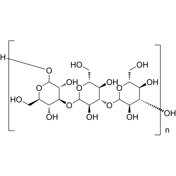
Zymosan (ZM), 95% is a yeast cell wall-derived carbohydrate-rich preparation and immunomodulator. Zymosan (ZM), 95% binds to and activates TLR-2, TLR-4, and Dectin-1 receptor to trigger downstream signaling pathways. Zymosan (ZM), 95% upregulates TLR-2, TLR-4, and TNF-α mRNA expression, increases serum TNF-α levels, and stimulates splenocyte number and viability in mice. Zymosan (ZM), 95% attenuates melanoma growth progression, modulates macrophage marker gene expression, and mediates phagocytosis, ROS generation, and cytokine production. Zymosan (ZM), 95% reduces Connexin 43 protein and mRNA levels, inhibits gap junctional intercellular communication, and induces proinflammatory factor production in human corneal cells. Zymosan (ZM), 95% induces peritoneal inflammation in mice, functions as a drug carrier, and supports fibroblast cell attachment in hydrogel formulations. Zymosan (ZM), 95% can be used for the research of melanoma, tumors, fungal keratitis, ocular surface inflammatory disorders, and peritoneal inflammation.
연구목적의 판매만을 진행합니다. 환자를 대상으로 한 판매는 하지 않습니다.
- Purity : 96.11%
- CAS No.: 9010-72-4
-
보관:Powder -20°C, 3 years , 4°C, 2 years ; In solvent -80°C, 6 months , -20°C, 1 month


To place orders, for customer services and technical support, please contact: MedChemExpress USA
Tel: 609-228-6898 E-mail: [email protected] [email protected]


-
 Biological Activity
-
Chemical Information
- Protocol
- 순도&문서
- References
-
 Help & FAQs




Biological Activity
제품 설명
IC50 & Target
|
Dectin-1 |
TLR4 |
TLR2 |
In Vitro
Zymosan (ZM), 95% (0.01-100 μg/mL; 72 h), enhances the viability and proliferation of mouse splenocytes in a concentration-dependent, bell-shaped curve manner, with the maximal effect observed at the concentration of 1 μg/mL after 72 h of incubation[1].
Zymosan (ZM), 95% (10-100 μg/mL; 24 h), is biocompatible with Raw 264.7 macrophages, and the cell viability remains above 90% after incubation at concentrations of 10-100 μg/mL for 24 h[2].
Zymosan (ZM), 95% (1 mg; 6 h), induces an anti-tumor M1 macrophage phenotype in differentiated human THP-1 macrophages, which is characterized by upregulated gene expression of TNFα, CXCL10 and TLR2, as well as downregulated expression of M2 marker genes, after 6 h of incubation with 1 mg ZM[3].
Zymosan (ZM), 95% (20-600 μg/mL; 24 h), reduces the expression of Cx43 protein in cultured human corneal fibroblasts in a concentration-dependent manner, with 200 μg/mL and 600 μg/mL (for 24 h) inducing 45% and 54% downregulation of expression, respectively[4].
The downregulation of Cx43 protein expression in cultured human corneal fibroblasts induced by 95% Zymosan (ZM) (600 μg/mL; 24 h) can be reversed in a concentration-dependent manner (with a reversal range of 6%-78%) by co-treatment with IKK2 inhibitor IV[4].
Zymosan (ZM), 95% (1-50 μg/mL; 4-24 h), induces concentration- and time-dependent increases in the mRNA expression of proinflammatory cytokines, chemokines, and MMPs in primary human corneal epithelial cells, with the peak effect observed at 4 h[5].
MedChemExpress (MCE) has not independently confirmed the accuracy of these methods. They are for reference only. Further protocols information, click here.
-
Cell Line:Cultured human corneal fibroblasts (HCFs)
-
Concentration:20-600 μg/mL; 20 μg/mL, 60 μg/mL (non-significant)
-
Incubation Time:24 h
-
Result:Induced a concentration-dependent reduction in Cx43 protein expression.
Reduced Cx43 protein levels by 45% relative to untreated controls at 200 μg/mL.
Reduced Cx43 protein levels by 54% relative to untreated controls at 600 μg/mL.
Showed relative Cx43 band intensities of 0.98 and 0.81 for 200 μg/mL and 600 μg/mL zymosan, respectively, compared to 1.78 for untreated controls.
-
Cell Line:Cultured human corneal fibroblasts (HCFs)
-
Concentration:600 μg/mL (co-treated with PD98059 at 1-10 μmol/L); 600 μg/mL (co-treated with JNK inhibitor II at 3-10 μmol/L)
-
Incubation Time:24 h
-
Result:Attenuated the zymosan-induced reduction in Cx43 protein expression by PD98059 in a concentration-dependent manner: 1 μmol/L PD98059 attenuated the inhibition by 13%, 3 μmol/L by 19%, and 10 μmol/L by 34%.
Attenuated the zymosan-induced reduction in Cx43 protein expression by JNK inhibitor II in a concentration-dependent manner: 3 μmol/L JNK inhibitor II attenuated the inhibition by 20%, and 10 μmol/L by 34%.
Showed relative Cx43 band intensities of 2.12, 2.37, and 3.00 for zymosan plus 1, 3, 10 μmol/L PD98059, respectively, compared to 1.60 for zymosan alone; and 2.55 and 3.12 for zymosan plus 3, 10 μmol/L JNK inhibitor II, respectively, compared to 1.72 for zymosan alone.
-
Cell Line:Cultured human corneal fibroblasts (HCFs)
-
Concentration:600 μg/mL (co-treated with IKK2 inhibitor IV at 1-10 μmol/L)
-
Incubation Time:24 h
-
Result:Attenuated the zymosan-induced reduction in Cx43 protein expression by IKK2 inhibitor IV in a concentration-dependent manner: 1 μmol/L IKK2 inhibitor IV attenuated the inhibition by 6%, 3 μmol/L by 39%, and 10 μmol/L by 78%.
Showed relative Cx43 band intensities of 0.63, 1.85, and 3.33 for zymosan plus 1, 3, 10 μmol/L IKK2 inhibitor IV, respectively, compared to 0.40 for zymosan alone.
In Vivo
Zymosan (ZM), 95% (10 μg; i.p.; daily; for 4 consecutive days) significantly upregulates the mRNA expression of TNF-α, TLR-2 and TLR-4 in peritoneal macrophages of healthy C57BL/6 mice, and increases their serum TNF-α levels[1].
MedChemExpress (MCE) has not independently confirmed the accuracy of these methods. They are for reference only.
-
Animal Model:C57BL/6 (female, 6-8 weeks old, average weight 18 g, melanoma induced by subcutaneous injection of 1×10⁶ B16F10 cells)[1]
-
Dosage:10 μg
-
Administration:i.p.; daily; 4 consecutive days
-
Result:Reduced mean tumor weight to approximately 0.6 g.
Elevated serum TNF-α levels compared to control and untreated melanoma-bearing mice.
Upregulated peritoneal macrophage TNF-α mRNA expression to 12.27-fold relative to control mice.
Upregulated peritoneal macrophage TLR-2 mRNA expression to 3.02-fold relative to control mice.
Increased peritoneal macrophage TLR-4 mRNA expression to 1.11-fold relative to control mice.
-
Animal Model:C57BL/6 (female, 6-8 weeks old, average weight 18 g)[1]
-
Dosage:10 μg
-
Administration:i.p.; daily; 4 consecutive days
-
Result:Increased serum TNF-α levels significantly greater than all other groups.
Upregulated peritoneal macrophage TNF-α mRNA expression to 39.85-fold relative to control mice.
Upregulated peritoneal macrophage TLR-2 mRNA expression to 12.95-fold relative to control mice.
Upregulated peritoneal macrophage TLR-4 mRNA expression to 2.18-fold relative to control mice.


Chemical Information
-
CAS No. 9010-72-4
-
Appearance Solid
-
Color White to light yellow
-
SMILES
[Zymosan (ZM), 95%]
-
선적
Room temperature in continental US; may vary elsewhere.
-
보관
Powder -20°C 3 years 4°C 2 years In solvent -80°C 6 months -20°C 1 month

Protocol
-
RT-PCR
Reverse transcription technology uses RNA as a template to synthesize DNA. RT-PCR is simple, specific and sensitive, and can be used to detect gene expression levels and expression differences in cells; detect RNA virus content; clone cDNA sequences of specific genes.
-
Phagocytosis Functional Assay
A phagocytosis functional assay measures the ability of phagocytic cells, such as neutrophils, macrophages, monocytes, or microglia/macrophages, to bind and internalize particulate targets including bacteria, yeast particles, beads, or myelin particles. Fluorescent flow-cytometry assays detect target uptake as fluorescence associated with gated phagocytes, while pH-sensitive dyes such as pHrodo increase signal in acidic phagosomal compartments and therefore preferentially report internalized particles rather than particles remaining outside the cell. Microscopy or high-content imaging can be used to confirm intracellular localization and, in some protocols, to follow uptake kinetics.
-
3D Hydrogel Synthetic Scaffold Culture
3D hydrogel synthetic scaffold culture embeds cells, spheroids, organoids, or tissue fragments inside a hydrated crosslinked polymer network so that cells receive matrix and cell-cell cues in three dimensions rather than from a flat plastic surface. A literature-supported model protocol is PEG-4MAL hydrogel culture, in which four-arm maleimide-terminated PEG is functionalized with cysteine-containing adhesive peptides such as RGD and crosslinked with protease-degradable peptides such as GPQ-W; this creates a defined, modular scaffold that supports human organoid generation and culture. The readouts are scaffold-supported growth, morphology, lumen formation, budding, viability, proliferation, lineage-marker expression, and matrix-dependent expansion or differentiation; reported assays include transmitted-light imaging, immunofluorescence, in situ hybridization, qRT-PCR, and rheological characterization.
-
RNA extraction experimental
By lysing cells, releasing RNA, and removing impurities such as proteins and DNA, high-purity RNA products are finally obtained. The commonly used traditional method is the guanidine isothiocyanate/phenol/chloroform method (Trizol), which is suitable for a variety of animal materials including animal tissues, microorganisms, cultured cells, etc., and most plant materials.
-
ROS/oxidative-stress fluorescent staining
ROS/oxidative-stress fluorescent staining uses cell-permeant fluorogenic probes that become fluorescent after oxidation inside cells or tissues; commonly used examples include DCFH-DA/DCFDA for broad cellular oxidant detection, DHE for superoxide-related signal detection, MitoSOX for mitochondrial superoxide-related signal detection, and CellROX probes for oxidative-stress-associated fluorescence readouts. The assay detects probe oxidation rather than a single ROS species unless the probe and analysis method have been chemically validated for that species. DCFH-DA enters cells, is deacetylated by intracellular esterases to DCFH, and produces fluorescent DCF after oxidation, so the readout is used as an operational measure of total cellular oxidative stress rather than a species-specific ROS measurement. DHE and MitoSOX can report superoxide-related oxidation, but red fluorescence alone can include non-specific ethidium-like oxidation products; HPLC or optimized spectral approaches are
-
Research Protocol for Inflammation-related Diseases
The NLRP3 inflammasome is a cytosolic innate immune signaling platform that integrates priming signals and danger-signal activation to promote caspase-1 activation, maturation of IL-1β and IL-18, and gasdermin D-mediated pyroptotic cell death. The core experimental logic is to determine whether inflammatory disease phenotypes are driven by increased NLRP3 expression, ASC-containing inflammasome assembly, caspase-1 cleavage, GSDMD cleavage, and extracellular release of IL-1β/IL-18 rather than by nonspecific cell injury alone. The pathway is strongly linked to inflammation-related disease phenotypes because monosodium urate crystals activate NALP3/NLRP3 inflammasome signaling in gout-like crystal inflammation, cholesterol crystals activate NLRP3 inflammasomes in atherogenesis models, and DSS-induced intestinal inflammation has been reported to involve NLRP3 inflammasome activity. However, experimental colitis studies also show context-dependent protective effects of NLRP3 inflammasome co
-
Real Time qPCR (Q-PCR)
Real-time quantitative PCR (qPCR) quantifies an amplifiable nucleic-acid target by monitoring fluorescence during PCR cycling rather than measuring product only after amplification. The increase in fluorescence tracks accumulation of PCR product, and the quantification cycle (Cq; historically also Ct/CP) is related to the initial amount of target: samples containing more starting target generally reach the defined fluorescence threshold in fewer cycles.
-
Cell Viability Determination by MTT Colorimetric Assay
The following protocol uses the MTT colorimetric assay as a classic literature-established method for assessing cell viability/metabolic activity in cultured mammalian cells. MTT[3-(4,5-dimethylthiazol-2-yl)-2,5-diphenyltetrazolium bromide] is reduced by metabolically active cells to a colored formazan product; the amount of formazan is quantified spectrophotometrically and provides an indirect measure of metabolically active viable cells. Importantly, MTT reduction reflects cellular oxidoreductase/metabolic activity rather than an absolute direct count of living cells, so changes in cellular metabolism can alter the signal independently of cell number.


순도&문서
-
Data Sheet (279 KB)
-
SDS (251 KB)
- English - EN (251 KB)
- Français - FR (251 KB)
- Deutsch - DE (251 KB)
- Norwegian - NO (251 KB)
- Español - ES (251 KB)
- Swedish - SV (251 KB)
- Italian - IT (251 KB)
- Korean - KR (251 KB)
- Portuguese - PT (251 KB)
-
Handling Instructions (2659 KB)
References
[1]. Taghavi M, et al. Zymosan attenuates melanoma growth progression, increases splenocyte proliferation and induces TLR-2/4 and TNF-α expression in mice. J Inflamm (Lond). 2018;15:5. Published 2018 Mar 22. [Content Brief]
[2]. Venkatachalam G, et al. Synthesis, Characterization, and Biological Activity of Aminated Zymosan. ACS Omega. 2020;5(26):15973-15982. Published 2020 Jun 23. [Content Brief]
[3]. Venkatachalam G, et al. Immunomodulatory zymosan/ι-carrageenan/ agarose hydrogel for targeting M2 to M1 macrophages (antitumoral). RSC Adv. 2024;14(17):11694-11705. Published 2024 Apr 11. [Content Brief]
[4]. Zheng XS, et al. Effect of zymosan on the expression and function of the gap-junction protein connexin 43 in human corneal fibroblasts. Int J Ophthalmol. 2021;14(3):341-348. Published 2021 Mar 18. [Content Brief]
[5]. Li DQ, et al. Suppressive effects of azithromycin on zymosan-induced production of proinflammatory mediators by human corneal epithelial cells. Invest Ophthalmol Vis Sci. 2010;51(11):5623-5629. [Content Brief]
[6]. Kolaczkowska E, et al. Critical role of mast cells in morphine-mediated impairment of zymosan-induced peritonitis in mice. Inflamm Res. 2001;50(8):415-421. [Content Brief]

Calculators
Concentration (start) × Volume (start) = Concentration (final) × Volume (final)


Zymosan (ZM), 95% Related Classifications
HY-159069 Related Classifications
 Powered by Bioz
Keywords