



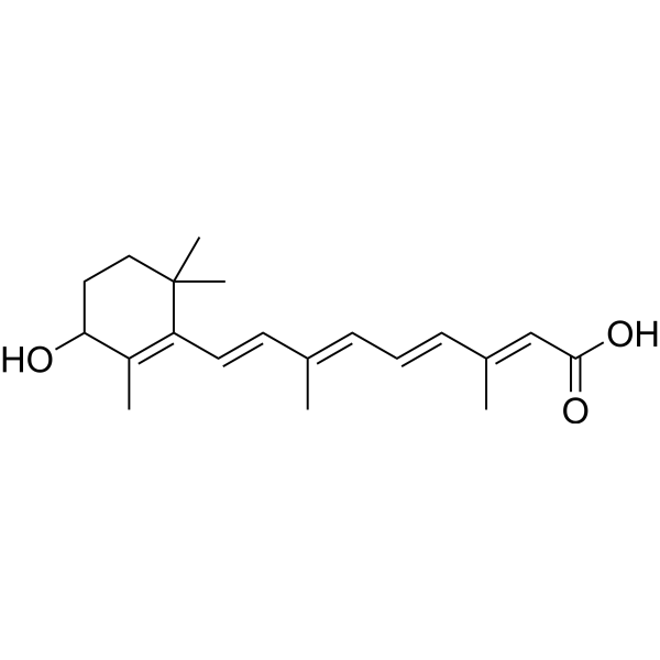
4-Hydroxyretinoic acid
Based on 1 publication(s) in Google Scholar
4-Hydroxyretinoic acid (4-HRA) is a derivative of Retinoic acid (HY-14649). 4-Hydroxyretinoic acid is formed via the catalysis of retinol by cytochrome P-450 isoenzymes. 4-Hydroxyretinoic acid also serves as a substrate for UDP-glucuronosyltransferase (s) and recombinant UGT2B7. 4-Hydroxyretinoic acid binds to the nuclear receptor RAR (Retinoic Acid Receptor), activates RAR and RXR-alpha, subsequently regulates gene expression and cell differentiation, and induces cancer cell apoptosis (Apoptosis). 4-Hydroxyretinoic acid also participates in multiple physiological processes such as immunoregulation, neuroprotection and antioxidation.
연구목적의 판매만을 진행합니다. 환자를 대상으로 한 판매는 하지 않습니다.
- Purity : 94.58%
- CAS No.: 66592-72-1
- 화학식: C20H28O3
- 분자량:316.43
-
보관:
-80°C, protect from light, stored under nitrogen

To place orders, for customer services and technical support, please contact: MedChemExpress USA
Tel: 609-228-6898 E-mail: [email protected] [email protected]


-
 Biological Activity
-
Chemical Information
- Protocol
- 순도&문서
- References
-
 Help & FAQs


-
HY-101106
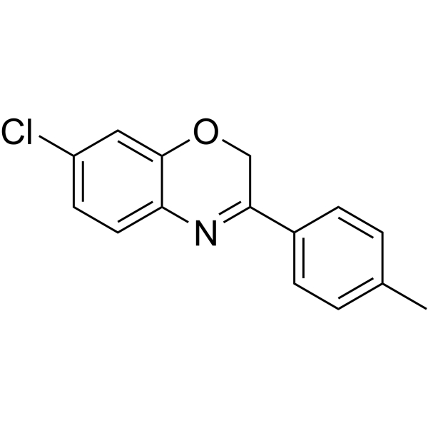
AR7RARA/RARα Antagonist -
HY-B1384
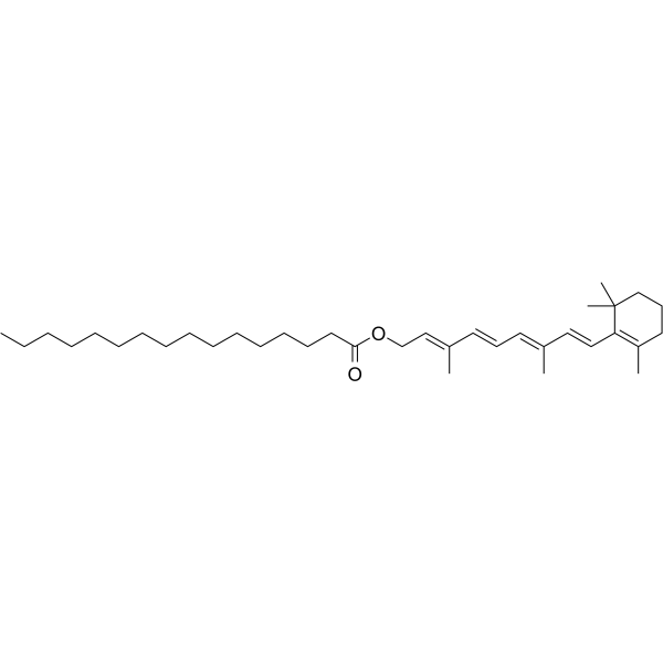
Retinyl palmitateEndogenous Metabolite -
HY-107494A
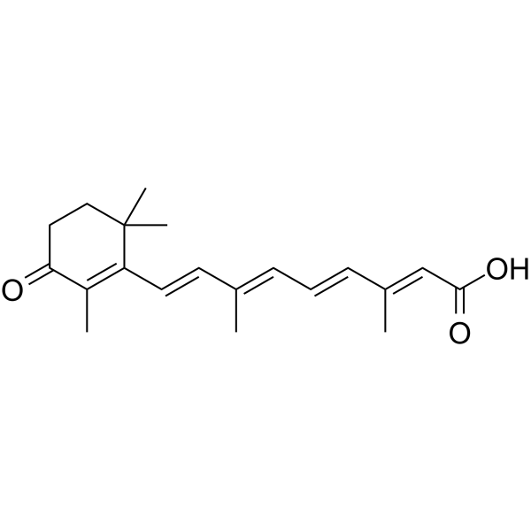
all-trans-4-Oxoretinoic acidEndogenous Metabolite -
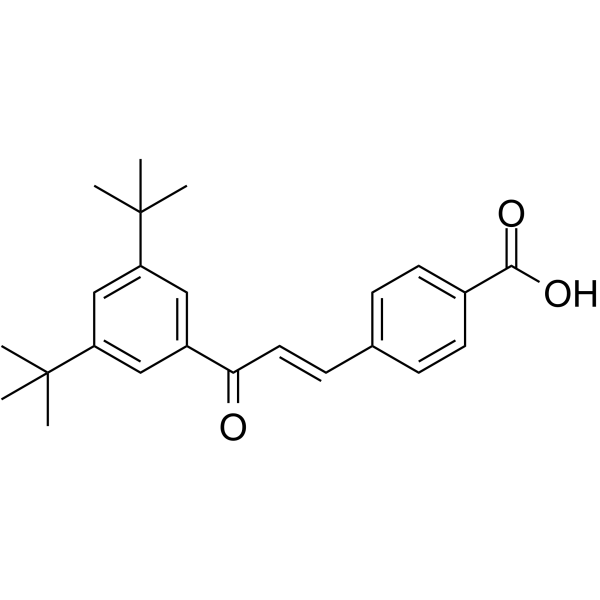
HY-107397
Ch55RAR-α/RAR-β Agonist -

HY-W019726
Dehydro NifedipineNifedipine Metabolite -
HY-A0183
PhosphatidylserineAnti-Inflammatory/Immunosuppressive Signal -

HY-137197
6β-Hydroxy dexamethasoneDexamethasone Metabolite -

HY-107494R
13-cis-4-Oxoretinoic acid (Standard)Analytical Standard -

HY-124542
5-HydroxydiclofenacMetabolite of Diclofenac -
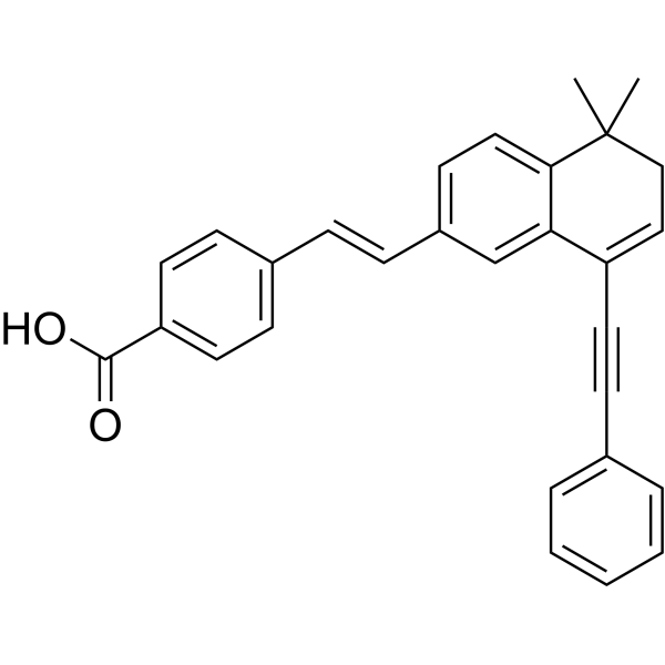
HY-108529
BMS493Inverse RAR Inhibitor -
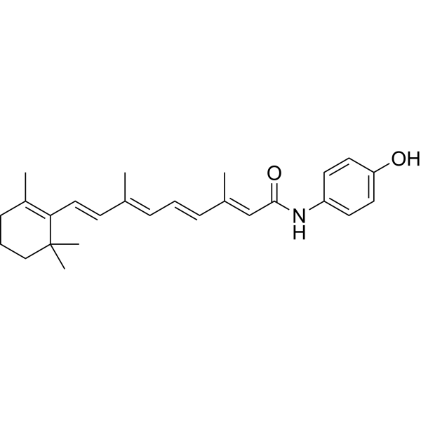
HY-15373
FenretinideAutophagy Inducer -

HY-107397R
Ch55 (Standard)Reference Standard
Publications Citing Use of MedChemExpress (MCE) 4-Hydroxyretinoic acid
More

Biological Activity
제품 설명
IC50 & Target
|
UGT2B7 |
In Vitro
4-Hydroxyretinoic acid (10-11-5×10⁻⁴ M) stimulates RAR-β, RAR-γ, RAR-α, and RXR-α mediated transcription of the TRE3-tk-CAT reporter gene in cotransfected CV-1 cells with ED50 values of 75 nM, 30 nM, >500 nM, and >500 nM, respectively[2].
MedChemExpress (MCE) has not independently confirmed the accuracy of these methods. They are for reference only.


Chemical Information
-
CAS No. 66592-72-1
-
Appearance Solid
-
분자량 316.43
-
화학식 C20H28O3
-
Color White to yellow
-
SMILES
CC(/C=C/C=C(/C=C/C1=C(C(O)CCC(C)1C)C)C)=C\C(O)=O
-
선적
Shipping with dry ice.
-
보관
-80°C, protect from light, stored under nitrogen


Publications (1)
-
Journal Impact Factor
-
Most Recent
-
J Biol Chem
A cell-based assay for retinaldehyde dehydrogenase activity: Retinoid quantification as an alternative to current fluorescence-based approaches. [Abstract]2026 Jan 29;302(3):111211. PMID: 41617028


순도&문서
-
Data Sheet (269 KB)
-
SDS (701 KB)
- English - EN (701 KB)
- Français - FR (701 KB)
- Deutsch - DE (701 KB)
- Norwegian - NO (701 KB)
- Español - ES (701 KB)
- Swedish - SV (701 KB)
- Italian - IT (701 KB)
- Korean - KR (701 KB)
- Portuguese - PT (701 KB)
-
Handling Instructions (2659 KB)
References
[1]. Samokyszyn VM, et al. 4-hydroxyretinoic acid, a novel substrate for human liver microsomal UDP-glucuronosyltransferase(s) and recombinant UGT2B7. J Biol Chem. 2000;275(10):6908-6914. [Content Brief]
[2]. Duell EA, et al. Human skin levels of retinoic acid and cytochrome P-450-derived 4-hydroxyretinoic acid after topical application of retinoic acid in vivo compared to concentrations required to stimulate retinoic acid receptor-mediated transcription in vitro. J Clin Invest. 1992;90(4):1269-1274. [Content Brief]

Calculators
Concentration (start) × Volume (start) = Concentration (final) × Volume (final)


4-Hydroxyretinoic acid Related Classifications
HY-125904 Related Classifications
 Powered by Bioz