



PP1
Based on 13 publication(s) in Google Scholar
PP1 is a potent, and Src family-selective tyrosine kinase inhibitor with IC50 of 5 and 6 nM for Lck and Fyn, respectively.
For research use only. We do not sell to patients.
- Purity : 99.57%
- CAS No.: 172889-26-8
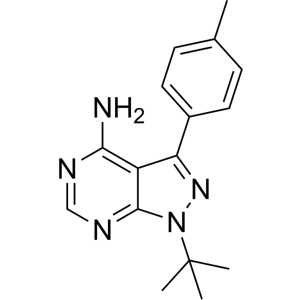
- Formula: C16H19N5
- Molecular Weight:281.36
-
Storage:Powder -20°C, 3 years , 4°C, 2 years ; In solvent -80°C, 2 years , -20°C, 1 year

To place orders, for customer services and technical support, please contact: MedChemExpress USA
Tel: 609-228-6898 E-mail: [email protected] [email protected]


-
 Biological Activity
-
Chemical Information
-
Solvent & Solubility
- Purity & Documentation
- References
-
 Help & FAQs


-
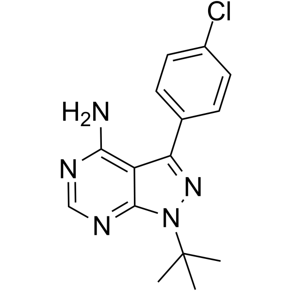
HY-13805
PP2Src Inhibitor -
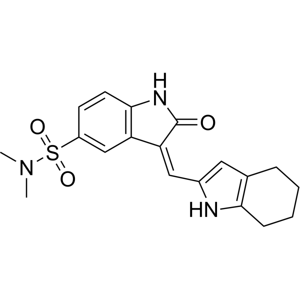
HY-B0789
SU6656Src Inhibitor -
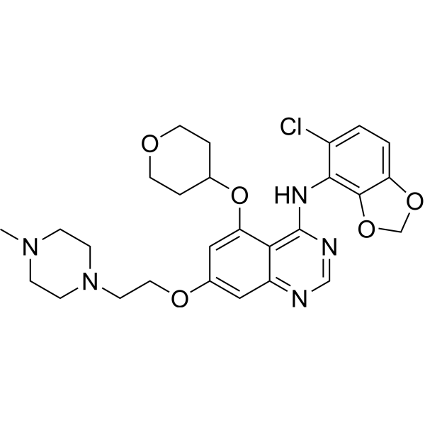
HY-10234
SaracatinibSrc Inhibitor -
HY-13941
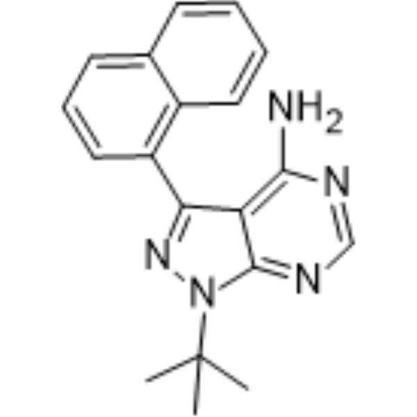
1-Naphthyl PP1Src Inhibitor -
HY-101053
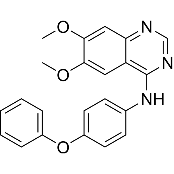
Src Inhibitor 1Src Inhibitor -
HY-10181
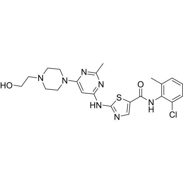
DasatinibSrc/Bcr-Abl Inhibitor -
HY-13942
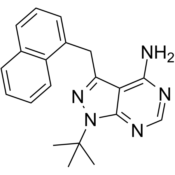
1-NM-PP1Src Inhibitor -
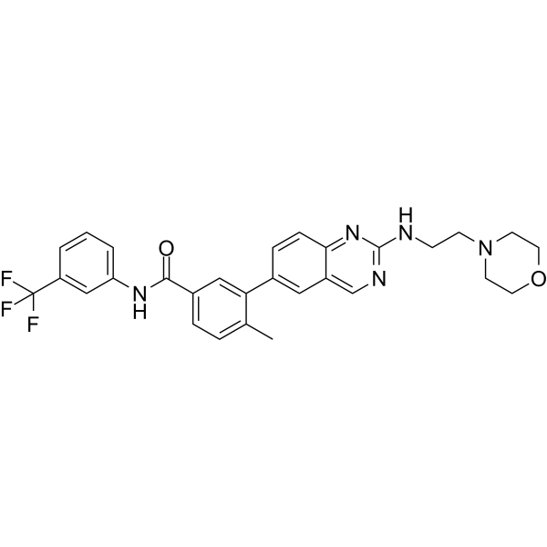
HY-18303
AMG-47aLck Inhibitor -
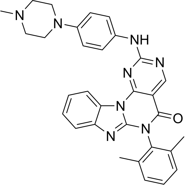
HY-12072
Lck InhibitorLck Inhibitor -

HY-15763
ErastinFerroptosis Inducer -
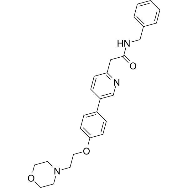
HY-10340
TirbanibulinSrc Inhibitor -

HY-13805R
PP2 (Standard)Src Inhibitor
Publications Citing Use of MedChemExpress (MCE) PP1
More- Nat Microbiol. 2025 Nov;10(11):2949-2965. [Abstract]
- Nat Commun. 2022 Sep 27;13(1):5675. [Abstract]
- Cell Rep Med. 2025 Feb 18;6(2):101922. [Abstract]
- Cell Rep Med. 2024 Jun 18;5(6):101592. [Abstract]
- Front Immunol. 2021 Nov 24;12:786602. [Abstract]
- J Thromb Haemost. 2021 Aug;19(8):2029-2043. [Abstract]
- Cell Signal. 2026 Feb:138:112204. [Abstract]
- J Cell Mol Med. 2019 Apr;23(4):2399-2409. [Abstract]
- BMC Cancer. 2022 Nov 24;22(1):1211. [Abstract]
- Reprod Toxicol. 2025 May:134:108899. [Abstract]
- Mol Reprod Dev. 2022 May;89(5-6):256-268. [Abstract]
- bioRxiv. 2025 Jul 7:2025.07.04.663030. [Abstract]
- Patent. US20180263995A1.
 Customer Validation & Images

-
IP
-
Cell Proliferation/Viability Assay
-
WB

-
RT-PCR

-
WB


Biological Activity
Description
IC50 & Target
IC50: 5 nM (Lck), 6 nM (Fyn), 250 nM (EGFR), >50 μM (JAK2)[1]
Cellular Effect
|
Cell Line

|
Type | Value | Description | References |
|---|---|---|---|---|
| A549 | IC50 |
0.01 μM
Compound: PP1
|
Growth inhibition of human A549 cells
Growth inhibition of human A549 cells
|
[PMID: 28814374] |
| DOHH-2 | GI50 |
3.7 μM
Compound: PP1
|
Growth inhibition of human DOHH-2 cells measured after 72 hrs by cell titer glo luminescent cell viability assay
Growth inhibition of human DOHH-2 cells measured after 72 hrs by cell titer glo luminescent cell viability assay
|
[PMID: 37163946] |
| Jurkat | IC50 |
0.4 μM
Compound: 1
|
Inhibition of MLR stimulated IL-2 production in Jurkat cells
Inhibition of MLR stimulated IL-2 production in Jurkat cells
|
[PMID: 11012021] |
| Jurkat | IC50 |
1.2 μM
Compound: 1
|
Inhibition of anti-CD3 stimulated IL-2 production by Jurkat cells
Inhibition of anti-CD3 stimulated IL-2 production by Jurkat cells
|
[PMID: 11012021] |
| Jurkat | IC50 |
5 nM
Compound: PP1
|
Inhibition of p56 Lck tyrosine kinase in Jurkat cells where p56lck autophosphorylation is inhibited.
Inhibition of p56 Lck tyrosine kinase in Jurkat cells where p56lck autophosphorylation is inhibited.
|
10.1016/S0960-894X(97)00034-6 |
| MCF7 | EC50 |
6.76 μM
Compound: PP1
|
Antiproliferative activity against human MCF7 cells assessed as reduction in cell viability after 5 days by PrestoBlue assay
Antiproliferative activity against human MCF7 cells assessed as reduction in cell viability after 5 days by PrestoBlue assay
|
[PMID: 27115835] |
| SUD4 | GI50 |
>10 μM
Compound: PP1
|
Growth inhibition of human SU-DHL-4 cells measured after 72 hrs by cell titer glo luminescent cell viability assay
Growth inhibition of human SU-DHL-4 cells measured after 72 hrs by cell titer glo luminescent cell viability assay
|
[PMID: 37163946] |
| SU-DHL-6 | GI50 |
4.7 μM
Compound: PP1
|
Growth inhibition of human SU-DHL-6 cells measured after 72 hrs by cell titer glo luminescent cell viability assay
Growth inhibition of human SU-DHL-6 cells measured after 72 hrs by cell titer glo luminescent cell viability assay
|
[PMID: 37163946] |
In Vitro
PP1 inhibits Lck (IC50=5 nM) and FynT (IC50=6 nM) in vitro at concentrations significantly lower than those required to inhibit ZAP-70 (IC50>100 μM), JAK2 (IC50>50 μM), the EGFR kinase, and protein kinase A. PP1 inhibits whole cell tyrosine phosphorylation and proliferation in T cells stimulated with anti-CD3 and mitogens. PP1 selectively inhibits IL-2 gene expression over GM-CSF and IL-2R gene induction in human T cells[1].
MedChemExpress (MCE) has not independently confirmed the accuracy of these methods. They are for reference only. .
Clinical Trial
| NCT Number | Sponsor | Condition | Start Date |
Phase
|
|---|---|---|---|---|
| NCT01329991 | Plexxikon| | 2011-05 | PHASE1 |


Chemical Information
-
CAS No. 172889-26-8
-
Appearance Solid
-
Molecular Weight 281.36
-
Formula C16H19N5
-
Color White to off-white
-
SMILES
NC1=C2C(N(C(C)(C)C)N=C2C3=CC=C(C)C=C3)=NC=N1
-
Synonyms
AGL 1872; EI 275
-
Shipping
Room temperature in continental US; may vary elsewhere.
-
Storage
Powder -20°C 3 years 4°C 2 years In solvent -80°C 2 years -20°C 1 year


Publications (13)
-
Journal Impact Factor
-
Most Recent
-
Nat Microbiol
Mycobacterium tuberculosis-derived linoleic acid increases regulatory T cell function to promote bacterial survival within macrophages. [Abstract]2025 Nov;10(11):2949-2965. PMID: 41073667 -
Nat Commun
Phosphorylation of Jhd2 by the Ras-cAMP-PKA(Tpk2) pathway regulates histone modifications and autophagy. [Abstract]2022 Sep 27;13(1):5675. PMID: 36167807
PP1 purchased from MedChemExpress. Usage Cited in: Nat Commun. 2022 Sep 27;13(1):5675. [Abstract]
PP1 (1NM-PP1; 25 μM; 0.5 h) directly inhibited Tpk2-catalyzed Jhd2 phosphorylation in yeast cells.
-
Cell Rep Med
MFGE8 induces anti-PD-1 therapy resistance by promoting extracellular vesicle sorting of PD-L1. [Abstract]2025 Feb 18;6(2):101922. PMID: 39842432 -
Cell Rep Med
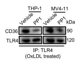
A CD36-dependent non-canonical lipid metabolism program promotes immune escape and resistance to hypomethylating agent therapy in AML. [Abstract]2024 Jun 18;5(6):101592. PMID: 38843841
PP1 purchased from MedChemExpress. Usage Cited in: Cell Rep Med. 2024 Jun 18;5(6):101592. [Abstract]
PP1 (10 μM; 5 h) decreased the OxLDL-induced association of TLR4 and CD36 in AML cells.
PP1 purchased from MedChemExpress. Usage Cited in: Cell Rep Med. 2024 Jun 18;5(6):101592. [Abstract]
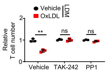
PP1 (10 μM; 3 days) failed to suppress T cell proliferation in MV4-11 cells. MV4-11 cells treated with LDM supplied with different reagents (OxLDL, 25 mg/mL; TAK-242, 10 μM; PP1, 10 μM) for 3 days were further used for T cell proliferation assay.
-
Front Immunol
Fpr2/CXCL1/2 Controls Rapid Neutrophil Infiltration to Inhibit Streptococcus agalactiae Infection. [Abstract]2021 Nov 24;12:786602. PMID: 34899755
PP1 purchased from MedChemExpress. Usage Cited in: Front Immunol. 2021 Nov 24;12:786602. [Abstract]
PP1 (10 μM; 1 h) decreased 2×10⁶ CFU/ml GBS for 1hour induced CXCL1 and CXCL2 mRNA expression in neutrophils.
-
J Thromb Haemost
Photobiomodulation therapy for thrombocytopenia by upregulating thrombopoietin expression via the ROS-dependent Src/ERK/STAT3 signaling pathway. [Abstract]2021 Aug;19(8):2029-2043. PMID: 33501731
PP1 purchased from MedChemExpress. Usage Cited in: J Thromb Haemost. 2021 Aug;19(8):2029-2043. [Abstract]
PP1 (5 μM) reversed PBMT-induceed the phosphorylation level of STAT3 in HepG2 cells.

PP1 purchased from MedChemExpress. Usage Cited in: J Thromb Haemost. 2021 Aug;19(8):2029-2043. [Abstract]
PP1 (5 μM) reversed PBMT-induced STAT3 phosphorylation and TPO expression in HepG2 cells.
-
Cell Signal
Phosphoproteomic analysis of successive Jurkat CD19-CAR generations reveals TCRζ-driven signalling. [Abstract]2026 Feb:138:112204. PMID: 41177417 -
J Cell Mol Med
Knockdown of Golgi phosphoprotein 73 blocks the trafficking of matrix metalloproteinase-2 in hepatocellular carcinoma cells and inhibits cell invasion. [Abstract]2019 Apr;23(4):2399-2409. PMID: 30677226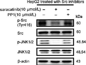
PP1 purchased from MedChemExpress. Usage Cited in: J Cell Mol Med. 2019 Apr;23(4):2399-2409. [Abstract]
Immunoblot analysis of p-JNK1/2 (T183/Y185) in HepG2 cells treated with saracatinib (10 μM, 12 h) and PP1 (10 μM, 2 h).
-
BMC Cancer
Artificial intelligence to guide precision anticancer therapy with multitargeted kinase inhibitors. [Abstract]2022 Nov 24;22(1):1211. PMID: 36434556 -
Reprod Toxicol
2025 May:134:108899. PMID: 40139512 -
Mol Reprod Dev
CD55 is upregulated by cAMP/PKA/AKT and modulates human decidualization via Src and ERK pathway and decidualization-related genes. [Abstract]2022 May;89(5-6):256-268. PMID: 35474595 -
bioRxiv
2025 Jul 7:2025.07.04.663030. PMID: 40672291 -


Solvent & Solubility
In Vitro:
DMSO : 28 mg/mL (99.52 mM; Need ultrasonic; Hygroscopic DMSO has a significant impact on the solubility of product, please use newly opened DMSO)
Please refer to the solubility information to select the appropriate solvent. Once prepared, please aliquot and store the solution to prevent product inactivation from repeated freeze-thaw cycles.
Storage method and period of stock solution: -80°C, 2 years; -20°C, 1 year. When stored at -80°C, please use it within 2 years. When stored at -20°C, please use it within 1 year.
Please refer to the solubility information to select the appropriate solvent. Once prepared, please aliquot and store the solution to prevent product inactivation from repeated freeze-thaw cycles.
Storage method and period of stock solution: -80°C, 2 years; -20°C, 1 year. When stored at -80°C, please use it within 2 years. When stored at -20°C, please use it within 1 year.
Concentration (start) × Volume (start) = Concentration (final) × Volume (final)
In Vivo:
Select the appropriate dissolution method based on your experimental animal and administration route.
- For the following dissolution methods, please ensure to first prepare a clear stock solution using an In Vitro approach and then sequentially add co-solvents:
- To ensure reliable experimental results, the clarified stock solution can be appropriately stored based on storage conditions. As for the working solution for In Vivo experiments, it is recommended to prepare freshly and use it on the same day.
- The percentages shown for the solvents indicate their volumetric ratio in the final prepared solution. If precipitation or phase separation occurs during preparation, heat and/or sonication can be used to aid dissolution.
Add each solvent one by one: 10% DMSO 40% PEG300 5% Tween-80 45% Saline
Solubility: ≥ 1.67 mg/mL (5.94 mM); Clear solution
This protocol yields a clear solution of ≥ 1.67 mg/mL (saturation unknown).
Taking 1 mL working solution as an example, add 100 μL DMSO stock solution (16.7 mg/mL) to 400 μL PEG300, and mix evenly; then add 50 μL Tween-80 and mix evenly; then add 450 μL Saline to adjust the volume to 1 mL.
Preparation of Saline: Dissolve 0.9 g sodium chloride in ddH₂O and dilute to 100 mL to obtain a clear Saline solution.
Add each solvent one by one: 10% DMSO 90% (20% SBE-β-CD in Saline)
Solubility: ≥ 1.67 mg/mL (5.94 mM); Clear solution
This protocol yields a clear solution of ≥ 1.67 mg/mL (saturation unknown).
Taking 1 mL working solution as an example, add 100 μL DMSO stock solution (16.7 mg/mL) to 900 μL 20% SBE-β-CD in Saline, and mix evenly.
Preparation of 20% SBE-β-CD in Saline (4°C, storage for one week): 2 g SBE-β-CD powder is dissolved in 10 mL Saline, completely dissolve until clear.
In Vivo Dissolution Calculator
Please enter the basic information of animal experiments:
-
-
-
-
Recommended: Prepare an additional quantity of animals to account for potential losses during experiments.
Please enter your animal formula composition:
-
%DMSO +
Recommended: Keep the proportion of DMSO in working solution below 2% if your animal is weak.
-
%+
-
+%Tween-80 + +
-
%Saline +
The co-solvents required include: DMSO, . All of co-solvents are available by MedChemExpress (MCE). , Tween 80. All of co-solvents are available by MedChemExpress (MCE).
Working solution concentration: 0.22 mg/mL
Method for preparing stock solution: mg drug dissolved in μL DMSO. Stock solution concentration: mg/mL.
1. Take μL DMSO stock solution;
2. Add μL .
μL , mix evenly;
3. Then add μL Tween 80, mix evenly;
4. Then add μL
Please ensure that the stock solution in the first step is dissolved to a clear state, and add co-solvents in sequence. You can use ultrasonic heating (ultrasonic cleaner, recommended frequency 20-40 kHz), vortexing, etc. to assist dissolution.


Purity & Documentation
-
Data Sheet (282 KB)
-
SDS (479 KB)
- English - EN (479 KB)
- Français - FR (479 KB)
- Deutsch - DE (479 KB)
- Norwegian - NO (479 KB)
- Español - ES (479 KB)
- Swedish - SV (479 KB)
- Italian - IT (479 KB)
- Korean - KR (479 KB)
- Portuguese - PT (479 KB)
-
Handling Instructions (2659 KB)
References


Complete Stock Solution Preparation Table
Please refer to the solubility information to select the appropriate solvent. Once prepared, please aliquot and store the solution to prevent product inactivation from repeated freeze-thaw cycles.
Storage method and period of stock solution: -80°C, 2 years; -20°C, 1 year. When stored at -80°C, please use it within 2 years. When stored at -20°C, please use it within 1 year.
| Optional Solvent | Concentration Solvent Mass | 1 mg | 5 mg | 10 mg | 25 mg |
|---|---|---|---|---|---|
| DMSO | 1 mM | 3.5542 mL | 17.7708 mL | 35.5417 mL | 88.8541 mL |
| 5 mM | 0.7108 mL | 3.5542 mL | 7.1083 mL | 17.7708 mL | |
| 10 mM | 0.3554 mL | 1.7771 mL | 3.5542 mL | 8.8854 mL | |
| 15 mM | 0.2369 mL | 1.1847 mL | 2.3694 mL | 5.9236 mL | |
| 20 mM | 0.1777 mL | 0.8885 mL | 1.7771 mL | 4.4427 mL | |
| 25 mM | 0.1422 mL | 0.7108 mL | 1.4217 mL | 3.5542 mL | |
| 30 mM | 0.1185 mL | 0.5924 mL | 1.1847 mL | 2.9618 mL | |
| 40 mM | 0.0889 mL | 0.4443 mL | 0.8885 mL | 2.2214 mL | |
| 50 mM | 0.0711 mL | 0.3554 mL | 0.7108 mL | 1.7771 mL | |
| 60 mM | 0.0592 mL | 0.2962 mL | 0.5924 mL | 1.4809 mL | |
| 80 mM | 0.0444 mL | 0.2221 mL | 0.4443 mL | 1.1107 mL |


PP1 Related Classifications
HY-13804 Related Classifications
 Powered by Bioz