



Apogossypolone

Based on 1 Customer Validation
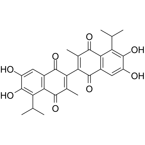
Apogossypolone (ApoG2) is an orally active Bcl-2 family proteins inhibitor with Ki values of 35, 25 and 660 nM for Bcl-2, Mcl-1 and Bcl-XL, respectively. Apogossypolone shows antitumor activities, induces cell apoptosis and autophagy. Apogossypolone also has antifungal activity.
For research use only. We do not sell to patients.
- Purity : 98.58%
- CAS No.: 886578-07-0
- Formula: C28H26O8
- Molecular Weight:490.50
-
Storage:Powder -20°C, 3 years , 4°C, 2 years ; In solvent -80°C, 6 months , -20°C, 1 month

To place orders, for customer services and technical support, please contact: MedChemExpress USA
Tel: 609-228-6898 E-mail: [email protected] [email protected]


-
 Biological Activity
-
Chemical Information
- Protocols
- Purity & Documentation
- References
-
 Help & FAQs


-
HY-15531
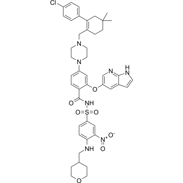
VenetoclaxBCL-2 Inhibitor -
HY-10087
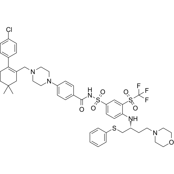
NavitoclaxBcl-2 Inhibitor -
HY-50907
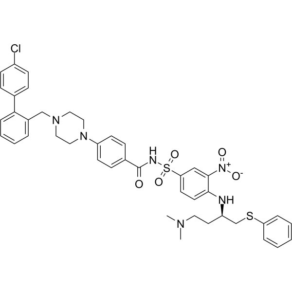
ABT-737Bcl-2 Family Inhibitor/BH3 Mimetic -
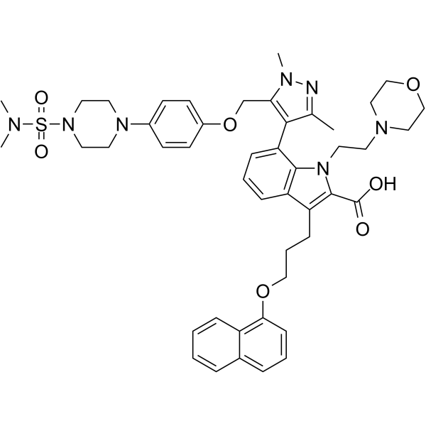
HY-12468
A-1210477Mcl-1 Inhibitor -

HY-15531R
Venetoclax (Standard)BCL-2 Inhibitor -
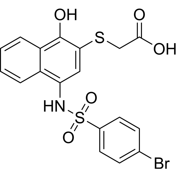
HY-18628
UMI-77Bcl-2 Family Inhibitor -

HY-10087R
Navitoclax (Standard)Reference Standard -
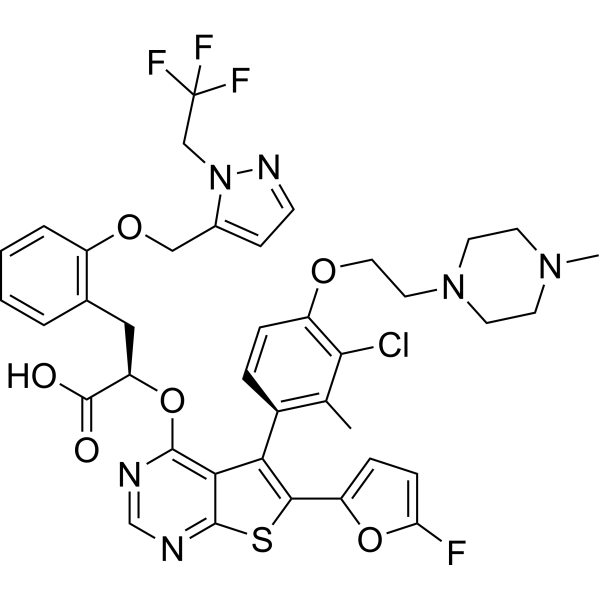
HY-100741
S63845MCL1 Inhibitor -
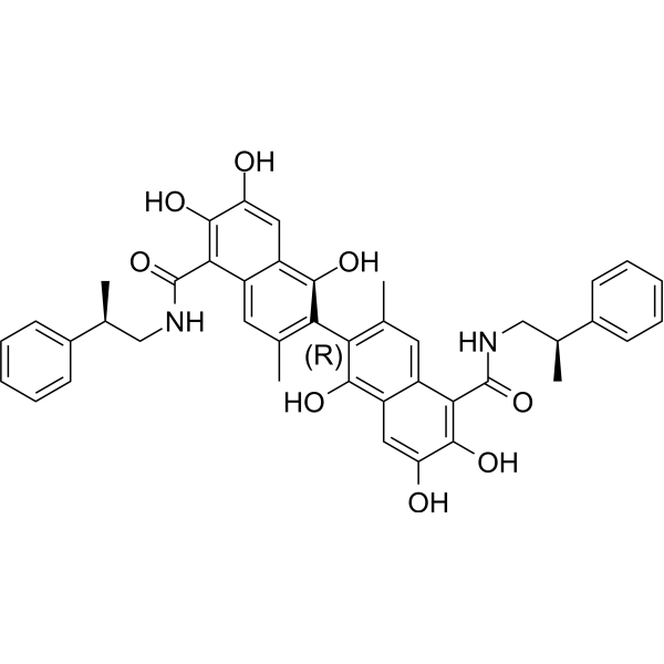
HY-15191
SabutoclaxBcl-2 Family Inhibitor -
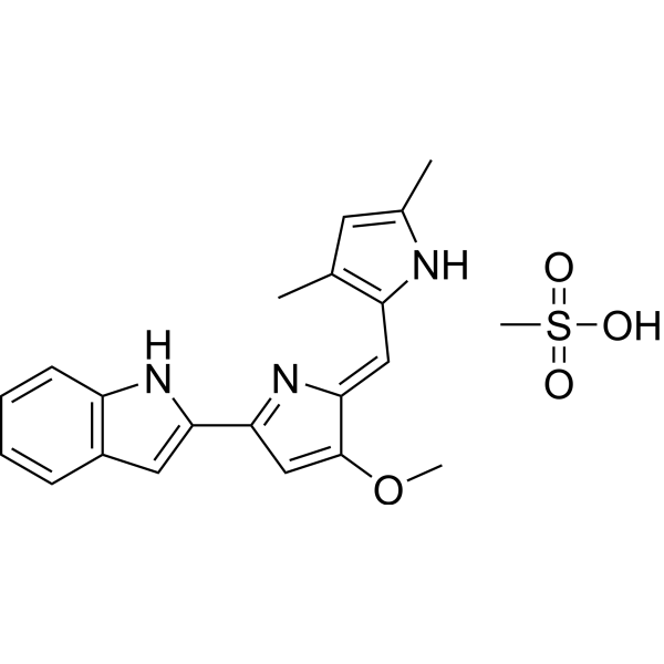
HY-10969
Obatoclax MesylateBcl-2 Family Inhibitor -
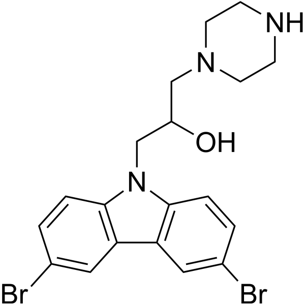
HY-103269
BAI1BAX Inhibitor -
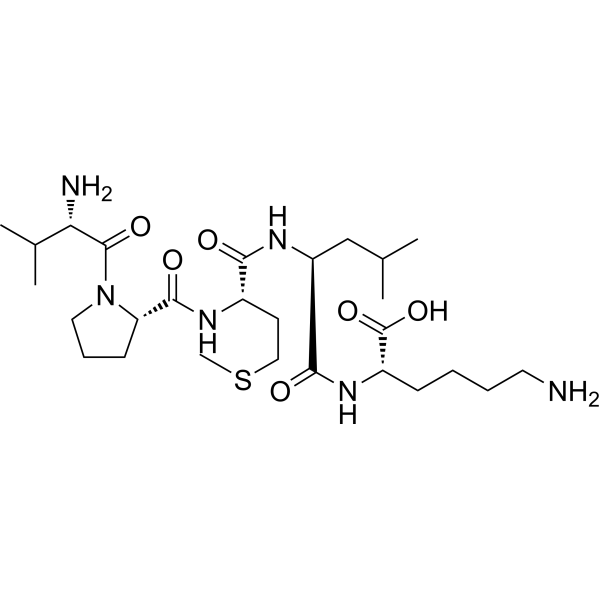
HY-P0081
Bax inhibitor peptide V5Bcl-2 Family Inhibitor


Biological Activity
Description
IC50 & Target
|
Mcl-1 25 nM (Ki) |
Bcl-2 35 nM (Ki) |
Bcl-xL 660 nM (Ki) |
Cellular Effect
|
Cell Line

|
Type | Value | Description | References |
|---|---|---|---|---|
| NCI-H1299 | EC50 |
2.76 μM
Compound: 6a
|
Cytotoxicity against human H1299 cells expressing high level of Mcl-1 after 72 hrs by ATP-LITE assay
Cytotoxicity against human H1299 cells expressing high level of Mcl-1 after 72 hrs by ATP-LITE assay
|
[PMID: 21033669] |
| NCI-H460 | EC50 |
0.4 μM
Compound: 6a
|
Cytotoxicity against human H460 cells expressing high level of Bcl-2 after 72 hrs by ATP-LITE assay
Cytotoxicity against human H460 cells expressing high level of Bcl-2 after 72 hrs by ATP-LITE assay
|
[PMID: 21033669] |
| PC-3 | EC50 |
1.46 μM
Compound: 6a
|
Cytotoxicity against human PC3 cells expressing high level of Bcl-xL after 72 hrs by ATP-LITE assay
Cytotoxicity against human PC3 cells expressing high level of Bcl-xL after 72 hrs by ATP-LITE assay
|
[PMID: 21033669] |
| RS4-11 | EC50 |
7.4 μM
Compound: 6a
|
Induction of apoptosis in human RS4:11 cells expressing high level of Bcl-2 and Bcl-xL after 24 hrs by annexin V-FITC and propidium iodide staining
Induction of apoptosis in human RS4:11 cells expressing high level of Bcl-2 and Bcl-xL after 24 hrs by annexin V-FITC and propidium iodide staining
|
[PMID: 21033669] |
| RS4-11 | EC50 |
7.47 μM
Compound: 6a
|
Cytotoxicity against human RS4:11 cells expressing high level of Bcl-2 and Bcl-xL after 72 hrs by annexin V-FITC and propidium iodide staining
Cytotoxicity against human RS4:11 cells expressing high level of Bcl-2 and Bcl-xL after 72 hrs by annexin V-FITC and propidium iodide staining
|
[PMID: 21033669] |
In Vitro
Apogossypolone (ApoG2) shows improved stability under stressed conditions[1].
Apogossypolone (0-1 μM, 72 or 96 h) inhibits WSU-DLCL2 cells growth in a dose-dependent manner[1].
Apogossypolone (0-5 μM, 24 or 48 h) interferes with the formation of heterodimers between anti-apoptotic and pro-apoptotic Bcl-2 family members, and leads to cleavage of caspase-3, caspase-9 and PARP[1].
Apogossypolone (0-8 μM, 0-72 h) induces apoptotic WSU-DLCL2 cell death in a time- and dose-dependent manner[1].
Apogossypolone (0-10 μM, 0-24 h) induces autophagy and promotes ROS generation in HCC cells[2].
MedChemExpress (MCE) has not independently confirmed the accuracy of these methods. They are for reference only.
-
Cell Line:WSU-DLCL2
-
Concentration:250, 350, 500 and 1000 nM
-
Incubation Time:96 h for cell counting, 72 h for MTT
-
Result:Inhibited growth in a dose-dependent manner. The 50% growth inhibition concentration (IC50) was approximately 350 nM.
-
Cell Line:WSU-DLCL2
-
Concentration:0.35, 0.5, 1 and 5 µM
-
Incubation Time:24 or 48 h
-
Result:Blocked the formation of heterodimers between Bcl-XL and Bim in a concentration-dependent manner. Resulted in the activation of cleavages of caspase-3, caspase-9 and PARP.
-
Cell Line:WSU-DLCL2
-
Concentration:0, 1, 2, 4 and 8 µM
-
Incubation Time:24, 48 and 72 h
-
Result:Induced cell apoptosis in a time- and dose-dependent manner.
-
Cell Line:HepG2 and Hep3B
-
Concentration:1.25, 2.5, 5 and 10 µM
-
Incubation Time:6, 12, 18 and 24 h
-
Result:Induced LC3 (Light chain 3)-II conversion in a dose- and time-dependent manner.
In Vivo
MedChemExpress (MCE) has not independently confirmed the accuracy of these methods. They are for reference only.
-
Animal Model:Four-week-old female ICR-SCID mice, each mouse received 107 WSU-DLCL2 cells (in serum-free RPMI 1640) subcutaneously (sc) in each flank area[1]
-
Dosage:120 mg/kg
-
Administration:Intravenous or administration per day for five days
-
Result:Inhibited the growth of WSU-DLCL2 and significantly decreased the tumor weight.
-
Animal Model:Non-tumor-bearing SCID mice[1]
-
Dosage:160 mg/kg
-
Administration:Intravenous or administration per day for five days
-
Result:Was well tolerated in mice up to 800 mg/kg. Displayed no gross signs of toxicity.


Chemical Information
-
CAS No. 886578-07-0
-
Appearance Solid
-
Molecular Weight 490.50
-
Formula C28H26O8
-
Color Orange to reddish brown
-
SMILES
O=C1C(C(C2=O)=C(C)C(C3=C2C=C(O)C(O)=C3C(C)C)=O)=C(C)C(C4=C1C=C(O)C(O)=C4C(C)C)=O
-
Synonyms
ApoG2
-
Shipping
Room temperature in continental US; may vary elsewhere.
-
Storage
Powder -20°C 3 years 4°C 2 years In solvent -80°C 6 months -20°C 1 month


Purity & Documentation
-
Data Sheet (279 KB)
-
SDS (393 KB)
- English - EN (393 KB)
- Français - FR (393 KB)
- Deutsch - DE (393 KB)
- Norwegian - NO (393 KB)
- Español - ES (393 KB)
- Swedish - SV (393 KB)
- Italian - IT (393 KB)
- Korean - KR (393 KB)
- Portuguese - PT (393 KB)
-
Handling Instructions (2659 KB)
References
[1]. Yuan Sun, et al. Apogossypolone, a nonpeptidic small molecule inhibitor targeting Bcl-2 family proteins, effectively inhibits growth of diffuse large cell lymphoma cells in vitro and in vivo. Cancer Biol Ther. 2008 Sep;7(9):1418-26. [Content Brief]
[2]. Cheng P, et al. The novel BH-3 mimetic apogossypolone induces Beclin-1- and ROS-mediated autophagy in human hepatocellular carcinoma [corrected] cells. Cell Death Dis. 2013 Feb 7;4(2):e489. [Content Brief]
[3]. Jay E Mellon, et al. Inhibitory effects of gossypol, gossypolone, and apogossypolone on a collection of economically important filamentous fungi. J Agric Food Chem. 2012 Mar 14;60(10):2740-5. [Content Brief]

Calculators
Concentration (start) × Volume (start) = Concentration (final) × Volume (final)


Apogossypolone Related Classifications
HY-19551 Related Classifications
 Powered by Bioz