



BET BD2-IN-1
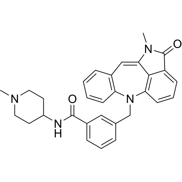
BET BD2-IN-1 (compound 45) is a potent and selective inhibitor of BET BD2 (IC50=1.6 nM). BET BD2-IN-1 inhibits the differentiation of Th17 cells by decreasing the activation of STAT3 and NF-κB. BET BD2-IN-1 is used in psoriasis and inflammatory bowel disease (IBD) research.
For research use only. We do not sell to patients.
- CAS No.: 2677039-24-4
- Formula: C30H30N4O2
- Molecular Weight:478.58
-
Storage:
Please store the product under the recommended conditions in the Certificate of Analysis.

To place orders, for customer services and technical support, please contact: MedChemExpress USA
Tel: 609-228-6898 E-mail: [email protected] [email protected]


-
 Biological Activity
-
Chemical Information
- Protocols
- Purity & Documentation
- References
-
 Help & FAQs




Biological Activity
Description
IC50 & Target
|
BRD2 BD1 570 nM (IC50) |
BRD2 BD2 5.9 nM (IC50) |
BRD3 BD1 465 nM (IC50) |
BRD3 BD2 6.0 mM (IC50) |
BRD4 BD1 524 nM (IC50) |
BRD4 BD2 1.6 nM (IC50) |
BRDT BD1 527 nM (IC50) |
In Vitro
BET BD2-IN-1 (500 nM) effectively inhibits Th17 cell differentiation and has excellent selectivity for BD2 over BD1[1].
BET BD2-IN-1 (4 nM) binds to BRD4 BD2 in intact cells and has stabilization effect on BRD4 BD2[1].
MedChemExpress (MCE) has not independently confirmed the accuracy of these methods. They are for reference only. Further protocols information, click here.
-
Cell Line:Th17 cells
-
Concentration:500 nM
-
Incubation Time:
-
Result:Showed the strongest inhibitory activity and reduced differentiation from 19.0 to 7.81%.
-
Cell Line:HEK 293 T cells
-
Concentration:4, 20, 100, 500, 2500, 10000 nM
-
Incubation Time:30 min
-
Result:Stabilized BRD4 BD2 at 50.4 °C.
In Vivo
BET BD2-IN-1 (20 mg/kg for i.v; once daily for seven days) significantly decreases the disease activity index (DAI) score in dextran sulfate sodium (DSS) induced IBD mouse model[1].
Pharmacokinetic Analysis of BET BD2-IN-1 in Sprague Dawley Rats Model[1]
MedChemExpress (MCE) has not independently confirmed the accuracy of these methods. They are for reference only.
-
Animal Model:Imiquimod (HY-B0180)-induced Psoriasis mouse model[1]
-
Dosage:10 and 20 mg/kg
-
Administration:Intravenous injection (i.v.) ; Once daily for seven days
-
Result:Significantly alleviated the Imiquimod-induced skin lesions in a dose-dependent manner.
Obviously reduced the enlarged spleen.
Significantly decreased the expression of p-STAT3 and p-NF-κB in mouse skin tissues.
-
Animal Model:DSS (HY-116282C)-induced IBD mouse model[1]
-
Dosage:10 and 20 mg/kg
-
Administration:Intravenous injection (i.v.) ; Once daily for seven days
-
Result:Effectively prevented colon shortening.
Effectively alleviated the DSS-induced weight loss.
Significantly alleviated the infiltration of inflammatory cells, areas of ulceration as well as loss of mucosal epithelium, and goblet cells caused by DSS.


Chemical Information
-
CAS No. 2677039-24-4
-
Molecular Weight 478.58
-
Formula C30H30N4O2
-
SMILES
O=C(C1=CC=CC(CN2C3=C(C=C4C5=C2C=CC=C5C(N4C)=O)C=CC=C3)=C1)NC6CCN(CC6)C
-
Shipping
Room temperature in continental US; may vary elsewhere.
-
Storage
Please store the product under the recommended conditions in the Certificate of Analysis.

Protocols
-
Cell Cytotoxicity Assay
Cytotoxicity assays are usually based on the assessment of cell membrane damage, which can also be indirectly detected by measuring cell viability. Detection methods include MTT assay, CKK-8 assay, LDH assay and ATP assay, etc.
-
Naïve CD4+ T-cell subset differentiation/polarization
Naïve CD4+ T-cell subset differentiation/polarization is an in vitro assay in which purified naïve CD4+ T cells are activated through TCR and CD28 costimulation and cultured with defined cytokines and neutralizing antibodies to generate Th0, Th1, Th2, Th17, or induced Treg-like populations. Differentiation is detected by subset-associated cytokines and transcription factors: IFN-γ/T-bet for Th1, IL-4/GATA3 for Th2, IL-17A/RORγt for Th17, and Foxp3 for induced Treg cells. The assay readout is usually generated by intracellular cytokine staining after restimulation, transcription-factor staining by flow cytometry, ELISA of secreted cytokines, or gene-expression analysis. The result reflects cytokine-directed lineage commitment or polarization rather than antigen-specific immune protection by itself.
-
Imiquimod-Induced Psoriasiform Dermatitis
Imiquimod (IMQ)-induced psoriasiform dermatitis is a widely used murine model in which topical application of IMQ, a Toll-like receptor 7 (TLR7) agonist, triggers innate immune activation in the skin and induces a psoriasis-like inflammatory cascade characterized by epidermal hyperplasia, immune cell infiltration, and cytokine production dominated by the IL-23/IL-17 axis. This inflammatory response is mediated through activation of dendritic cells and downstream induction of IL-23, IL-17A, IL-22, and related pro-inflammatory mediators, recapitulating key features of human plaque psoriasis and enabling mechanistic and therapeutic studies. The model is commonly induced using Aldara (5% IMQ cream) applied topically to murine skin, resulting in rapid onset of erythema, scaling, and thickening that can be quantified as disease severity indices and validated histologically.
-
DSS-Induced Colitis
Dextran sulfate sodium (DSS)-induced colitis is generated by administering DSS in mouse drinking water, producing epithelial injury, barrier disruption, weight loss, diarrhea, fecal blood, colon shortening, histologic mucosal damage, and inflammatory mediator changes; the model is mainly used to study acute or chronic intestinal inflammation resembling selected features of ulcerative colitis. DSS injury is interpreted through clinical and tissue readouts rather than a single molecular endpoint: daily body weight, stool consistency, and bleeding are combined into a disease activity index, while colon length, histology, cytokines, myeloperoxidase activity, intestinal permeability, and tight-junction markers provide complementary measures of inflammation and barrier damage.
-
TNBS-Induced Colitis
TNBS-induced colitis is produced by intrarectal delivery of 2,4,6-trinitrobenzene sulfonic acid in ethanol, where ethanol disrupts the mucosal barrier and TNBS haptenates colonic proteins, generating immune-mediated colonic inflammation with weight loss, diarrhea, ulceration, transmural injury, inflammatory-cell infiltration, and cytokine responses. The model is used as an experimental intestinal inflammation model with Crohn’s disease–like features, especially when Th1-type responses, IL-12–dependent inflammation, chronic relapsing inflammation, or fibrosis-related endpoints are studied.
-
Research Protocol for Inflammation-related Diseases
The NLRP3 inflammasome is a cytosolic innate immune signaling platform that integrates priming signals and danger-signal activation to promote caspase-1 activation, maturation of IL-1β and IL-18, and gasdermin D-mediated pyroptotic cell death. The core experimental logic is to determine whether inflammatory disease phenotypes are driven by increased NLRP3 expression, ASC-containing inflammasome assembly, caspase-1 cleavage, GSDMD cleavage, and extracellular release of IL-1β/IL-18 rather than by nonspecific cell injury alone. The pathway is strongly linked to inflammation-related disease phenotypes because monosodium urate crystals activate NALP3/NLRP3 inflammasome signaling in gout-like crystal inflammation, cholesterol crystals activate NLRP3 inflammasomes in atherogenesis models, and DSS-induced intestinal inflammation has been reported to involve NLRP3 inflammasome activity. However, experimental colitis studies also show context-dependent protective effects of NLRP3 inflammasome co


Purity & Documentation
References

Calculators
Concentration (start) × Volume (start) = Concentration (final) × Volume (final)


BET BD2-IN-1 Related Classifications
HY-155680 Related Classifications
 Powered by Bioz