



Cefatrizine
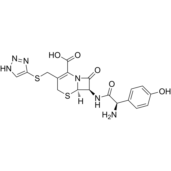
Cefatrizine (BL-S-640) is an orally active and broad-spectrum cephalosporin antibiotic. Cefatrizine is also a eEF2K inhibitor, with anti-proliferative activity in human breast cancer cells, which could induce ER stress, leading to cell death. Cefatrizine can be used in studies of cancer and bacterial infection.
Nur für Forschungszwecke. Wir verkaufen nicht an Patienten.
- CAS. Nr.: 51627-14-6
- Formel: C18H18N6O5S2
- Molecular Weight:462.50
-
Speicherung:
Please store the product under the recommended conditions in the Certificate of Analysis.

To place orders, for customer services and technical support, please contact: MedChemExpress USA
Tel: 609-228-6898 E-mail: [email protected] [email protected]


-
 Biologische Aktivität
-
Chemical Information
-
Lösungsmittel & Löslichkeit
- Protokoll
- Reinheit & Dokumentation
- Verweise
-
 Help & FAQs


Alle Antibiotic Isoform-spezifische Produkte anzeigen
More

Biologische Aktivität
Beschreibung
IC50 & Target
|
β-lactam |
Cellular Effect
|
Cell Line

|
Type | Value | Description | References |
|---|---|---|---|---|
| MDA-MB-436 | IC50 |
30 μM
Compound: 40
|
Anticancer activity against human MDA-MB-436 cells assessed as cell growth inhibition incubated for 24 hrs by MTT assay
Anticancer activity against human MDA-MB-436 cells assessed as cell growth inhibition incubated for 24 hrs by MTT assay
|
[PMID: 33650861] |
In Vitro
Cefatrizine (0-100 µM; 24 h) causes a remarkable anti-proliferative effect on MCF-7 and MDA-MB-436 cell growth in a dose-dependent manner[1].
Cefatrizine (30 µM; 12 h) induces ER stress in breast cancer cells[1].
Cefatrizine (12, 24, 36 h) increases level of CHOP (marker of ER stress induced apoptosis) and promotes expressions of core proteins in eEF2K-modulated ER stress pathways (Bip, p-PERK, XBP-1S and p-JNK) in MCF-7 and MDA-MB-436 cells[1].
MedChemExpress (MCE) has not independently confirmed the accuracy of these methods. They are for reference only. Further protocols information, click here.
-
Cell Line:MCF-7, MDA-MB-436 cells
-
Concentration:0-100 µM
-
Incubation Time:24 h
-
Result:Led to a remarkable anti-proliferative effect on cells and resulted in almost 50% inhibition in the MCF-7 and MDA-MB-436 cells when at 33 µM and 29 µM.
-
Cell Line:MCF-7, MDA-MB-436 cells
-
Concentration:30 µM
-
Incubation Time:12 h
-
Result:Led to massive cytoplasmic vacuolization.
In Vivo
MedChemExpress (MCE) has not independently confirmed the accuracy of these methods. They are for reference only.
-
Animal Model:MaleSwiss-Webster mice (19-21 g; P. mirabilis intracystically infected model)[2].
-
Dosage:0.2, 1, 5, 25 mg/kg
-
Administration:Oral administration; 4 times daily for 3 days.
-
Result:Reduced the number of infecting organisms to less than 1,000 in the bladder when 1 mg/kg, and in the kidneys when 0.2 mg/kg.


Chemical Information
-
CAS. Nr. 51627-14-6
-
Molecular Weight 462.50
-
Formel C18H18N6O5S2
-
SMILES
O=C(O)C(N1[C@@]([H])([C@@H](C1=O)NC([C@@H](C2=CC=C(C=C2)O)N)=O)SC3)=C3CSC4=CNN=N4
-
Synonyms
BL-S-640; SK&F 60771
-
Versand
Room temperature in continental US; may vary elsewhere.
-
Speicherung
Please store the product under the recommended conditions in the Certificate of Analysis.


Lösungsmittel & Löslichkeit
In Vitro:
H2O : 250 mg/mL (540.54 mM; Need ultrasonic)
* Note: If you choose water as the stock solution, please dilute it to the working solution, then filter and sterilize it with a 0.22 μm filter before use.
* Note: If you choose water as the stock solution, please dilute it to the working solution, then filter and sterilize it with a 0.22 μm filter before use.
Konzentration (Stammlösung) × Volumen (Stammlösung) = Konzentration (Ziellösung) × Volumen (Ziellösung)

Protokoll
-
Research Protocol for Infectious Diseases
Infectious-disease experiments test how pathogens interact with host barriers, innate immune receptors, inflammatory signaling, pathogen replication, and tissue injury; pattern-recognition receptors such as TLRs, RIG-I-like receptors, NOD-like receptors, and inflammasomes detect microbial molecules and activate NF-κB, interferon, and cytokine responses. The central hypothesis is that infection severity reflects the balance between pathogen burden and host response: protective inflammation restricts pathogen growth, whereas excessive or mislocalized inflammation contributes to tissue damage and disease phenotype. Unresolved questions include which host pathways are protective versus pathogenic, why some infection models fail to translate to human disease, and which combined readouts best predict clinically relevant infection outcomes.
-
Cell Cytotoxicity Assay
Cytotoxicity assays are usually based on the assessment of cell membrane damage, which can also be indirectly detected by measuring cell viability. Detection methods include MTT assay, CKK-8 assay, LDH assay and ATP assay, etc.
-
Mammalian live/dead viability and cytotoxicity staining
Live/dead viability and cytotoxicity staining assays are based on the simultaneous detection of intracellular esterase activity in metabolically active (viable) cells and membrane integrity loss in non-viable cells. In commonly used dual-staining approaches, membrane-permeant fluorogenic substrates are converted by intracellular esterases into fluorescent products in live cells, while impermeant DNA-binding dyes selectively enter cells with compromised plasma membranes and label nucleic acids in dead or dying cells, enabling discrimination between viable and non-viable populations by fluorescence microscopy or flow cytometry.
-
Apoptosis Solutions
Apoptosis is a regulated, generally non-lytic cell-death pathway that removes unwanted, damaged, infected, or abnormal cells through coordinated morphological changes, caspase activation, DNA fragmentation, and membrane remodeling. The intrinsic apoptosis pathway is controlled mainly by mitochondrial outer membrane permeabilization, BCL-2 family proteins, cytochrome c release, apoptosome formation, caspase-9 activation, and downstream executioner caspase-3/7 activation. The extrinsic apoptosis pathway is initiated by death receptors such as Fas, TNFR, and TRAIL receptors, which recruit adaptor proteins and activate caspase-8 before engaging executioner caspases or mitochondrial amplification through BID cleavage. Apoptosis is linked to many phenotypes, including cancer cell killing, tissue homeostasis, immune regulation, neurodegeneration, infection response, and treatment-induced cytotoxicity; unresolved questions include how apoptosis interacts with necroptosis, pyroptosis, ferroptos
-
Breast Cancer Modeling
Breast cancer is a heterogeneous cancer, and it has been distinguished into four subtypes: luminal A, luminal B, HER2-positive and basal-like. Molecular mutations, epigenetic alterations, hormone exposure and immune microenvironment are related to the progression of breast cancer.
-
Cell Viability Determination by MTT Colorimetric Assay
The following protocol uses the MTT colorimetric assay as a classic literature-established method for assessing cell viability/metabolic activity in cultured mammalian cells. MTT[3-(4,5-dimethylthiazol-2-yl)-2,5-diphenyltetrazolium bromide] is reduced by metabolically active cells to a colored formazan product; the amount of formazan is quantified spectrophotometrically and provides an indirect measure of metabolically active viable cells. Importantly, MTT reduction reflects cellular oxidoreductase/metabolic activity rather than an absolute direct count of living cells, so changes in cellular metabolism can alter the signal independently of cell number.


Reinheit & Dokumentation
Verweise
[1]. Yao Z, et al. Integrative bioinformatics and proteomics-based discovery of an eEF2K inhibitor (cefatrizine) with ER stress modulation in breast cancer cells. Mol Biosyst. 2016 Mar;12(3):729-36. [Content Brief]
[2]. Leitner F, et al. BL-S640, a cephalosporin with a broad spectrum of antibacterial activity: bioavailability and therapeutic properties in rodents. Antimicrob Agents Chemother. 1975 Mar;7(3):306-10. [Content Brief]


Complete Stock Solution Preparation Table
| Optional Solvent | Concentration Solvent Mass | 1 mg | 5 mg | 10 mg | 25 mg |
|---|---|---|---|---|---|
| H2O | 1 mM | 2.1622 mL | 10.8108 mL | 21.6216 mL | 54.0541 mL |
| 5 mM | 0.4324 mL | 2.1622 mL | 4.3243 mL | 10.8108 mL | |
| 10 mM | 0.2162 mL | 1.0811 mL | 2.1622 mL | 5.4054 mL | |
| 15 mM | 0.1441 mL | 0.7207 mL | 1.4414 mL | 3.6036 mL | |
| 20 mM | 0.1081 mL | 0.5405 mL | 1.0811 mL | 2.7027 mL | |
| 25 mM | 0.0865 mL | 0.4324 mL | 0.8649 mL | 2.1622 mL | |
| 30 mM | 0.0721 mL | 0.3604 mL | 0.7207 mL | 1.8018 mL | |
| 40 mM | 0.0541 mL | 0.2703 mL | 0.5405 mL | 1.3514 mL | |
| 50 mM | 0.0432 mL | 0.2162 mL | 0.4324 mL | 1.0811 mL | |
| 60 mM | 0.0360 mL | 0.1802 mL | 0.3604 mL | 0.9009 mL | |
| 80 mM | 0.0270 mL | 0.1351 mL | 0.2703 mL | 0.6757 mL | |
| 100 mM | 0.0216 mL | 0.1081 mL | 0.2162 mL | 0.5405 mL |
* Note: If you choose water as the stock solution, please dilute it to the working solution, then filter and sterilize it with a 0.22 μm filter before use.


Cefatrizine Related Classifications
HY-123024 Related Classifications
 Powered by Bioz