



Fludrocortisone

Based on 1 Customer Validation
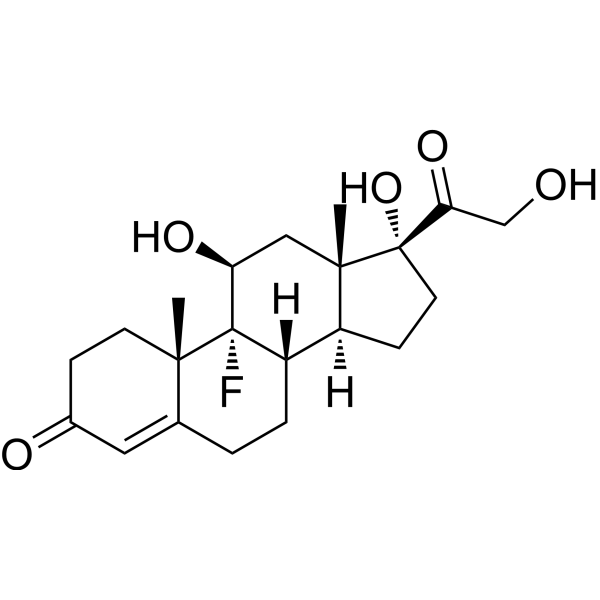
Fludrocortisone (9α-Fludrocortisone) is an orally active mineralocorticoid and glucocorticoid receptor agonist. Fludrocortisone suppresses pro-inflammatory cytokine expression, reduces CCL2, IL-6, IL-8 levels, upregulates mineralocorticoid receptor (MR) expression, induces PI3K/Akt, GSK-3β, CREB, ERK1/2, mTOR phosphorylation, blocks Tau hyperphosphorylation, prevents apoptosis, promotes survival and proliferation, enhances renal sodium and water transport, increases plasma volume and blood pressure, reduces plasma potassium and renin activity, stimulates erythropoietin expression, modulates uterine receptivity genes, and reverses PP242-induced MUC1 upregulation. Fludrocortisone can be used for the research of congenital adrenal hyperplasia, postural hypotension, and adrenal insufficiency.
For research use only. We do not sell to patients.
- Purity: 98.99%
- CAS No.: 127-31-1
- Formula: C21H29FO5
- Molecular Weight:380.45
-
Storage:Powder -20°C, 3 years , 4°C, 2 years ; In solvent -80°C, 6 months , -20°C, 1 month

To place orders, for customer services and technical support, please contact: MedChemExpress USA
Tel: 609-228-6898 E-mail: [email protected] [email protected]


-
 Biological Activity
-
Chemical Information
-
Solvent & Solubility
- Purity & Documentation
- References
-
 Help & FAQs


-
HY-113414
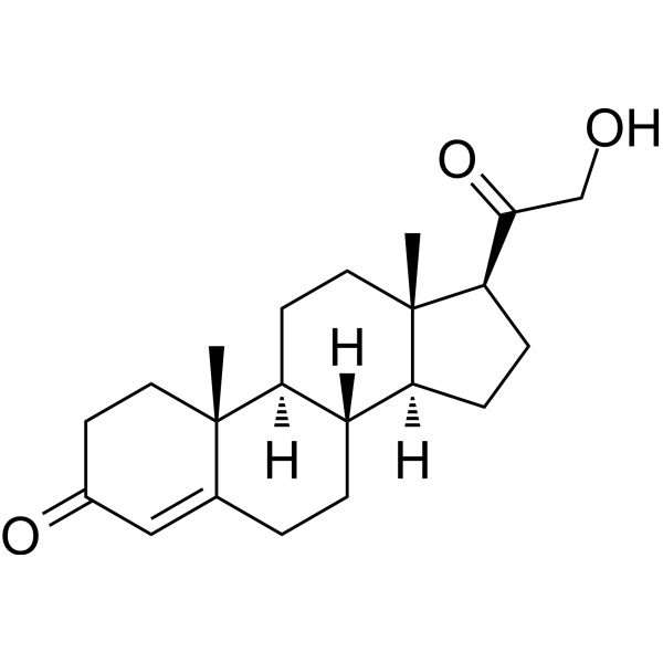
Deoxycorticosterone
-

HY-14648
Dexamethasone
-

HY-100558
Bafilomycin A1
-
HY-B0328
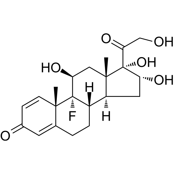
Triamcinolone
-
HY-13580
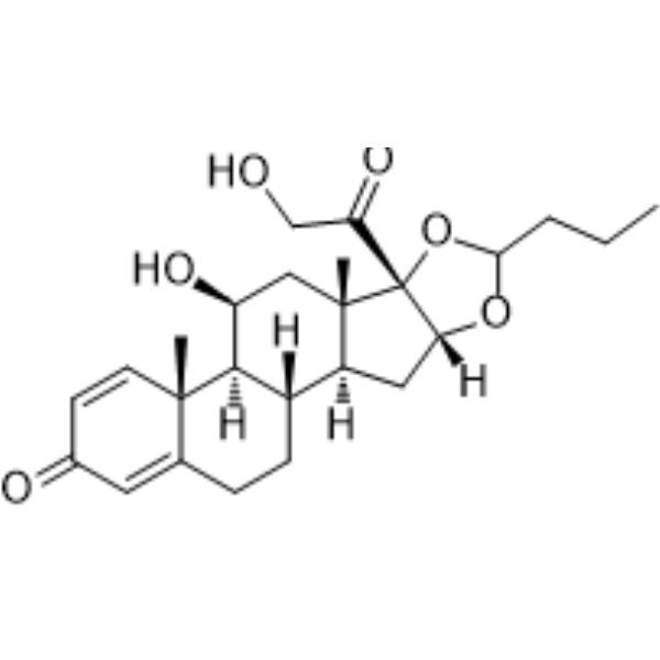
Budesonide
-
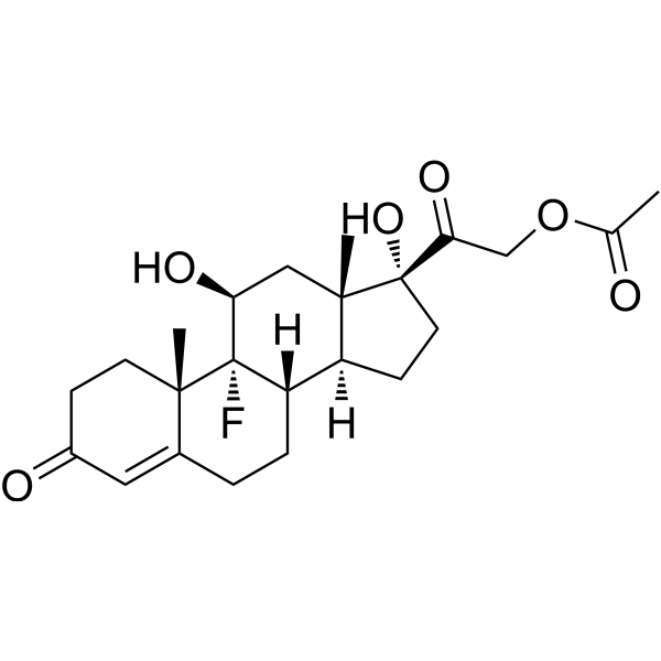
HY-B1203AR
Fludrocortisone acetate (Standard)
-

HY-B1203R
Fludrocortisone (Standard)
-
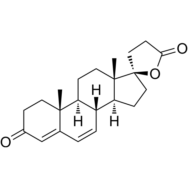
HY-B1438
Canrenone
-
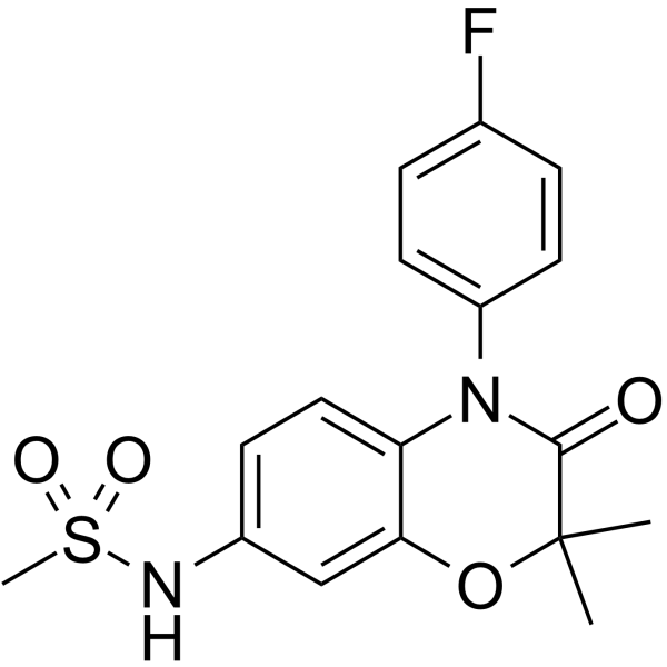
HY-109002
Apararenone
-
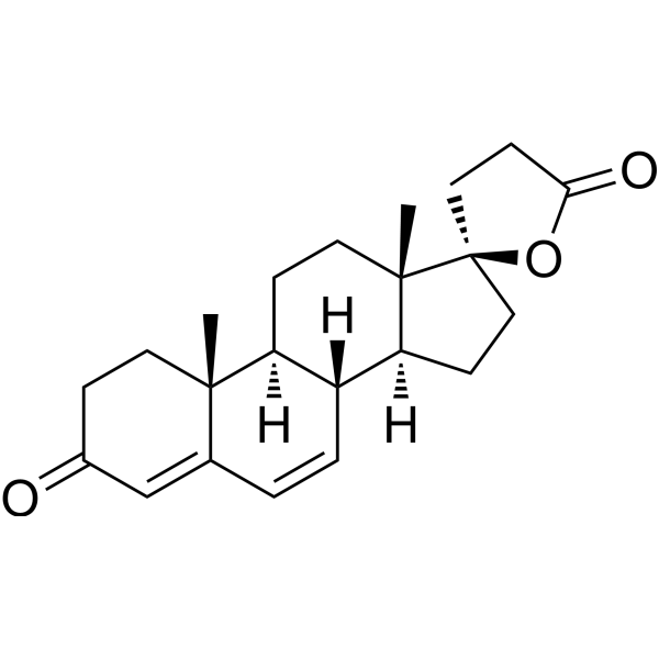
HY-B1438R
Canrenone (Standard)
-
HY-B0251
Eplerenone
-
HY-100471
Esaxerenone


Biological Activity
Fludrocortisone (0.01-10 μg/μL; 12 h) is non-cytotoxic to MIO-M1, ARPE19, and 661W human retinal cell lines at concentrations up to 1 μg/μL, with reduced viability only seen at 10 μg/μL in MIO-M1 and 661W cells[1].
Fludrocortisone (1 μg/μL; 12 h) potently suppresses IL-1β- and TNF-α-induced CCL2, IL-6, and IL-8 mRNA expression in MIO-M1 human Müller glial cells[1].
Fludrocortisone (1 μM; 24 h) increases mineralocorticoid receptor
(MR) gene expression in AHPs without altering glucocorticoid receptor (GR) expression[2].
Fludrocortisone (0.5-2 μM; 24 h) promotes survival, proliferation, and reduces apoptosis in growth factor-deprived AHPs, with 1 μM being the optimal concentration[2].
Fludrocortisone (1 μM) activates cAMP/PKA/CREB signaling in AHPs, which is required for its pro-survival and pro-proliferative effects[2].
Fludrocortisone (1 μM) activates PI3K/Akt and mTOR/p70S6K signaling, and inactivates GSK-3β, which is required for its protective effects in growth factor-deprived AHPs[2].
Fludrocortisone (1 μM) protects AHPs from Aβ1-42-induced toxicity by promoting survival, proliferation, reducing apoptosis, restoring Akt/GSK-3β phosphorylation, and blocking Tau hyperphosphorylation[2].
Fludrocortisone (4-6 h) stimulates Epo, HIF2α, HIF1α, and PHD2 mRNA expression in microdissected rat nephron segments and mouse kidney nephron segments and peritubular cells, with significant increases relative to basal conditions[5].
Fludrocortisone (6 h, 72 h) significantly increases Epo protein expression in rat renal cortex but not in rat liver, relative to basal conditions[5].
Fludrocortisone (4-6 h) stimulates Epo protein production in mouse MAL, CCD, and OMCD, with specific upregulation in type A intercalated cells of the collecting ducts, and effects diminish to basal levels by 72 h[5].
MedChemExpress (MCE) has not independently confirmed the accuracy of these methods. They are for reference only.
-
Cell Line:human MIO-M1 Müller glial cells, ARPE19 retinal pigment epithelial cells, 661W cone photoreceptor-like cells
-
Concentration:0.01-10 μg/μL
-
Incubation Time:12 h
-
Result:Did not significantly alter cell viability in ARPE19 cells at all tested concentrations. Showed unchanged viability in MIO-M1 and 661W cells at 0.01, 0.1, and 1 μg/μL; caused a slight but statistically significant reduction in viability only at the highest concentration of 10 μg/μL in MIO-M1 and 661W cells.
-
Cell Line:human MIO-M1 Müller glial cells
-
Concentration:1 μg/μL
-
Incubation Time:12 h (co-incubated with IL-1β stimulation)
-
Result:Completely abrogated IL-1β-induced significant increases in CCL2, IL-6, and IL-8 mRNA expression, suppressing expression to near baseline levels.
-
Cell Line:human MIO-M1 Müller glial cells
-
Concentration:1 μg/μL
-
Incubation Time:12 h (co-incubated with TNF-α stimulation)
-
Result:Sharply reduced TNF-α-induced significant increases in CCL2, IL-6, and IL-8 mRNA expression, suppressing expression to near baseline levels.
-
Cell Line:Human MIO-M1 Müller glial cells
-
Concentration:1 μg/μL
-
Incubation Time:12 h (co-incubated with IL-1β stimulation)
-
Result:Had its suppressive effect on IL-1β-induced CCL2, IL-6, and IL-8 expression completely abrogated by co-incubation with glucocorticoid receptor antagonist RU486.
-
Cell Line:Adult rat hippocampal progenitor cells (AHPs)
-
Concentration:1 μM
-
Incubation Time:24 h
-
Result:Strongly increased mineralocorticoid receptor (MR) gene expression; showed no significant change in glucocorticoid receptor (GR) gene expression.
-
Cell Line:Adult rat hippocampal progenitor cells (AHPs)
-
Concentration:0.01, 0.1, 0.5, 1, 2 μM
-
Incubation Time:24 h
-
Result:Progressively increased cell survival and proliferation at 0.5, 1, and 2 μM; strongly reduced serum starvation-induced apoptosis (via decreased caspase-3 activity) at 0.5, 1, and 2 μM; identified 1 μM as the optimal concentration for subsequent experiments.
-
Cell Line:adult rat hippocampal progenitor cells (AHPs)
-
Concentration:1 μM (cAMP/CREB phosphorylation assays); 1 μM (functional validation with KT5720)
-
Incubation Time:5, 15, 30, 60, 90 min (cAMP/CREB phosphorylation assays); 24 h (functional validation with KT5720)
-
Result:Increased intracellular cAMP levels, with a peak at 15 min and sustained elevation through 90 min; promoted CREB phosphorylation (Ser133), peaking at 15-30 min and returning to near basal levels by 90 min; had induced survival and proliferation reduced by co-treatment with PKA inhibitor KT5720.
-
Cell Line:adult rat hippocampal progenitor cells (AHPs)
-
Concentration:1 μM (phosphorylation assays); 1 μM (functional validation with wortmannin/rapamycin)
-
Incubation Time:5, 15, 30, 60, 90 min (phosphorylation assays); 24 h (functional validation with wortmannin/rapamycin)
-
Result:Induced time-dependent phosphorylation of Akt (peaking at 30 min, sustained through 90 min), GSK-3β (sustained elevation from 5 to 90 min), and p70S6K (progressive elevation through 90 min); had induced survival, proliferation, and antiapoptotic effects reduced by co-treatment with Wortmannin or Rapamycin.
Fludrocortisone (1.5 mg/kg; i.p.; twice on Days 4 and 5 of pregnancy) modulates uterine receptivity in pregnant BALB/c mice by upregulating LIF, HB-EGF, Msx.1, and miRNA Let-7a, and activating the ERK1/2-mTOR pathway without altering MUC1 or miRNA 223-3p expression[4].
Fludrocortisone (1.5 mg/kg; i.p.; twice on Days 4 and 5 of pregnancy) co-administered with PP242 (30 mg/kg) reverses PP242-induced MUC1 upregulation and restores upregulation of key uterine receptivity genes (LIF, HB-EGF, Msx.1, miRNA Let-7a) in pregnant BALB/c mice[4].
Fludrocortisone (2.5 mg/100 g BW/day; i.p.; daily) stimulates renal Epo mRNA expression (peak at 4 hours) and Epo protein expression (at 6 and 72 hours) in male Sprague Dawley rats, with a statistically significant increase in plasma Epo concentration at 6 hours post-administration[5].
MedChemExpress (MCE) has not independently confirmed the accuracy of these methods. They are for reference only.
-
Animal Model:C57BL/6J (60 post-natal days old)[1]
-
Dosage:1 μg/μL
-
Administration:intravitreal injection; single dose
-
Result:Preserved outer nuclear layer thickness at locations 1-2 mm and 2-3 mm superior to the optic nerve compared to controls; Achieved significantly higher a-wave and b-wave ERG responses; Reduced the number of TUNEL+ apoptotic photoreceptors significantly compared to vehicle-treated mice; Decreased the number of subretinal IBA1+ macrophages significantly compared to vehicle-treated mice.
-
Animal Model:BALB/c (8-week-old virgin female; pregnancy model, estrous induced, mated with fertile males, vaginal plug confirmed)[4]
-
Dosage:1.5 mg/kg
-
Administration:i.p.; twice on Days 4 and 5 of pregnancy at 8:00 a.m.
-
Result:Did not significantly alter mRNA expression of mucin-1 (MUC1) or miRNA 223-3p; significantly upregulated mRNA expression of leukemia inhibitory factor (LIF), heparin-binding epidermal growth factor (HB-EGF, Msx.1, and miRNA Let-7a; significantly increased phosphorylated ERK1/2 (p-ERK1/2) and phosphorylated mammalian target of rapamycin (p-mTOR) levels; did not change phosphorylated eukaryotic translation initiation factor 4E-binding protein 1 (p-4E-BP1) levels significantly; induced round epithelial cell nuclei, cuboidal endometrial epithelium, no detectable glycoprotein layer on the epithelial surface, a thin continuous basement membrane beneath epithelial cells, and proliferating fibroblast-like stromal cells.
-
Animal Model:BALB/c (8-week-old virgin female; pregnancy model, estrous induced, mated with fertile males, vaginal plug confirmed)[4]
-
Dosage:1.5 mg/kg (fludrocortisone); 30 mg/kg (PP242)
-
Administration:i.p.; twice on Days 4 and 5 of pregnancy at 8:00 a.m. (fludrocortisone); co-administered with PP242
-
Result:Significantly upregulated mRNA expression of LIF, HB-EGF, Msx.1, and miRNA Let-7a compared to PP242-only group; significantly downregulated mRNA expression of MUC1 compared to PP242-only group; did not change miRNA 223-3p expression significantly compared to PP242-only group.
-
Animal Model:Sprague Dawley (male)[5]
-
Dosage:2.5 mg/100 g BW/day
-
Administration:i.p.; daily
-
Result:Increased Epo, HIF2α, and HIF1α mRNA expression in renal cortex at 4 hours post-injection; the stimulating effect disappeared by 72 hours. Increased Epo protein expression in renal cortex at 6 and 72 hours. Increased plasma Epo concentration at 6 hours (statistically significant elevation vs. vehicle), but not at 72 hours. Did not alter Epo mRNA or protein expression in the liver.
| NCT Number | Sponsor | Condition | Start Date |
Phase

|
|---|---|---|---|---|
| NCT01329991 | Plexxikon| | 2011-05 | PHASE1 |


Chemical Information
-
CAS No. 127-31-1
-
Appearance Solid
-
Molecular Weight 380.45
-
Formula C21H29FO5
-
Color White to off-white
-
SMILES
C[C@@]12[C@](C(CO)=O)(O)CC[C@@]1([H])[C@]3([H])CCC4=CC(CC[C@]4(C)[C@@]3(F)[C@@H](O)C2)=O
-
Synonyms
9α-Fludrocortisone; 9α-Fluorcortisol
-
Shipping
Room temperature in continental US; may vary elsewhere.
-
Storage
Powder -20°C 3 years 4°C 2 years In solvent -80°C 6 months -20°C 1 month


Solvent & Solubility
DMSO : 100 mg/mL (262.85 mM; Need ultrasonic; Hygroscopic DMSO has a significant impact on the solubility of product, please use newly opened DMSO)
Please refer to the solubility information to select the appropriate solvent. Once prepared, please aliquot and store the solution to prevent product inactivation from repeated freeze-thaw cycles.
Storage method and period of stock solution: -80°C, 6 months; -20°C, 1 month. When stored at -80°C, please use it within 6 months. When stored at -20°C, please use it within 1 month.
Please refer to the solubility information to select the appropriate solvent. Once prepared, please aliquot and store the solution to prevent product inactivation from repeated freeze-thaw cycles.
Storage method and period of stock solution: -80°C, 6 months; -20°C, 1 month. When stored at -80°C, please use it within 6 months. When stored at -20°C, please use it within 1 month.
Concentration (start) × Volume (start) = Concentration (final) × Volume (final)
Select the appropriate dissolution method based on your experimental animal and administration route.
- For the following dissolution methods, please ensure to first prepare a clear stock solution using an In Vitro approach and then sequentially add co-solvents:
- To ensure reliable experimental results, the clarified stock solution can be appropriately stored based on storage conditions. As for the working solution for In Vivo experiments, it is recommended to prepare freshly and use it on the same day.
- The percentages shown for the solvents indicate their volumetric ratio in the final prepared solution. If precipitation or phase separation occurs during preparation, heat and/or sonication can be used to aid dissolution.
Add each solvent one by one: 10% DMSO 90% (20% SBE-β-CD in Saline)
Solubility: ≥ 2.5 mg/mL (6.57 mM); Clear solution
This protocol yields a clear solution of ≥ 2.5 mg/mL (saturation unknown).
Taking 1 mL working solution as an example, add 100 μL DMSO stock solution (25.0 mg/mL) to 900 μL 20% SBE-β-CD in Saline, and mix evenly.
Preparation of 20% SBE-β-CD in Saline (4°C, storage for one week): 2 g SBE-β-CD powder is dissolved in 10 mL Saline, completely dissolve until clear.
Please enter the basic information of animal experiments:
-
-
-
-
Recommended: Prepare an additional quantity of animals to account for potential losses during experiments.
Please enter your animal formula composition:
-
%DMSO +
Recommended: Keep the proportion of DMSO in working solution below 2% if your animal is weak.
-
%+
-
+%Tween-80 + +
-
%Saline +
The co-solvents required include: DMSO, . All of co-solvents are available by MedChemExpress (MCE). , Tween 80. All of co-solvents are available by MedChemExpress (MCE).
Working solution concentration: 0.22 mg/mL
Method for preparing stock solution: mg drug dissolved in μL DMSO. Stock solution concentration: mg/mL.
1. Take μL DMSO stock solution;
2. Add μL .
μL , mix evenly;
3. Then add μL Tween 80, mix evenly;
4. Then add μL
Please ensure that the stock solution in the first step is dissolved to a clear state, and add co-solvents in sequence. You can use ultrasonic heating (ultrasonic cleaner, recommended frequency 20-40 kHz), vortexing, etc. to assist dissolution.


Purity & Documentation
-
Data Sheet (294 KB)
-
SDS (558 KB)
- English - EN (558 KB)
- Français - FR (558 KB)
- Deutsch - DE (558 KB)
- Norwegian - NO (558 KB)
- Español - ES (558 KB)
- Swedish - SV (558 KB)
- Italian - IT (558 KB)
- Korean - KR (558 KB)
- Portuguese - PT (558 KB)
-
Handling Instructions (2659 KB)
References
[1]. Racic T, et al. Anti-inflammatory and neuroprotective properties of the corticosteroid fludrocortisone in retinal degeneration. Exp Eye Res. 2021;212:108765. [Content Brief]
[2]. Gesmundo I, et al. The Mineralocorticoid Agonist Fludrocortisone Promotes Survival and Proliferation of Adult Hippocampal Progenitors. Front Endocrinol (Lausanne). 2016;7:66. Published 2016 Jun 16. [Content Brief]
[3]. Veazie S, et al, Helfand M. Fludrocortisone for orthostatic hypotension. Cochrane Database Syst Rev. 2021 May 17;5(5):CD012868. [Content Brief]
[4]. Hesam Shariati MB, et al. The effect of fludrocortisone on the uterine receptivity partially mediated by ERK1/2-mTOR pathway. J Cell Physiol. 2019;234(11):20098-20110. [Content Brief]
[5]. Yasuoka Y, et al. Fludrocortisone stimulates erythropoietin production in the intercalated cells of the collecting ducts. Biochem Biophys Res Commun. 2018;503(4):3121-3127. [Content Brief]
[6]. Jaal J, et al. Small Cell Lung Cancer Patient with Profound Hyponatremia and Acute Neurological Symptoms: An Effective Treatment with Fludrocortisone. Case Rep Oncol Med. 2015;2015:286029. [Content Brief]


Complete Stock Solution Preparation Table
Please refer to the solubility information to select the appropriate solvent. Once prepared, please aliquot and store the solution to prevent product inactivation from repeated freeze-thaw cycles.
Storage method and period of stock solution: -80°C, 6 months; -20°C, 1 month. When stored at -80°C, please use it within 6 months. When stored at -20°C, please use it within 1 month.
| Optional Solvent | Concentration Solvent Mass | 1 mg | 5 mg | 10 mg | 25 mg |
|---|---|---|---|---|---|
| DMSO | 1 mM | 2.6285 mL | 13.1423 mL | 26.2847 mL | 65.7117 mL |
| 5 mM | 0.5257 mL | 2.6285 mL | 5.2569 mL | 13.1423 mL | |
| 10 mM | 0.2628 mL | 1.3142 mL | 2.6285 mL | 6.5712 mL | |
| 15 mM | 0.1752 mL | 0.8762 mL | 1.7523 mL | 4.3808 mL | |
| 20 mM | 0.1314 mL | 0.6571 mL | 1.3142 mL | 3.2856 mL | |
| 25 mM | 0.1051 mL | 0.5257 mL | 1.0514 mL | 2.6285 mL | |
| 30 mM | 0.0876 mL | 0.4381 mL | 0.8762 mL | 2.1904 mL | |
| 40 mM | 0.0657 mL | 0.3286 mL | 0.6571 mL | 1.6428 mL | |
| 50 mM | 0.0526 mL | 0.2628 mL | 0.5257 mL | 1.3142 mL | |
| 60 mM | 0.0438 mL | 0.2190 mL | 0.4381 mL | 1.0952 mL | |
| 80 mM | 0.0329 mL | 0.1643 mL | 0.3286 mL | 0.8214 mL | |
| 100 mM | 0.0263 mL | 0.1314 mL | 0.2628 mL | 0.6571 mL |


Fludrocortisone Related Classifications
HY-B1203 Related Classifications
 Powered by Bioz