



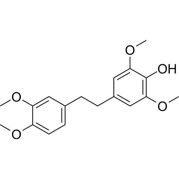
Chrysotoxine
Based on 1 publication(s) in Google Scholar
Chrysotoxine is a dual inhibitor of Src/Akt. Chrysotoxine suppresses cancer stem cells (CSCs) phenotypes by down-regulating Src/Akt signaling. Chrysotoxine reduces cell viability and increases apoptosis level in H460 and H23 cells instead of non-tumor cell lines. Chrysotoxine shows rapid excretion and low bioavailability in rats. Chrysotoxine is used in cancer research.
Nos produits utilisent uniquement pour la recherche. Nous ne vendons pas aux patients.
- Pureté: 99.52%
- CAS No.: 156951-82-5
- Formule: C18H22O5
- Masse moléculaire:318.36
-
Stockage:Powder -20°C, 3 years ; In solvent -80°C, 6 months , -20°C, 1 month

To place orders, for customer services and technical support, please contact: MedChemExpress USA
Tel: 609-228-6898 E-mail: [email protected] [email protected]


-
 Activité biologique
-
Chemical Information
-
Solvant et solubilité
- Pureté et documentation
- Références
-
 Help & FAQs



Publications Citing Use of MedChemExpress (MCE) Chrysotoxine
More

Activité biologique
|
Cell Line

|
Type | Value | Description | References |
|---|---|---|---|---|
| NCI-H23 | IC50 |
145.47 μM
Compound: 3
|
Anti-cancer activity against human NCI-H23 cells
Anti-cancer activity against human NCI-H23 cells
|
[PMID: 32711292] |
| NCI-H460 | IC50 |
127.34 μM
Compound: 3
|
Cytotoxicity against human NCI-H460 cells assessed as cell viability by MTT assay
Cytotoxicity against human NCI-H460 cells assessed as cell viability by MTT assay
|
[PMID: 32711292] |
Chrysotoxine (50 nM, 24 h) reduces cell viability and increases apoptosis level in H460 and H23 cells[1].
Chrysotoxine (5-20 nM, 72 h) suppresses the CSC populations in H460 and H23 cells[1].
Chrysotoxine (0-20 nM, overnight) decreases the stemness of H460 and H23 cells by suppressing
the Src-Akt activating mechanism[1].
MedChemExpress (MCE) has not independently confirmed the accuracy of these methods. They are for reference only.
-
Cell Line:460, H23 cells
-
Concentration:0-20 µM
-
Incubation Time:Overnight
-
Result:Significant decreased the p-Src and p-Akt expression in a dose-dependent manner instead of Src and Akt.
Significantly reduced the down-stream stem cell transcription factor Sox2 as the decline of p-Src in H460 and H23 cells.
-
Cell Line:460, H23 cells
-
Concentration:0, 1, 5, 10, 20 and 50 µM
-
Incubation Time:24 h
-
Result:Significantly reduced cell viability and increased H460 and H23 cells cell apoptosis at 50 µM with IC50s of 127.34 and 145.47 µM, respectively.
Showed no cytotoxic effect on non-tumor cell lines at all tested concentrations.
-
Cell Line:460, H23 cells
-
Concentration:5-20 µM
-
Incubation Time:72 h
-
Result:Decreased approximately 30, 60, 90 and 95% of the H460 CSC spheroid size at day 7 with treated 1, 5,10 and 20 µM Chrysotoxine, respectively.
Decreased approximately 40, 60, 80 and 92% of the H23 CSC spheroid size at day 7 with treated 1, 5,10 and 20 µM Chrysotoxine, respectively.
| Parameters | Intravenous | Oral |
| AUC0–t (μg h/L) | 1257.6 ± 570.7 | 172.8 ± 118.9 |
| AUC0–∞ (μg h/L) | 1270.1 ± 560.6 | 202.5 ± 123.8 |
| MRT0–t (μg h/L) | 0.467 ± 0.056 | 1.2 ± 0.46 | MRT0–∞ (μg h/L) | 0.59 ± 0.21 | 2.4 ± 1.8 |
| t1/2Z ( h) | 1.4 ± 0.76 | 1.7 ± 1.1 |
| Tmax ( h) | / | 0.098 ± 0.040 |
| CLZ /F (L/h/kg) | 22.9 ± 11.2 | 668.7 ± 396.9 |
| VZ /F (L/kg) | 55.3 ± 54.1 | 1443.2 ± 943.0 |
| Cmax (μg/L) | 4961.2 ± 3254.8 | 408.8 ± 160.5 |
| F (%) | / | 3.4 ± 2.4 |