



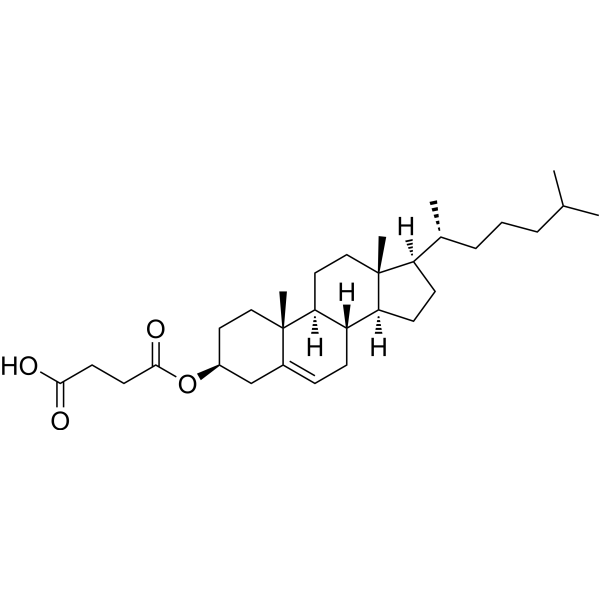
Cholesteryl hemisuccinate

Based on 1 Customer Validation
Cholesteryl hemisuccinate is a with hepatoprotective an anticancer activity. Cholesteryl hemisuccinate inhibits Acetaminophen (AAP, HY-66005) hepatotoxicity, and prevents AAP-induced hepatic apoptosis and necrosis. Cholesteryl hemisuccinate inhibits DNA polymerase and DNA topoisomerase to inhibit DNA replication and repair and cell division. Thus, Cholesteryl hemisuccinate inhibits tumor growth.
Nur für Forschungszwecke. Wir verkaufen nicht an Patienten.
- Reinheit : 99.94%
- CAS. Nr.: 1510-21-0
- Formel: C31H50O4
- Molecular Weight:486.73
-
Speicherung:Powder -20°C, 3 years , 4°C, 2 years ; In solvent -80°C, 6 months , -20°C, 1 month

To place orders, for customer services and technical support, please contact: MedChemExpress USA
Tel: 609-228-6898 E-mail: [email protected] [email protected]


-
 Biologische Aktivität
-
Chemical Information
-
Lösungsmittel & Löslichkeit
- Protokoll
- Reinheit & Dokumentation
- Verweise
-
 Help & FAQs


-
HY-128974
N-Dodecyl-β-D-maltosideNonionic Detergent -
HY-112005
DOPEFerroptosis Inhibitor -
HY-128699
D-DesthiobiotinEndogenous Metabolite -

HY-W010800R
Cholesteryl hemisuccinate (Standard)Topoisomerase Inhibitor -
HY-34477
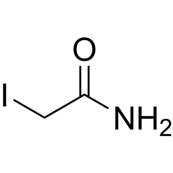
2-IodoacetamideAlkylating Agent -
HY-W011500
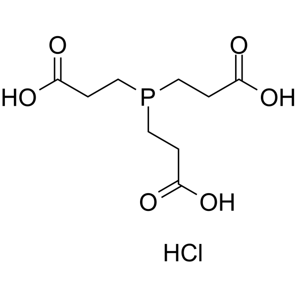
TCEP hydrochlorideNon-Thiol Reducing Agent -
HY-W040193
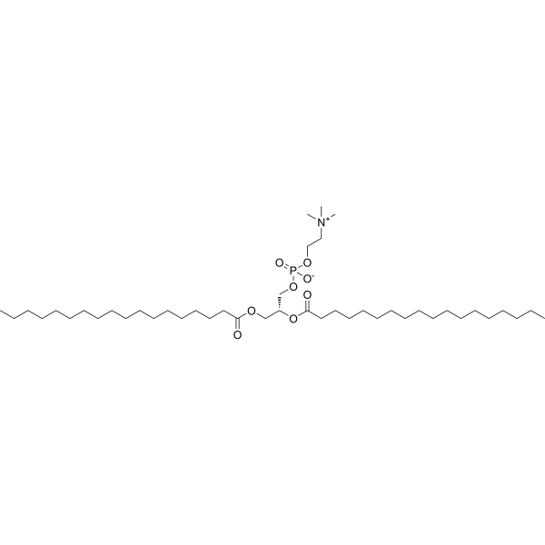
DSPCLiposome Compound -
HY-109506
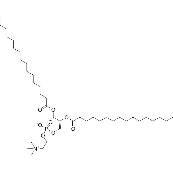
DPPCEndogenous Metabolite -
HY-N0322
Cholesterol (from animal)ERRα Agonist -

HY-100579
Ferrostatin-1Ferroptosis Inhibitor -

HY-15763
ErastinFerroptosis Inducer -

HY-Y0320
Dimethyl sulfoxide, meets analytical specification of Ch.P.Aprotic Solvent
Alle Topoisomerase Isoform-spezifische Produkte anzeigen
MoreAlle DNA/RNA Synthesis Isoform-spezifische Produkte anzeigen
More

Biologische Aktivität
Beschreibung
IC50 & Target
|
DNA Polymerase |
In Vivo
MedChemExpress (MCE) has not independently confirmed the accuracy of these methods. They are for reference only.
- Detection of Apoptosis
- TUNEL staining for apoptotic DNA fragmentation
- PCNA Immunodetection Proliferation Assay
- Annexin V plus membrane-impermeant dye apoptosis staining
- CFSE Dye Dilution Proliferation Assay
- Patient-Derived Xenograft (PDX)
- Apoptosis Solutions
- Dye-dilution cell tracking and proliferation staining
- Patient-Derived Orthotopic Xenograft (PDOX)
- Hepatotoxicity Study
- Subcutaneous Cell-Line-Derived Xenograft
- Orthotopic Cell-Line Xenograft


Chemical Information
-
CAS. Nr. 1510-21-0
-
Appearance Solid
-
Molecular Weight 486.73
-
Formel C31H50O4
-
Color White to off-white
-
SMILES
C[C@@]12[C@]3([H])[C@](CC=C1C[C@H](CC2)OC(CCC(O)=O)=O)([H])[C@@]4([H])[C@](CC3)([C@@](CC4)([H])[C@H](C)CCCC(C)C)C
-
Versand
Room temperature in continental US; may vary elsewhere.
-
Speicherung
Powder -20°C 3 years 4°C 2 years In solvent -80°C 6 months -20°C 1 month


Lösungsmittel & Löslichkeit
In Vitro:
Ethanol : 25 mg/mL (51.36 mM; Need ultrasonic)
DMSO : 8.33 mg/mL (17.11 mM; ultrasonic and warming and heat to 60°C; Hygroscopic DMSO has a significant impact on the solubility of product, please use newly opened DMSO)
Please refer to the solubility information to select the appropriate solvent. Once prepared, please aliquot and store the solution to prevent product inactivation from repeated freeze-thaw cycles.
Storage method and period of stock solution: -80°C, 6 months; -20°C, 1 month. When stored at -80°C, please use it within 6 months. When stored at -20°C, please use it within 1 month.
Please refer to the solubility information to select the appropriate solvent. Once prepared, please aliquot and store the solution to prevent product inactivation from repeated freeze-thaw cycles.
Storage method and period of stock solution: -80°C, 6 months; -20°C, 1 month. When stored at -80°C, please use it within 6 months. When stored at -20°C, please use it within 1 month.
Konzentration (Stammlösung) × Volumen (Stammlösung) = Konzentration (Ziellösung) × Volumen (Ziellösung)
In Vivo:
Select the appropriate dissolution method based on your experimental animal and administration route.
- For the following dissolution methods, please ensure to first prepare a clear stock solution using an In Vitro approach and then sequentially add co-solvents:
- To ensure reliable experimental results, the clarified stock solution can be appropriately stored based on storage conditions. As for the working solution for In Vivo experiments, it is recommended to prepare freshly and use it on the same day.
- The percentages shown for the solvents indicate their volumetric ratio in the final prepared solution. If precipitation or phase separation occurs during preparation, heat and/or sonication can be used to aid dissolution.
Add each solvent one by one: 10% EtOH 90% Corn Oil
Solubility: ≥ 2.5 mg/mL (5.14 mM); Clear solution
This protocol yields a clear solution of ≥ 2.5 mg/mL (saturation unknown). If the continuous dosing period exceeds half a month, please choose this protocol carefully.
Taking 1 mL working solution as an example, add 100 μL EtOH stock solution (25.0 mg/mL) to 900 μL Corn oil, and mix evenly.
In Vivo Dissolution Calculator
Please enter the basic information of animal experiments:
-
-
-
-
Recommended: Prepare an additional quantity of animals to account for potential losses during experiments.
Please enter your animal formula composition:
-
%DMSO +
Recommended: Keep the proportion of DMSO in working solution below 2% if your animal is weak.
-
%+
-
+%Tween-80 + +
-
%Saline +
The co-solvents required include: DMSO, . All of co-solvents are available by MedChemExpress (MCE). , Tween 80. All of co-solvents are available by MedChemExpress (MCE).
Working solution concentration: 0.22 mg/mL
Method for preparing stock solution: mg drug dissolved in μL DMSO. Stock solution concentration: mg/mL.
1. Take μL DMSO stock solution;
2. Add μL .
μL , mix evenly;
3. Then add μL Tween 80, mix evenly;
4. Then add μL
Please ensure that the stock solution in the first step is dissolved to a clear state, and add co-solvents in sequence. You can use ultrasonic heating (ultrasonic cleaner, recommended frequency 20-40 kHz), vortexing, etc. to assist dissolution.


Reinheit & Dokumentation
-
Data Sheet (277 KB)
-
SDS (396 KB)
- English - EN (396 KB)
- Français - FR (396 KB)
- Deutsch - DE (396 KB)
- Norwegian - NO (396 KB)
- Español - ES (396 KB)
- Swedish - SV (396 KB)
- Italian - IT (396 KB)
- Korean - KR (396 KB)
- Portuguese - PT (396 KB)
-
Handling Instructions (2659 KB)
Verweise
[1]. Ray SD, et al. Protection of acetaminophen-induced hepatocellular apoptosis and necrosis by cholesteryl hemisuccinate pretreatment. J Pharmacol Exp Ther. 1996 Dec;279(3):1470-83. [Content Brief]
[2]. Varshosaz J, et al. Folated synperonic-cholesteryl hemisuccinate polymeric micelles for the targeted delivery of docetaxel in melanoma. Biomed Res Int. 2015;2015:746093. [Content Brief]


Complete Stock Solution Preparation Table
Please refer to the solubility information to select the appropriate solvent. Once prepared, please aliquot and store the solution to prevent product inactivation from repeated freeze-thaw cycles.
Storage method and period of stock solution: -80°C, 6 months; -20°C, 1 month. When stored at -80°C, please use it within 6 months. When stored at -20°C, please use it within 1 month.
| Optional Solvent | Concentration Solvent Mass | 1 mg | 5 mg | 10 mg | 25 mg |
|---|---|---|---|---|---|
| DMSO / Ethanol | 1 mM | 2.0545 mL | 10.2726 mL | 20.5453 mL | 51.3632 mL |
| 5 mM | 0.4109 mL | 2.0545 mL | 4.1091 mL | 10.2726 mL | |
| 10 mM | 0.2055 mL | 1.0273 mL | 2.0545 mL | 5.1363 mL | |
| 15 mM | 0.1370 mL | 0.6848 mL | 1.3697 mL | 3.4242 mL | |
| Ethanol | 20 mM | 0.1027 mL | 0.5136 mL | 1.0273 mL | 2.5682 mL |
| 25 mM | 0.0822 mL | 0.4109 mL | 0.8218 mL | 2.0545 mL | |
| 30 mM | 0.0685 mL | 0.3424 mL | 0.6848 mL | 1.7121 mL | |
| 40 mM | 0.0514 mL | 0.2568 mL | 0.5136 mL | 1.2841 mL | |
| 50 mM | 0.0411 mL | 0.2055 mL | 0.4109 mL | 1.0273 mL |


Cholesteryl hemisuccinate Related Classifications
HY-W010800 Related Classifications
 Powered by Bioz