



Catalase Antibody (YA811)

Based on 1 Customer Validation
Catalase Antibody (YA811) is a Mouse-derived and non-conjugated IgG1 monoclonal antibody, targeting to Catalase.
-
Host:
Mouse
-
Isotype:
IgG1
-
Application:
WB, ICC/IF, IHC-P, FC
-
Reactivity :
Human, Mouse, Rat
-
Formulation:
Supplied in 1*PBS (pH7.4), 0.2% BSA and 40% Glycerol. Preservative: 0.05% Sodium Azide.
-
Conjugation:
Non-conjugated


To place orders, for customer services and technical support, please contact: MedChemExpress USA
Tel: 609-228-6898 E-mail: [email protected] [email protected]





Applications
| Application |
ICC/IF

ICC/IF: Immunocytochemistry/
Immunofluorescence |
WB
WB: Western Blot
|
IHC-P
IHC-P: Immunohistochemistry-Paraffin
|
FC
FC: Flow Cytometry
|
|---|---|---|---|---|
| Dilution Ratio | 1:100 | 1:2000-1:5000 | 1:100-1:400 | 1:50 |


Product Details
Catalase Antibody (YA811) is a Mouse-derived and non-conjugated IgG1 monoclonal antibody, targeting to Catalase.
-
Host Mouse

-
Clonality Monoclonal

-
Species ReactivityHuman, Mouse, Rat
-
Observed Molecular WeightObserved band size: 60 kDa
Note: Due to possible protein modifications or aggregation, the molecular weight should be confirmed by actual measurement, and the predicted value is for reference only.

-
Calculated Molecular Weight Predicted band size: 60 kDa

Entrez Gene: 847 Human ; 12359 Mouse ; 24248 Rat
SwissProt: P04040 Human ; P24270 Mouse ; P04762 Rat
OMIM: 614097 Human
Synthetic peptide corresponding to Human Catalase.AA range:475-527.
Endogenous
Protein A affinity purified.
Non-conjugated
Unmodified
IgG1


Product Properties
-
Appearance
Liquid
-
Formulation
Supplied in 1*PBS (pH7.4), 0.2% BSA and 40% Glycerol. Preservative: 0.05% Sodium Azide.
-
Concentration
Batch-dependent, Please check the COA for the concentration of each lot. Check Lot Concentration
-
Storage & Stability
Stored at -20°C for 1 year. Avoid repeated freeze / thaw cycles.
-
Shipping
Shipping with blue ice.


Verification Images
-
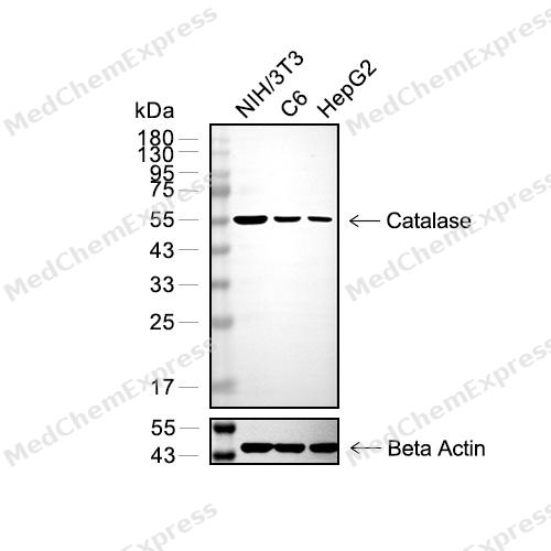 Western blot analysis of extracts from NIH/3T3 (lane 2(20μg) , C6 (lane 3(20μg) , HepG2 (lane 4(20μg),using Catalase Antibody (HY-P80052). Proteins were transferred to a PVDF membrane and blocked with 5% BSA in TBST for 2 hour at room temperature. The primary antibody and Loading control antibody (Beta Actin, HY-P80438, 1/3000) was used in 5% BSA in TBST at 4°C overnight. Goat Anti-Mouse/Rabbit IgG-HRP Secondary Antibody (HY-P8004/HY-P8001, 1/10,000) was used for 1 hour at room temperature.
 -
 Immunohistochemical analysis of paraffin-embedded human liver cancer using Catalase antibody. The section was pre-treated using heat mediated antigen retrieval with EDTA (pH 9.0) for 14 minutes. The tissues were blocked in 5% BSA for 30 minutes at room temperature, washed with TBST, and then probed with the primary antibody (HY-P80052, 1/200) for 30 minutes at room temperature. The detection was performed using Polymer HRP-conjugated Goat Anti-Mouse/Rabbit lgG(H&L) secondary antibody (HY-P83652). DAB was used as the chromogen. Tissues were counterstained with hematoxylin and mounted with DPX.
 -
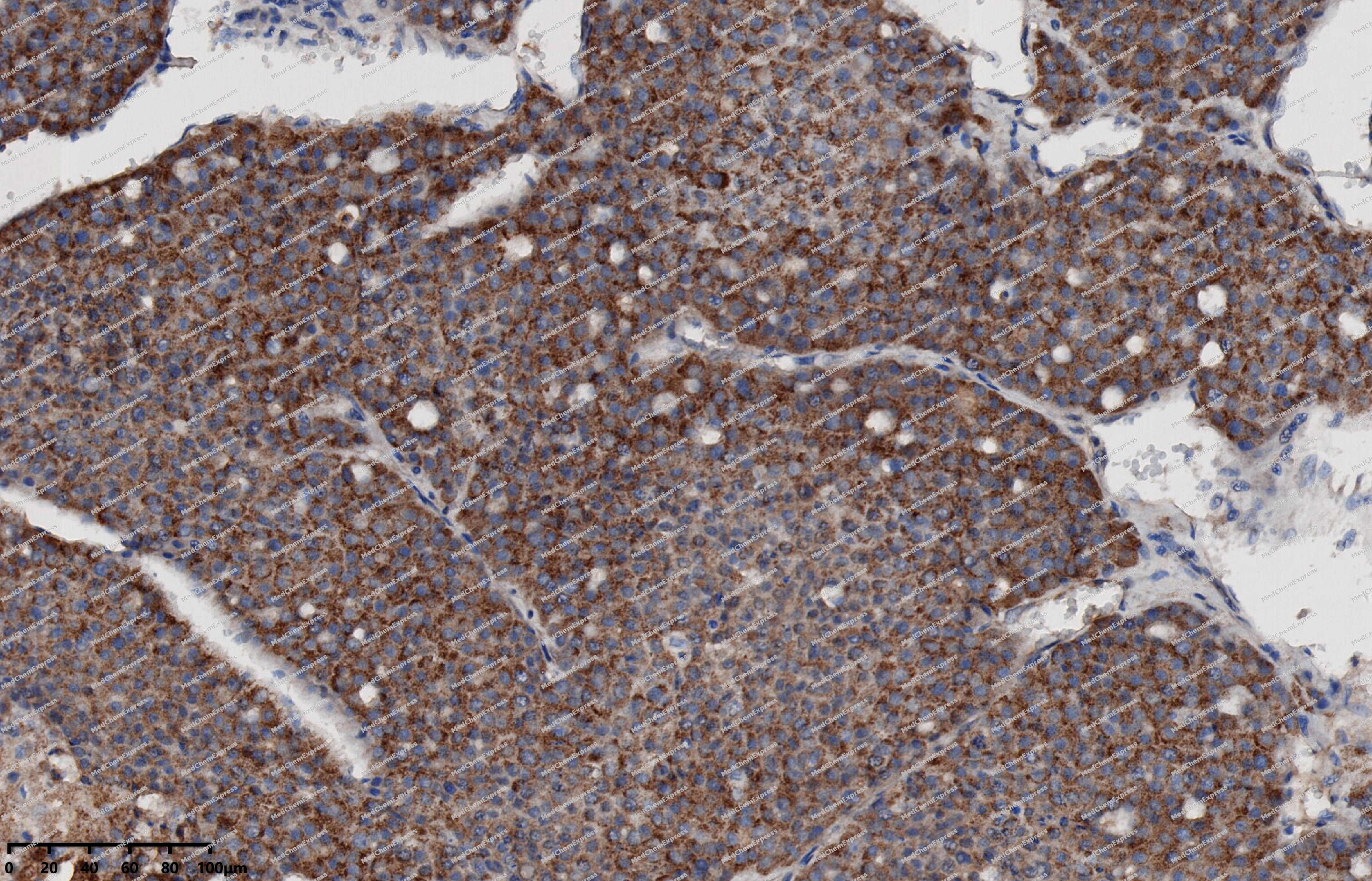 Immunohistochemical analysis of paraffin-embedded human liver using Catalase antibody. The section was pre-treated using heat mediated antigen retrieval with EDTA (pH 9.0) for 14 minutes. The tissues were blocked in 5% BSA for 30 minutes at room temperature, washed with TBST, and then probed with the primary antibody (HY-P80052, 1/200) for 30 minutes at room temperature. The detection was performed using Polymer HRP-conjugated Goat Anti-Mouse/Rabbit lgG(H&L) secondary antibody (HY-P83652). DAB was used as the chromogen. Tissues were counterstained with hematoxylin and mounted with DPX.
 -
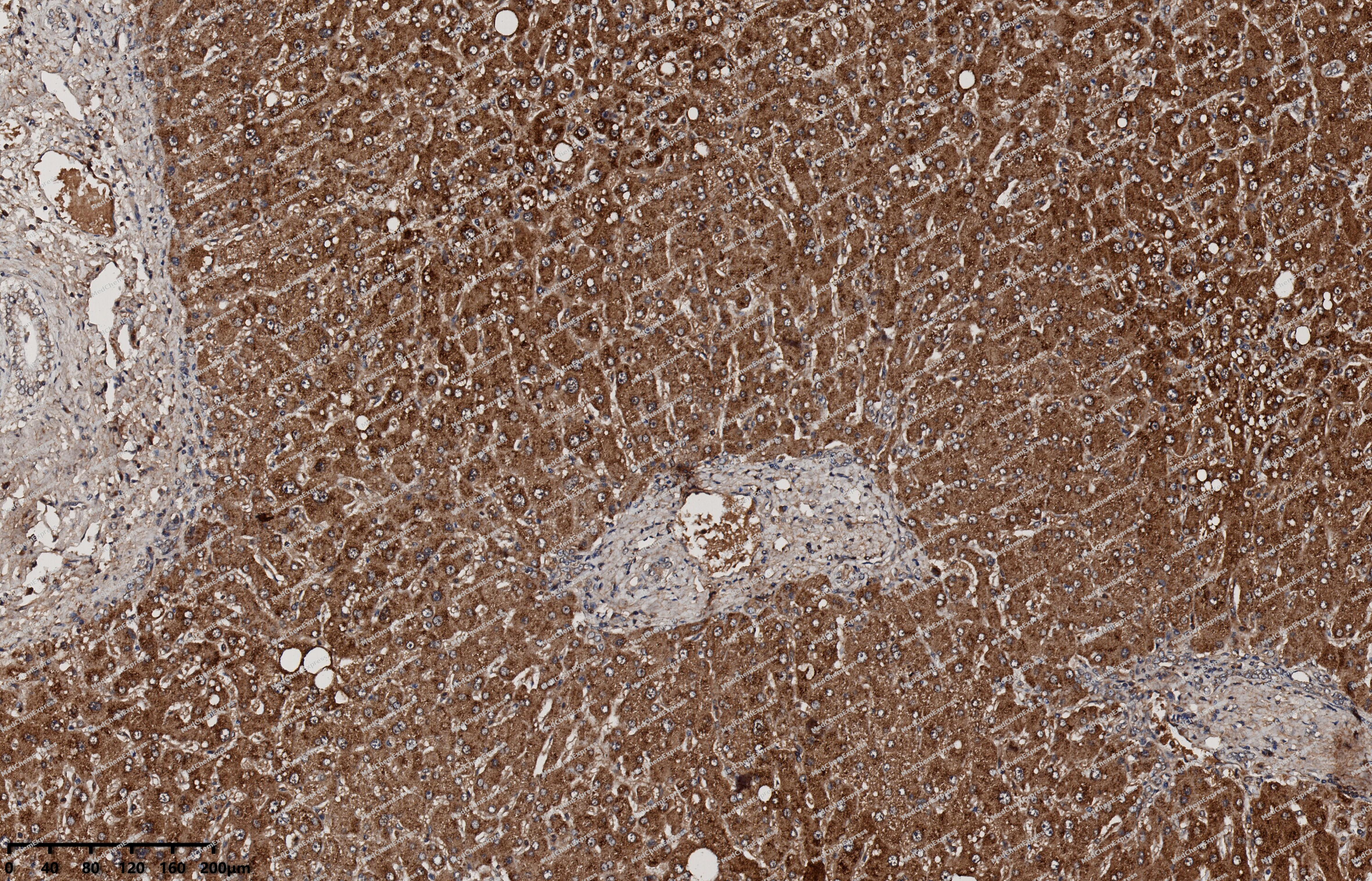 Immunohistochemical analysis of paraffin-embedded human kidney using Catalase antibody. The section was pre-treated using heat mediated antigen retrieval with EDTA (pH 9.0) for 14 minutes. The tissues were blocked in 5% BSA for 30 minutes at room temperature, washed with TBST, and then probed with the primary antibody (HY-P80052, 1/200) for 30 minutes at room temperature. The detection was performed using Polymer HRP-conjugated Goat Anti-Mouse/Rabbit lgG(H&L) secondary antibody (HY-P83652). DAB was used as the chromogen. Tissues were counterstained with hematoxylin and mounted with DPX.
 -
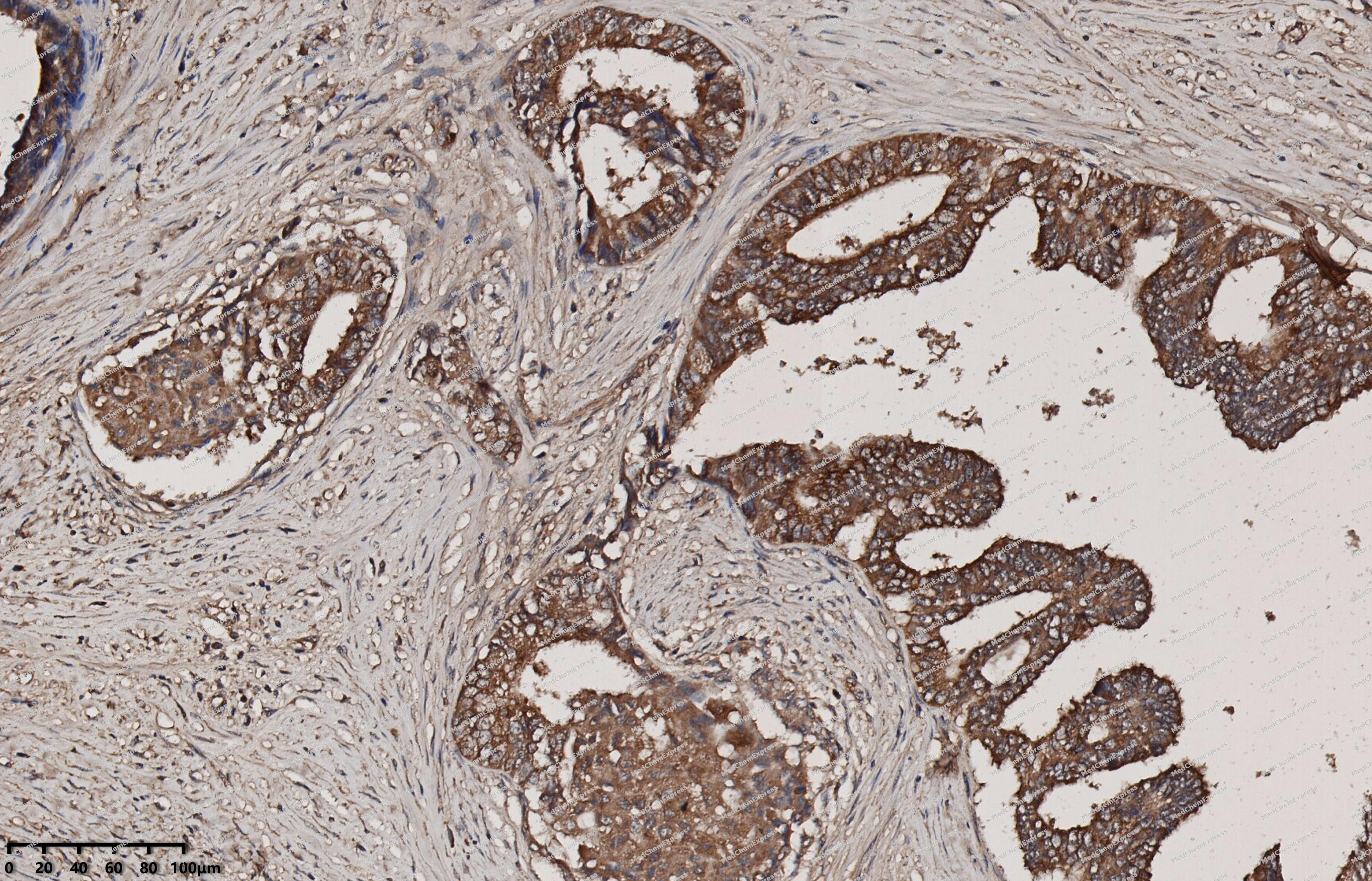 Immunohistochemical analysis of paraffin-embedded human cervical cancer using Catalase antibody. The section was pre-treated using heat mediated antigen retrieval with EDTA (pH 9.0) for 14 minutes. The tissues were blocked in 5% BSA for 30 minutes at room temperature, washed with TBST, and then probed with the primary antibody (HY-P80052, 1/200) for 30 minutes at room temperature. The detection was performed using Polymer HRP-conjugated Goat Anti-Mouse/Rabbit lgG(H&L) secondary antibody (HY-P83652). DAB was used as the chromogen. Tissues were counterstained with hematoxylin and mounted with DPX.
 -
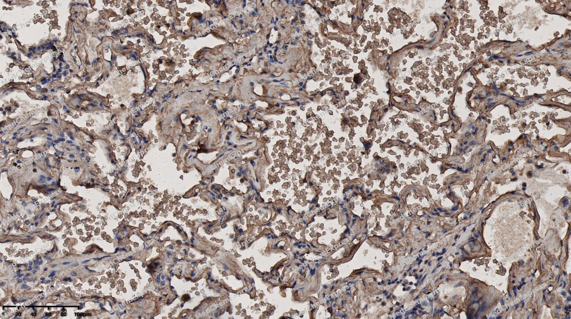 Immunohistochemical analysis of paraffin-embedded human lung cancer using Catalase antibody. The section was pre-treated using heat mediated antigen retrieval with EDTA (pH 9.0) for 14 minutes. The tissues were blocked in 5% BSA for 30 minutes at room temperature, washed with TBST, and then probed with the primary antibody (HY-P80052, 1/200) for 30 minutes at room temperature. The detection was performed using Polymer HRP-conjugated Goat Anti-Mouse/Rabbit lgG(H&L) secondary antibody (HY-P83652). DAB was used as the chromogen. Tissues were counterstained with hematoxylin and mounted with DPX.
 -
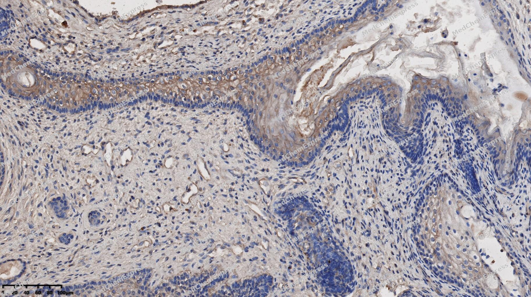 Immunohistochemical analysis of paraffin-embedded human ovarian cancer using Catalase antibody. The section was pre-treated using heat mediated antigen retrieval with EDTA (pH 9.0) for 14 minutes. The tissues were blocked in 5% BSA for 30 minutes at room temperature, washed with TBST, and then probed with the primary antibody (HY-P80052, 1/200) for 30 minutes at room temperature. The detection was performed using Polymer HRP-conjugated Goat Anti-Mouse/Rabbit lgG(H&L) secondary antibody (HY-P83652). DAB was used as the chromogen. Tissues were counterstained with hematoxylin and mounted with DPX.
 -
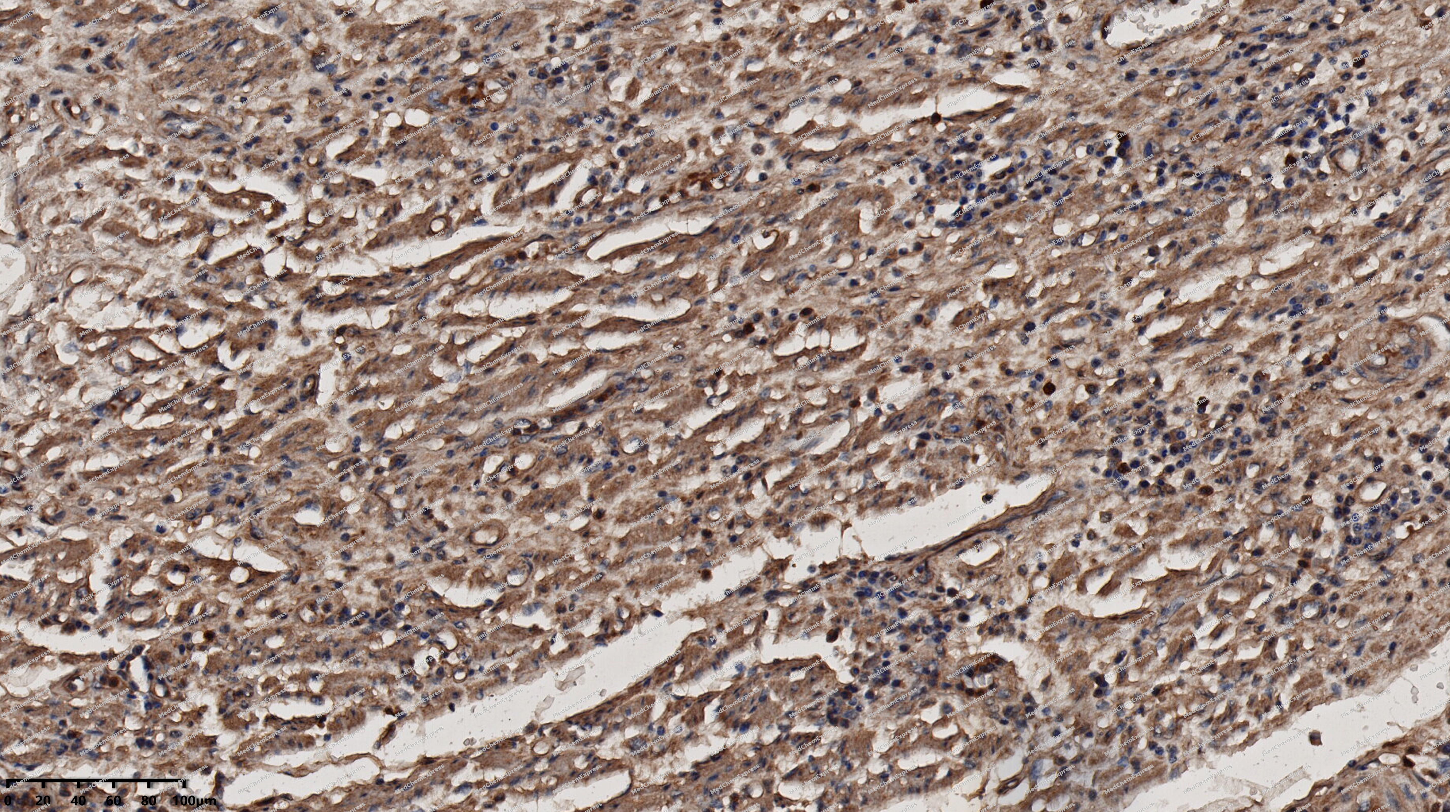 Immunohistochemical analysis of paraffin-embedded human stomach cancer using Catalase antibody. The section was pre-treated using heat mediated antigen retrieval with EDTA (pH 9.0) for 14 minutes. The tissues were blocked in 5% BSA for 30 minutes at room temperature, washed with TBST, and then probed with the primary antibody (HY-P80052, 1/200) for 30 minutes at room temperature. The detection was performed using Polymer HRP-conjugated Goat Anti-Mouse/Rabbit lgG(H&L) secondary antibody (HY-P83652). DAB was used as the chromogen. Tissues were counterstained with hematoxylin and mounted with DPX.
 -
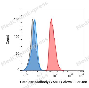 Flow cytometric analysis of 1X10^6 HepG2 cells labeling Catalase Antibody (YA811) (HY-P80052, red). Cells were fixed with 4% paraformaldehyde and permeabilised with 90% methanol. Then stained with the primary antibody at 1/50 dilution for an hour at 4℃. Alexa Fluor® 488-conjugated AffiniPure Goat Anti-Mouse IgG H&L (HY-P8005) was used as the secondary antibody at 1/1,000 dilution for 30 minutes at 4℃. Mouse IgG Isotype Control (HY-P80757, blue) was used as the isotype control, cells without incubation with primary antibody were used as the unlabeled control (black).
 -
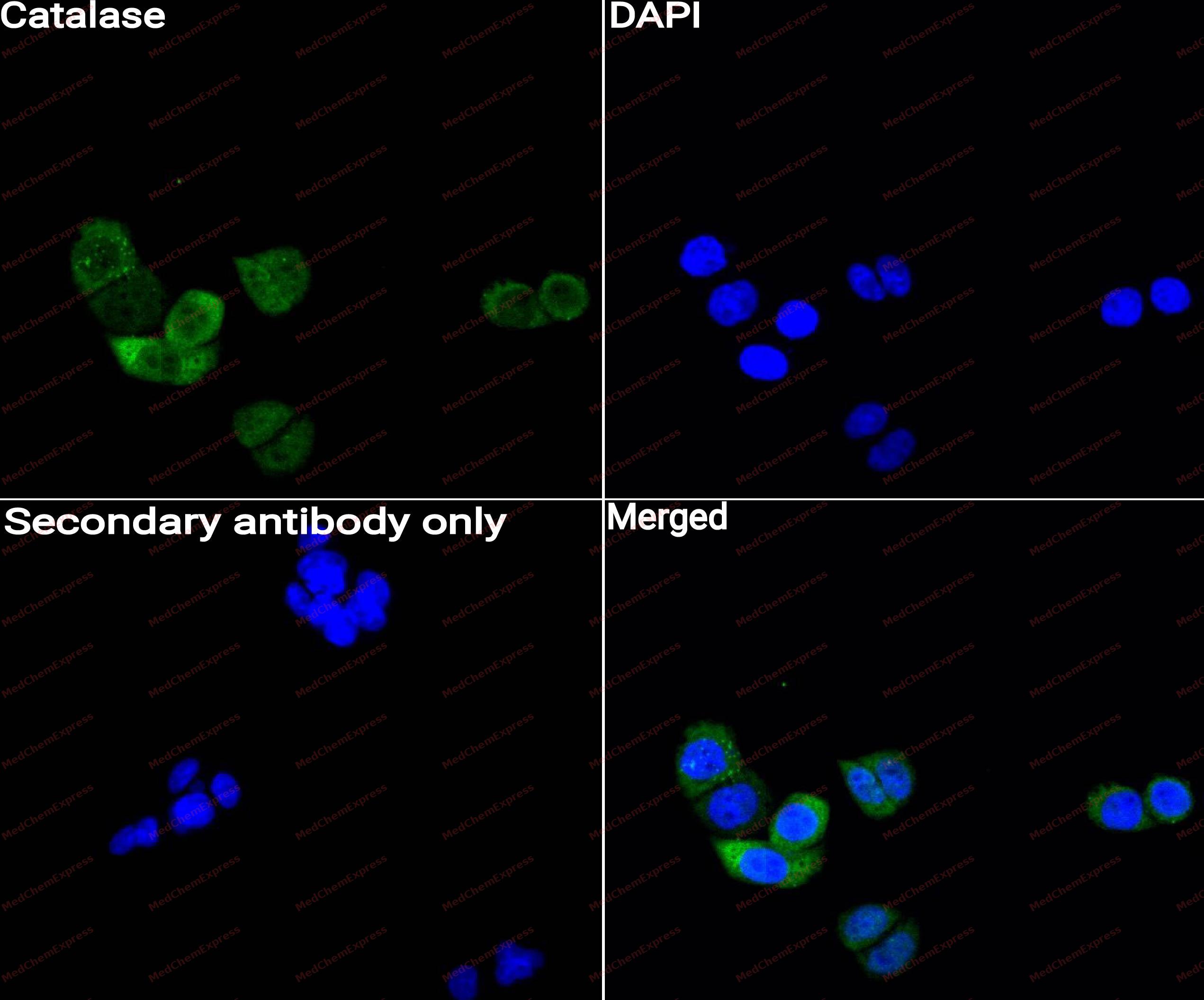 Immunocytochemistry analysis of HepG2 cells labeling Catalase with Catalase Antibody (HY-P80052)at 1/100 dilution. Cells were fixed in 4% paraformaldehyde for 15 minutes at room temperature, permeabilized with 0.1% Triton X-100 for 10 minutes at room temperature, then blocked with QuickBlock™ Blocking Buffer for Immunol Staining for 10 min at room temperature. Cells were then incubated with Catalase Antibody (HY-P80052) at 1/100 dilution in QuickBlock™ Blocking Buffer for Immunol Staining at 4 ℃. Alexa Fluor® 488-conjugated AffiniPure Goat Anti-Mouse IgG H&L(HY-P8005, Green) was used as the secondary antibody at 1/1,000 dilution. PBS instead of the primary antibody was used as the secondary antibody only control. The Nuclear counterstain was DAPI (Blue).
 -
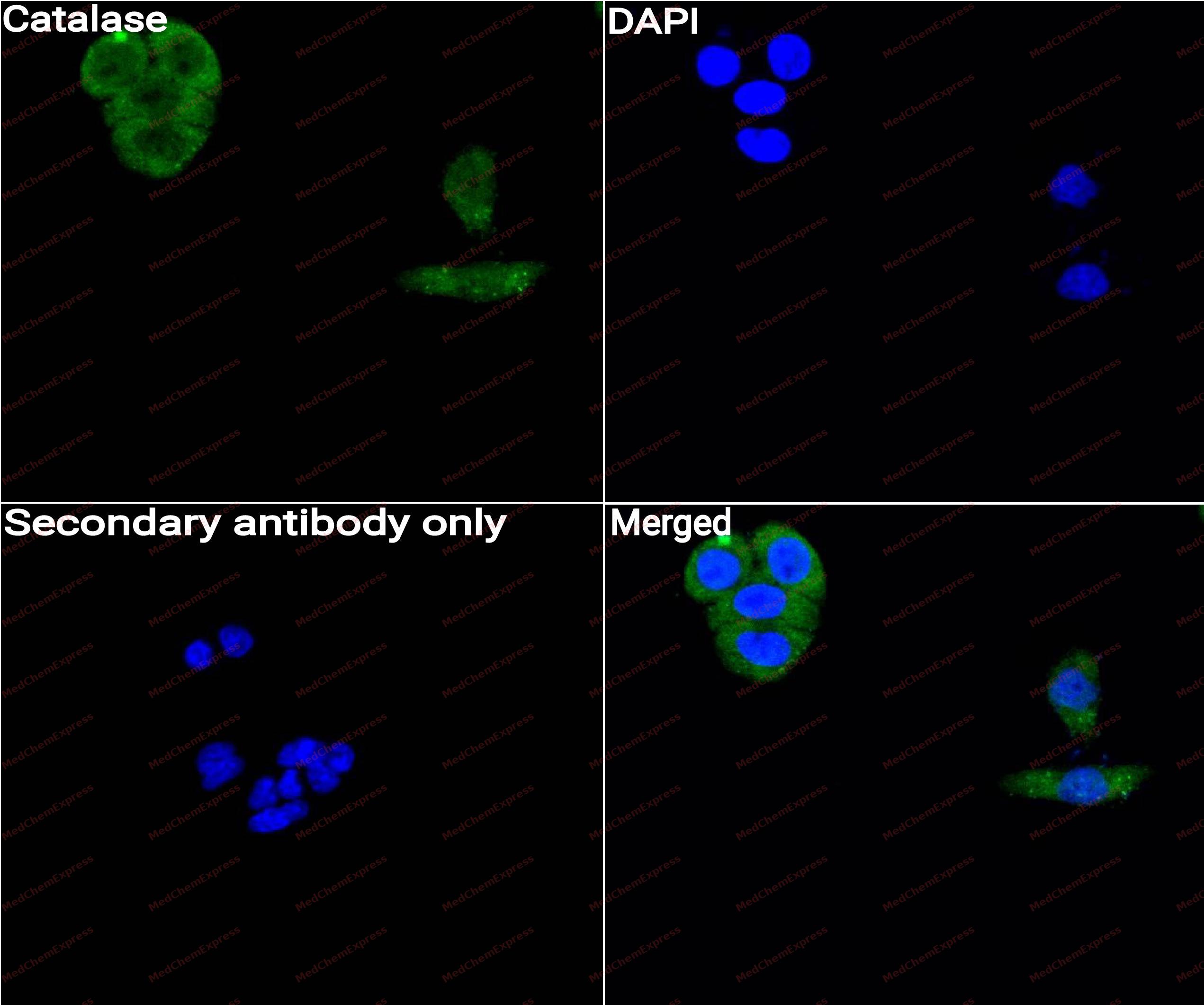 Immunocytochemistry analysis of HepG2 cells labeling Catalase with Catalase Antibody (HY-P80052) at 1/250 dilution. Cells were fixed in 4% paraformaldehyde for 15 minutes at room temperature, permeabilized with 0.1% Triton X-100 for 10 minutes at room temperature, then blocked with QuickBlock™ Blocking Buffer for Immunol Staining for 10 min at room temperature. Cells were then incubated with Catalase Antibody (HY-P80052)at 1/250 dilution in QuickBlock™ Blocking Buffer for Immunol Staining at 4 ℃. Alexa Fluor® 488-conjugated AffiniPure Goat Anti-Mouse IgG H&L(HY-P8005,Green) was used as the secondary antibody at 1/1,000 dilution. PBS instead of the primary antibody was used as the secondary antibody only control. The Nuclear counterstain was DAPI (Blue).


Background
-
Function
Catalase catalyzes the degradation of hydrogen peroxide (H(2)O(2)) generated by peroxisomal oxidases to water and oxygen, thereby protecting cells from the toxic effects of hydrogen peroxide. Promotes growth of cells including T-cells, B-cells, myeloid leukemia cells, melanoma cells, mastocytoma cells and normal and transformed fibroblast cells[1].
-
Subcellular Localization
Peroxisome matrix
-
Subunit
Homotetramer (PubMed:10656833, PubMed:10666617, PubMed:21976670). Interacts (via microbody targeting signal) with PEX5, monomeric form interacts with PEX5, leading to its translocation into peroxisomes (PubMed:21976670)
-
SwissProt ID
-
Research Field
Tags & Cell Markers


Documentation
-
Data Sheet (234 KB)
-
SDS (251 KB)
- English - EN (251 KB)
- Français - FR (251 KB)
- Deutsch - DE (251 KB)
- Norwegian - NO (251 KB)
- Español - ES (251 KB)
- Swedish - SV (251 KB)
- Italian - IT (251 KB)
- Korean - KR (251 KB)
- Portuguese - PT (251 KB)
-
User Guide for Antibodies (1077 KB)

 Powered by Bioz
