


Dexpramipexole
Based on 2 publication(s) in Google Scholar
Dexpramipexole ((R)-Pramipexole) is an orally active, blood-brain barrier permeable mitochondrial protective agent. Dexpramipexole upregulates the expression of Parkin, PINK1, GPX4 and FSP1; binds to mitochondrial F1/Fo-ATP synthase; blocks the Nav1.8 sodium channel; and inhibits the activation of the NLRP3 inflammasome. Dexpramipexole induces mitophagy, inhibits ferroptosis, pyroptosis, apoptosis, neuroinflammation and eosinophilopoiesis; maintains mitochondrial function and redox homeostasis; reduces reactive oxygen species production; and decreases myocardial infarct size. Dexpramipexole is applicable to studies on eosinophilic asthma, myocardial ischemia/reperfusion injury, sepsis-associated encephalopathy, analgesia, and more.
For research use only. We do not sell to patients.
- Purity: 99.56%
- CAS No.: 104632-28-2
- Formula: C10H17N3S
- Molecular Weight:211.33
-
Storage:Powder -20°C, 3 years ; In solvent -80°C, 6 months , -20°C, 1 month

To place orders, for customer services and technical support, please contact: MedChemExpress USA
Tel: 609-228-6898 E-mail: [email protected] [email protected]


-
 Biological Activity
-
Chemical Information
-
Solvent & Solubility
- Purity & Documentation
- References
-
 Help & FAQs
-


Apoptosis Compound Library
HY-L003
-
Immunology/Inflammation Compound Library
HY-L007
-
Membrane Transporter/Ion Channel Compound Library
HY-L011
-
Metabolism/Protease Compound Library
HY-L012
-
Neuronal Signaling Compound Library
HY-L013
-
NF-κB Signaling Compound Library
HY-L014
-
FDA-Approved Drug Library
HY-L022
-
CNS-Penetrant Compound Library
HY-L028
-
Autophagy Compound Library
HY-L029
-
Anti-Aging Compound Library
HY-L034
-
Drug Repurposing Compound Library
HY-L035
-
Antioxidant Compound Library
HY-L037
-
Anti-diabetic Compound Library
HY-L040
-
Oxygen Sensing Compound Library
HY-L045
-
Anti-Cardiovascular Disease Compound Library
HY-L046
-
Ferroptosis Compound Library
HY-L051
-
Pyroptosis Compound Library
HY-L059
-
Orally Active Compound Library
HY-L061
-
FDA Approved & Pharmacopeial Drug Library
HY-L066
-
Neuroprotective Compound Library
HY-L070
-
Anti-Cancer Metabolism Compound Library
HY-L083
-
Mitochondria-Targeted Compound Library
HY-L089
-
Glucose Metabolism Compound Library
HY-L092
-
Rare Diseases Drug Library
HY-L102
-
Antidepressant Compound Library
HY-L108
-
Sodium Channel Blocker Library
HY-L118
-
Off-patent Drug Library
HY-L141
-
Metabolic Enzyme Compound Library
HY-L146
-
Membrane Protein-targeted Compound Library
HY-L149
-
Membrane Receptor-targeted Compound Library
HY-L150
-
Autoimmune Disease Compound Library
HY-L156
-
Cell Death Library
HY-L162
-
Ion Channel Compound Library
HY-L166
-
Inflammasomes related Compound Library
HY-L175
-
Multi-Target Compound Library
HY-L176
-
Radioprotector Library
HY-L178
-
Mitophagy Compound Library
HY-L180
-
Bioactive Compound Library Max
HY-L181
-
MCE Bioactive Compound Library
HY-L001V
-
Drug Repurposing Compound Library Plus
HY-L035P
-
FDA-Approved Drug Library Plus
HY-L022P
-
FDA-Approved Drug Library Mini
HY-L022M
-
Bioactive Compound Library
HY-L001
-
Anti-Brain Cancer Compound Library
HY-L188
-
Non-Alcoholic Fatty Liver Disease (NAFLD) Compound Library
HY-L199
-
High-Throughput Bioactive Compound Library
HY-L205
-
Pattern Recognition Receptors Library
HY-L237
-
Lactylation Compound Library
HY-L249


Publications Citing Use of MedChemExpress (MCE) Dexpramipexole
More Customer Validation & Images

-
IF
-
IF


Biological Activity
|
Nav1.8 |
GPX4 |
PINK1 |
NLRP3 inflammasome |
Dexpramipexole (5-200 μM; 24 h) shows no cytotoxicity to primary cardiomyocytes isolated from neonatal rats at concentrations below 100 μM, while the compound at 200 μM reduces cell viability[1].
Dexpramipexole (2-50 μM) protects primary neonatal rat cardiomyocytes against hypoxia/reoxygenation (H/R)-induced injury by enhancing cell viability and reducing LDH release[1].
Dexpramipexole (2-50 μM) upregulates the expression level of PINK1 protein in primary cardiomyocytes from neonatal rats treated with H/R[1].
Dexpramipexole protects primary cardiomyocytes from neonatal rats against H/R-induced mitochondrial dysfunction by reducing mPTP opening, decreasing ROS production and restoring mitochondrial membrane potential[1].
Dexpramipexole enhances ATG7-dependent mitophagy in primary neonatal rat cardiomyocytes subjected to H/R, which is characterized by increased formation of mitophagosomes, enhanced autophagic mitochondrial sequestration, and improved mitochondrial clearance[1].
Dexpramipexole activates PINK1 and Parkin and promotes mitochondrial fission in primary neonatal rat cardiomyocytes exposed to H/R[1].
Dexpramipexole protects primary cardiomyocytes from neonatal rats against H/R injury by upregulating PINK1- and Parkin-dependent mitophagy[1].
Dexpramipexole (10 μM; 5 min) inhibits tetrodotoxin-resistant sodium currents in primary cultured rat dorsal root ganglion neurons with an IC50 of 294.4 nM, and does not alter the voltage-dependent activation or inactivation properties of the channels[5].
Dexpramipexole (10 μM) selectively inhibits Nav1.8-mediated sodium currents in primary cultured dorsal root ganglion neurons from wild-type mice[5].
MedChemExpress (MCE) has not independently confirmed the accuracy of these methods. They are for reference only.
Dexpramipexole (5-20 mg/kg; i.p., once daily for 14 consecutive days) improves survival rate, body weight gain, spontaneous activity, motor coordination and forelimb muscle strength, reduces hematoma volume, alleviates white matter injury, decreases iron and ROS accumulation, and inhibits ferroptosis by upregulating GPX4 and FSP1 in mice with intracerebral hemorrhage[2].
Dexpramipexole (3 mg/kg; i.p.; once daily for 6 consecutive days) improves lipopolysaccharide (LPS)-induced cognitive impairment in sepsis-associated encephalopathy (SAE), preserves the morphology and function of hippocampal mitochondria in mice, and inhibits mitochondria-mediated pyroptosis and apoptosis[3].
Dexpramipexole (3-20 mg/kg; 20-140 μg/20 μL; p.o.; i.p.; hind paw s.c.; single administration) exerts effective analgesic effects in various mouse models of nociceptive pain (e.g., inflammatory pain, visceral pain) and neuropathic pain (e.g., chemotherapy-induced pain, nerve compression pain, diabetic neuropathic pain)[5].
MedChemExpress (MCE) has not independently confirmed the accuracy of these methods. They are for reference only.
-
Animal Model:C57BL/6 (male, 9-10 weeks old, 24-26 g; intracerebral hemorrhage induced by stereotactic injection of autologous tail vein blood)[2]
-
Dosage:5 mg/kg; 10 mg/kg; 20 mg/kg
-
Administration:i.p., once daily for 14 days
-
Result:Significantly increased the survival rate of mice after cerebral hemorrhage.
Slowed down the postoperative weight loss and promoted the recovery of motor function, motor coordination ability, muscle strength and comprehensive neurological function (BMS).
Reduced iron deposition around the hematoma and the accumulation of reactive oxygen species.
Increased the expression of GPX4 and FSP1.
-
Animal Model:C57BL/6 (male, 3 months old, 20-28 g, LPS-induced sepsis-associated encephalopathy)[3]
-
Dosage:3 mg/kg
-
Administration:i.p.; daily; 6 consecutive days
-
Result:Improved the long-term cognitive dysfunction caused by LPS, particularly spatial working memory and hippocampus-dependent memory.
Protected the mitochondria of hippocampal neurons, improved their morphology (reduced swelling) and function (maintained ATP levels, reduced ROS).
Inhibited pyroptosis and apoptosis of cells
-
Animal Model:CD-1 (male, 20-25 g, intraplantar formalin injection-induced inflammatory pain)[5]
-
Dosage:3 mg/kg; 10 mg/kg; 20 μg/20 μL; 70 μg/20 μL; 140 μg/20 μL
-
Administration:p.o.; single dose 1 h before test; i.p.; single dose 1 h before test; s.c. in hind paw; single dose 10 min before test
-
Result:Reduced time spent in pain-like behaviors during the early formalin phase (10 mg/kg p.o.).
Reduced time spent in pain-like behaviors dose-dependently during the late formalin phase (3 mg/kg, 10 mg/kg p.o.).
Reduced time spent in pain-like behaviors dose-dependently during the late formalin phase (3 mg/kg, 10 mg/kg i.p.).
Reduced time spent in pain-like behaviors during the late formalin phase (70 μg/20 μL, 140 μg/20 μL s.c.).
Caused no effect on pain-like behaviors in the early phase at any tested subdermal dose.
-
Animal Model:CD-1 (male, 20-25 g; female, intraperitoneal acetic acid injection-induced visceral pain)[5]
-
Dosage:10 mg/kg; 20 mg/kg
-
Administration:i.p.; single dose 1 h before test
-
Result:Reduced the number of abdominal writhings dose-dependently in male mice
. Reduced the number of abdominal writhings in female mice.
-
Animal Model:CD-1 (male, 20-25 g, ankle complete Freund adjuvant injection-induced chronic inflammatory arthritis pain)[5]
-
Dosage:70 μg/20 μL
-
Administration:s.c. in hind paw; single dose 10 min before each test on day 7, 14, 21
-
Result:Increased mechanical thresholds at day 7, 14, and 21 post CFA injection.
-
Animal Model:CD-1 (subcutaneous oxaliplatin injection-induced acute chemotherapy-induced neuropathic pain)[5]
-
Dosage:10 mg/kg; 70 μg/20 μL
-
Administration:i.p.; single dose 30 min post oxaliplatin; s.c. in hind paw; single dose 50 min post oxaliplatin
-
Result:Reduced cold pain-related behaviors at 1, 2, 3, 4, and 5 h post oxaliplatin injection (10 mg/kg i.p.).
Reduced cold pain-related behaviors at 1 hour post oxaliplatin injection (70 μg/20 μL, s.c.).
-
Animal Model:CD-1 (intraperitoneal oxaliplatin injection-induced chronic chemotherapy-induced neuropathic pain)[5]
-
Dosage:10 mg/kg
-
Administration:i.p.; single dose 1 h before test; p.o.; single dose 1 h before test
-
Result:Reduced cold allodynia at day 21 post initial oxaliplatin injection (10 mg/kg i.p.).
Reduced cold allodynia at 1, 3, and 6 hours post dosing, with effects diminishing by 9 hours (10 mg/kg p.o.).
-
Animal Model:CD-1 (male, 20-25 g, chronic constriction injury of right sciatic nerve-induced neuropathic pain)[5]
-
Dosage:10 mg/kg
-
Administration:i.p.; single dose 1 h before test
-
Result:Reduced mechanical allodynia in the injured hind paw.
-
Animal Model:CD-1 (intraperitoneal streptozotocin injection-induced diabetic neuropathic pain, blood glucose > 250 mg/dl confirmed 3 weeks post-injection)[5]
-
Dosage:10 mg/kg
-
Administration:i.p.; single dose 1 h before test
-
Result:Increased latency to pain-related behaviors in diabetic mice.


Chemical Information
-
CAS No. 104632-28-2
-
Appearance Solid
-
Molecular Weight 211.33
-
Formula C10H17N3S
-
Color White to off-white
-
SMILES
NC1=NC(CC[C@@H](NCCC)C2)=C2S1
-
Synonyms
(R)-Pramipexole; R-(+)-Pramipexole; KNS-760704
-
Shipping
Room temperature in continental US; may vary elsewhere.
-
Storage
Powder -20°C 3 years In solvent -80°C 6 months -20°C 1 month


Publications (2)
-
Journal Impact Factor
-
Most Recent
-
Neuroreport
Dexpramipexole ameliorates cognitive deficits in sepsis-associated encephalopathy through suppressing mitochondria-mediated pyroptosis and apoptosis. [Abstract]2023 Mar 1;34(4):220-231. PMID: 36719835
Dexpramipexole purchased from MedChemExpress. Usage Cited in: Neuroreport. 2023 Mar 1;34(4):220-231. [Abstract]
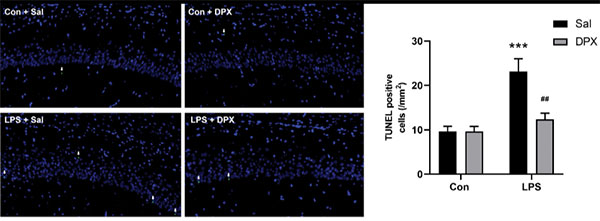
Dexpramipexole (DPX; 3 mg/kg; i.p.; once daily for 6 consecutive days) attenuates the LPS-induced increase in the number of TUNEL-positive cells in the hippocampus in SAE mice.
Dexpramipexole purchased from MedChemExpress. Usage Cited in: Neuroreport. 2023 Mar 1;34(4):220-231. [Abstract]
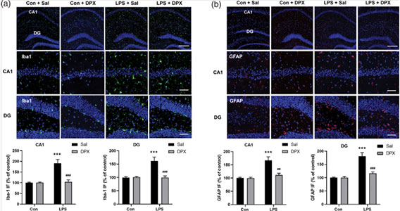
Dexpramipexole (DPX; 3 mg/kg; i.p.; once daily for 6 consecutive days) attenuates LPS-induced activation of microglia and astrocytes in the hippocampus in SAE mice. (a) Representative images of Iba-1 staining and quantification of Iba-1 (green) fluorescence intensity in the hippocampal CA1 and DG regions. (b) Representative images of GFAP staining and quantification of GFAP (red) fluorescence intensity in the hippocampal CA1 and DG regions. DAPI staining is shown in blue.
-
Oxid Med Cell Longev
Dexpramipexole Attenuates White Matter Injury to Facilitate Locomotion and Motor Coordination Recovery via Reducing Ferroptosis after Intracerebral Hemorrhage. [Abstract]2022 Aug 4;2022:6160701. PMID: 35965685


Solvent & Solubility
DMSO : 10 mg/mL (47.32 mM; ultrasonic and warming and heat to 60°C; Hygroscopic DMSO has a significant impact on the solubility of product, please use newly opened DMSO)
Please refer to the solubility information to select the appropriate solvent. Once prepared, please aliquot and store the solution to prevent product inactivation from repeated freeze-thaw cycles.
Storage method and period of stock solution: -80°C, 6 months; -20°C, 1 month. When stored at -80°C, please use it within 6 months. When stored at -20°C, please use it within 1 month.
Concentration (start) × Volume (start) = Concentration (final) × Volume (final)
Select the appropriate dissolution method based on your experimental animal and administration route.
- For the following dissolution methods, please ensure to first prepare a clear stock solution using an In Vitro approach and then sequentially add co-solvents:
- To ensure reliable experimental results, the clarified stock solution can be appropriately stored based on storage conditions. As for the working solution for In Vivo experiments, it is recommended to prepare freshly and use it on the same day.
- The percentages shown for the solvents indicate their volumetric ratio in the final prepared solution. If precipitation or phase separation occurs during preparation, heat and/or sonication can be used to aid dissolution.
Add each solvent one by one: 10% DMSO 40% PEG300 5% Tween-80 45% Saline
Solubility: ≥ 1 mg/mL (4.73 mM); Clear solution
This protocol yields a clear solution of ≥ 1 mg/mL (saturation unknown).
Taking 1 mL working solution as an example, add 100 μL DMSO stock solution (10.0 mg/mL) to 400 μL PEG300, and mix evenly; then add 50 μL Tween-80 and mix evenly; then add 450 μL Saline to adjust the volume to 1 mL.
Preparation of Saline: Dissolve 0.9 g sodium chloride in ddH₂O and dilute to 100 mL to obtain a clear Saline solution.
Add each solvent one by one: 10% DMSO 90% (20% SBE-β-CD in Saline)
Solubility: ≥ 1 mg/mL (4.73 mM); Clear solution
This protocol yields a clear solution of ≥ 1 mg/mL (saturation unknown).
Taking 1 mL working solution as an example, add 100 μL DMSO stock solution (10.0 mg/mL) to 900 μL 20% SBE-β-CD in Saline, and mix evenly.
Preparation of 20% SBE-β-CD in Saline (4°C, storage for one week): 2 g SBE-β-CD powder is dissolved in 10 mL Saline, completely dissolve until clear.
Please enter the basic information of animal experiments:
-
-
-
-
Recommended: Prepare an additional quantity of animals to account for potential losses during experiments.
Please enter your animal formula composition:
-
%DMSO +
Recommended: Keep the proportion of DMSO in working solution below 2% if your animal is weak.
-
%+
-
+%Tween-80 + +
-
%Saline +
The co-solvents required include: DMSO, . All of co-solvents are available by MedChemExpress (MCE). , Tween 80. All of co-solvents are available by MedChemExpress (MCE).
Working solution concentration: 0.22 mg/mL
Method for preparing stock solution: mg drug dissolved in μL DMSO. Stock solution concentration: mg/mL.
1. Take μL DMSO stock solution;
2. Add μL .
μL , mix evenly;
3. Then add μL Tween 80, mix evenly;
4. Then add μL
Please ensure that the stock solution in the first step is dissolved to a clear state, and add co-solvents in sequence. You can use ultrasonic heating (ultrasonic cleaner, recommended frequency 20-40 kHz), vortexing, etc. to assist dissolution.


Purity & Documentation
-
Data Sheet (288 KB)
-
SDS (396 KB)
- English - EN (396 KB)
- Français - FR (396 KB)
- Deutsch - DE (396 KB)
- Norwegian - NO (396 KB)
- Español - ES (396 KB)
- Swedish - SV (396 KB)
- Italian - IT (396 KB)
- Korean - KR (396 KB)
- Portuguese - PT (396 KB)
-
Handling Instructions (2659 KB)
References
[1]. Tang L, et al. Dexpramipexole attenuates myocardial ischemia/reperfusion injury through upregulation of mitophagy. Eur J Pharmacol. 2021;899:173962. [Content Brief]
[2]. Wang B, et al. Dexpramipexole Attenuates White Matter Injury to Facilitate Locomotion and Motor Coordination Recovery via Reducing Ferroptosis after Intracerebral Hemorrhage. Oxid Med Cell Longev. 2022;2022:6160701. Published 2022 Aug 4. [Content Brief]
[3]. Zhang Y, et al. Dexpramipexole ameliorates cognitive deficits in sepsis-associated encephalopathy through suppressing mitochondria-mediated pyroptosis and apoptosis. Neuroreport. 2023;34(4):220-231. [Content Brief]
[4].
Cusack RP, Sulaiman I, Gauvreau GM. Refashioning dexpramipexole: A new horizon in eosinophilic asthma? J Allergy Clin Immunol. 2023 Nov;152(5):1092-1094.
[Content Brief]
[5].
Urru M, et al. Dexpramipexole blocks Nav1.8 sodium channels and provides analgesia in multiple nociceptive and neuropathic pain models. Pain. 2020 Apr;161(4):831-841.
[Content Brief]
[6]. Panch SR, et al. Dexpramipexole as an oral steroid-sparing agent in hypereosinophilic syndromes. Blood. 2018;132(5):501-509. [Content Brief]


Complete Stock Solution Preparation Table
Please refer to the solubility information to select the appropriate solvent. Once prepared, please aliquot and store the solution to prevent product inactivation from repeated freeze-thaw cycles.
Storage method and period of stock solution: -80°C, 6 months; -20°C, 1 month. When stored at -80°C, please use it within 6 months. When stored at -20°C, please use it within 1 month.
| Optional Solvent | Concentration Solvent Mass | 1 mg | 5 mg | 10 mg | 25 mg |
|---|---|---|---|---|---|
| DMSO | 1 mM | 4.7319 mL | 23.6597 mL | 47.3194 mL | 118.2984 mL |
| 5 mM | 0.9464 mL | 4.7319 mL | 9.4639 mL | 23.6597 mL | |
| 10 mM | 0.4732 mL | 2.3660 mL | 4.7319 mL | 11.8298 mL | |
| 15 mM | 0.3155 mL | 1.5773 mL | 3.1546 mL | 7.8866 mL | |
| 20 mM | 0.2366 mL | 1.1830 mL | 2.3660 mL | 5.9149 mL | |
| 25 mM | 0.1893 mL | 0.9464 mL | 1.8928 mL | 4.7319 mL | |
| 30 mM | 0.1577 mL | 0.7887 mL | 1.5773 mL | 3.9433 mL | |
| 40 mM | 0.1183 mL | 0.5915 mL | 1.1830 mL | 2.9575 mL |


Dexpramipexole Related Classifications
HY-17355B Related Classifications
 Powered by Bioz
- Dexpramipexole
- 104632-28-2
- (R)-Pramipexole
- R-(+)-Pramipexole
- KNS-760704
- KNS760704
- KNS 760704
- KNS-760704
- PINK1/Parkin
- Glutathione Peroxidase
- Sodium Channel
- ATP Synthase
- NOD-like Receptor (NLR)
- Mitophagy
- Ferroptosis
- Autophagy
- Apoptosis
- Reactive Oxygen Species (ROS)
- Parkin
- PINK1
- Na?1.8 sodium channels
- neonatal rat primary cardiomyocytes
- FSP1
- F1/F0 ATP synthase b-subunits
- oligomycin sensitivity-conferring protein
- NLRP3 inflammasome
- GPX4
- dorsal root ganglion neurons
- Inhibitor
- inhibitor
- inhibit