



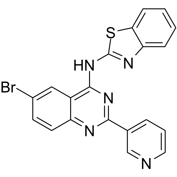
EGFR-IN-63
EGFR-IN-63 is an EGFR inhibition (IC50: 0.096 μM) and it has anticancer activity in MCF-7 cells (IC50: 2.49 μM).
Para uso exclusivo en investigación. No vendemos a pacientes.
- No. CAS: 2414635-72-4
- Fòrmula: C20H12BrN5S
- Peso molecular:434.31
-
Almacenamiento:
Please store the product under the recommended conditions in the Certificate of Analysis.

To place orders, for customer services and technical support, please contact: MedChemExpress USA
Tel: 609-228-6898 E-mail: [email protected] [email protected]


-
 Actividad biológica
-
Chemical Information
- Pureza y Documentación
- Referencias
-
 Help & FAQs


Ver todos los productos específicos de isoformas EGFR
More

Actividad biológica
Descripciòn
In Vitro
EGFR- IN-63 (2-4 hours) has superior EGFR inhibition (IC50 = 0.096 μM) compared to Gefitinib (IC50 = 0.166 μM)and anticancer activity against MCF-7 cell line (IC50 = 2.49 μM) compared to Gefitinib (IC50 = 4.972 μM). EGFR- IN-63 leads to pre G1 apoptosis with cell growth arrest at G2/M phase in MCF-7 cells after 24 h, the percentage of DNA content (41.45 %) shows increase compared to the control cells (7.1 %). EGFR- IN-63 arrest cell growth at G2/M phase and induces its apoptotic effect. The percentage of the total apoptotic cells in MCF-7 cell line increases after treatment with EGFR- IN-63(24.19 %) relative to control cells (1.47 %)[1].
MedChemExpress (MCE) has not independently confirmed the accuracy of these methods. They are for reference only.
-
Cell Line:A549, MCF-7,WI38, PC9 and HCC827 cell lines
-
Concentration:10% of the culture medium volume
-
Incubation Time:2 - 4 hours
-
Result:Displayed superior EGFR inhibitory、anticancer activity and low cytotoxicity.
-
Cell Line:MCF-7
-
Concentration:0.01 μM
-
Incubation Time:24 hour
-
Result:Led to pre G1 apoptosis with cell growth arrest at G2/M phase in MCF-7 cells.
-
Cell Line:MCF-7
-
Concentration:0.01 μM
-
Incubation Time:24 hour
-
Result:Revealed cell growth arrest at G2/M phase and inducted apoptotic effect.


Chemical Information
-
No. CAS 2414635-72-4
-
Peso molecular 434.31
-
Fòrmula C20H12BrN5S
-
SMILES
BrC1=CC2=C(NC3=NC4=CC=CC=C4S3)N=C(C5=CC=CN=C5)N=C2C=C1
-
Envío
Room temperature in continental US; may vary elsewhere.
-
Almacenamiento
Please store the product under the recommended conditions in the Certificate of Analysis.


Pureza y Documentación
Referencias

Calculators
Concentration (start) × Volume (start) = Concentration (final) × Volume (final)


EGFR-IN-63 Related Classifications
HY-147967 Related Classifications
 Powered by Bioz