



H122

Based on 1 Customer Validation
H122 is a PROTAC degrader for TEAD that degrades TEAD1 with a DC50 of 3 nM. H122 exhibits good affinity to TEAD2, TEAD3, and TEAD4 with Ki values of 2.0, 3.6 and 1.6 nM, respectively. H122 relies on binding to TEAD1 and CRBN, functional E3 ligase activity, and a functional proteasome for degradation.H122 induces degradation of TEAD3 and forms a ternary complex with TEAD4 and CRBN/DDB1 to mediate degradation.H122 downregulates expression of Myc target genes. H122 exhibits antitumor efficacy in MSTO-211H mouse xenograft models.H122 can be used for the research of malignant mesothelioma.
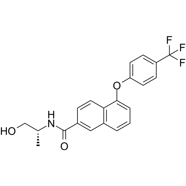
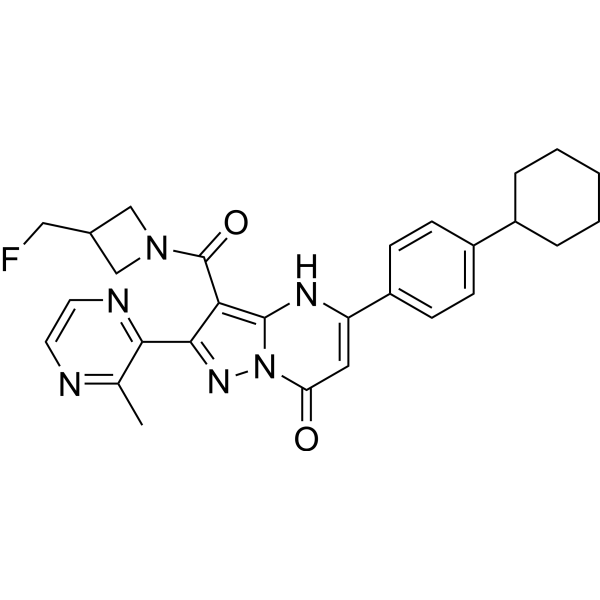
(Pink: TEAD ligand (HY-151525); Blue: Cereblon ligand (HY-W087383); Black: linker).
For research use only. We do not sell to patients.
- Purity : 98.33%
- Formula: C45H45ClFN5O8
- Molecular Weight:838.32
-
Storage:Powder -20°C, 3 years , 4°C, 2 years ; In solvent -80°C, 6 months , -20°C, 1 month


To place orders, for customer services and technical support, please contact: MedChemExpress USA
Tel: 609-228-6898 E-mail: [email protected] [email protected]


-
 Biological Activity
-
Chemical Information
-
Solvent & Solubility
- Protocols
- Purity & Documentation
- References
-
 Help & FAQs




Biological Activity
Description
In Vitro
H122 (Compound 40) binds potently to human TEAD1, TEAD2, TEAD3, and TEAD4 proteins in vitro, with highest affinity for TEAD1 (Ki=0.3 nM)[1].
H122 promotes the formation of a ternary complex between TEAD4 and the CRBN/DDB1 E3 ligase complex in vitro, a key step for PROTAC-mediated degradation[1].
H122 (4 days) potently inhibits the growth of MSTO-211H and NCI-H226 cancer cell lines in vitro, with higher activity in NCI-H226 cells (IC50=0.6 nM)[1].
H122 (10-100 nM; 1-24 h) potently and rapidly degrades TEAD1, TEAD3, and TEAD4 proteins in MSTO-211H and NCI-H226 cancer cells, with TEAD1 degradation DC50=3.0 nM in MSTO-211H cells[1].
H122 (0.1-1000 nM; 4 h) suppresses the expression of TEAD target genes in MSTO-211H cells in vitro in a dose-dependent manner, confirming inhibition of TEAD transcriptional activity[1].
H122 (100 nM; 4 h) induces widespread transcriptional reprogramming in MSTO-211H cells, with significant downregulation of Myc target genes and other TEAD-associated oncogenic genes[1].
H122 (1-1000 nM; 24 h (cell cycle), 48 h (apoptosis)) induces G1 cell cycle arrest and apoptosis in MSTO-211H cancer cells, contributing to its antiproliferative activity[1].
MedChemExpress (MCE) has not independently confirmed the accuracy of these methods. They are for reference only.
-
Cell Line:MSTO-211H and NCI-H226 cells
-
Concentration:10 nM (both cell lines); 10, 100 nM (MSTO-211H cells); 100 nM (NCI-H226 cells)
-
Incubation Time:1, 2, 4, 6, 8, 12, 24 h (10 nM treatment)
-
Result:Induced rapid degradation of TEAD1, TEAD3, and TEAD4 in both cell lines.
Achieved degradation half-life (t1/2) values: MSTO-211H (TEAD1): 3.52 h, NCI-H226 (TEAD1): 1.83 h.
Reached TEAD1 degradation DC50 in MSTO-211H cells: 3.0 nM.
-
Cell Line:MSTO-211H cells
-
Concentration:0.1 nM, 1 nM, 10 nM, 100 nM, 1000 nM
-
Incubation Time:4 h
-
Result:Dose-dependently downregulated mRNA levels of TEAD target genes CYR61, CTGF, ANKRD1, and NPPB.
-
Cell Line:MSTO-211H cells
-
Concentration:1 nM, 10 nM, 100 nM, 1000 nM
-
Incubation Time:48 h
-
Result:Induced significant apoptosis at 100 nM after 48 h treatment.
-
Cell Line:MSTO-211H cells
-
Concentration:1 nM, 10 nM, 100 nM, 1000 nM
-
Incubation Time:24 h
-
Result:Arrested cell cycle at the G1 phase at 10 nM after 24 h treatment.
Parmacokinetics
| Species | Dose | Route | T1/2 | Cmax | AUC0-t | AUC0-∞ | MRT | Bioavailability | C0 | Vz | CL | Vss |
|---|---|---|---|---|---|---|---|---|---|---|---|---|
| Mice[1] | 5 mg/kg | i.v. | 1.94 h | / | 1215 ng·h/mL | 1237 ng·h/mL | 0.69 h | / | 5691 ng/mL | 11.5 L/kg | 70.6 mL/min/kg | 3.79 L/kg |
| Mice[1] | 10 mg/kg | i.p. | 2.68 h | 1157 ng/mL | 2960 ng·h/mL | 3001 ng·h/mL | 2.76 h | 121 % | / | / | / | / |
| Mice[1] | 5 mg/kg | p.o. | / | 7.86 ng/mL | 5.39 ng·h/mL | / | 0.51 h | 0.44 % | / | / | / | / |
In Vivo
MedChemExpress (MCE) has not independently confirmed the accuracy of these methods. They are for reference only.
-
Animal Model:BALB/c nude (female, 5-6 weeks old, MSTO-211H cell xenograft model)[1]
-
Dosage:10 mg/kg; 20 mg/kg; 50 mg/kg
-
Administration:i.p.; daily for 21 days (10, 20 mg/kg); single injection (50 mg/kg)
-
Result:Significantly reduced TEAD1 levels in MSTO-211H tumor tissues (TEAD3/4 degradation was less pronounced, TEAD2 was undetectable) with a single 50 mg/kg i.p.
dose.
Resulted in a tumor volume T/C value of 37.7% (markedly reduced tumor growth compared to control) with daily 20 mg/kg i.p.
treatment for 21 days.
Inhibited tumor growth, with less robust efficacy than the 20 mg/kg dose, with daily 10 mg/kg i.p.
treatment for 21 days.
Observed no loss of body weight in treated mice, indicating good tolerance.
Led to persistent reduction of TEAD1 levels in tumor tissues with prolonged 20 mg/kg treatment.
Clinical Trial
| NCT Number | Sponsor | Condition | Start Date |
Phase

|
|---|---|---|---|---|
| NCT01329991 | 2011-05 | PHASE1 |


Chemical Information
-
Appearance Solid
-
Molecular Weight 838.32
-
Formula C45H45ClFN5O8
-
Color Light yellow to yellow
-
SMILES
COCCOC1=C([C@@]([C@@]2=C(C=CC3=C2C[C@@](C4=CC=CC=C4)(O3)CNCCC5CCN(CC5)C6=CC=C(C7=C6)C(N(C7=O)C8C(NC(CC8)=O)=O)=O)Cl)=C(C=C1)C(N)=O)F
-
Shipping
Room temperature in continental US; may vary elsewhere.
-
Storage
Powder -20°C 3 years 4°C 2 years In solvent -80°C 6 months -20°C 1 month


Solvent & Solubility
In Vitro:
DMSO : 100 mg/mL (119.29 mM; Need ultrasonic; Hygroscopic DMSO has a significant impact on the solubility of product, please use newly opened DMSO)
Please refer to the solubility information to select the appropriate solvent. Once prepared, please aliquot and store the solution to prevent product inactivation from repeated freeze-thaw cycles.
Storage method and period of stock solution: -80°C, 6 months; -20°C, 1 month. When stored at -80°C, please use it within 6 months. When stored at -20°C, please use it within 1 month.
Please refer to the solubility information to select the appropriate solvent. Once prepared, please aliquot and store the solution to prevent product inactivation from repeated freeze-thaw cycles.
Storage method and period of stock solution: -80°C, 6 months; -20°C, 1 month. When stored at -80°C, please use it within 6 months. When stored at -20°C, please use it within 1 month.
Concentration (start) × Volume (start) = Concentration (final) × Volume (final)
In Vivo:
Select the appropriate dissolution method based on your experimental animal and administration route.
- For the following dissolution methods, please ensure to first prepare a clear stock solution using an In Vitro approach and then sequentially add co-solvents:
- To ensure reliable experimental results, the clarified stock solution can be appropriately stored based on storage conditions. As for the working solution for In Vivo experiments, it is recommended to prepare freshly and use it on the same day.
- The percentages shown for the solvents indicate their volumetric ratio in the final prepared solution. If precipitation or phase separation occurs during preparation, heat and/or sonication can be used to aid dissolution.
Add each solvent one by one: 10% DMSO 40% PEG300 5% Tween-80 45% Saline
Solubility: ≥ 2.5 mg/mL (2.98 mM); Clear solution
This protocol yields a clear solution of ≥ 2.5 mg/mL (saturation unknown).
Taking 1 mL working solution as an example, add 100 μL DMSO stock solution (25.0 mg/mL) to 400 μL PEG300, and mix evenly; then add 50 μL Tween-80 and mix evenly; then add 450 μL Saline to adjust the volume to 1 mL.
Preparation of Saline: Dissolve 0.9 g sodium chloride in ddH₂O and dilute to 100 mL to obtain a clear Saline solution.
In Vivo Dissolution Calculator
Please enter the basic information of animal experiments:
-
-
-
-
Recommended: Prepare an additional quantity of animals to account for potential losses during experiments.
Please enter your animal formula composition:
-
%DMSO +
Recommended: Keep the proportion of DMSO in working solution below 2% if your animal is weak.
-
%+
-
+%Tween-80 + +
-
%Saline +
The co-solvents required include: DMSO, . All of co-solvents are available by MedChemExpress (MCE). , Tween 80. All of co-solvents are available by MedChemExpress (MCE).
Working solution concentration: 0.22 mg/mL
Method for preparing stock solution: mg drug dissolved in μL DMSO. Stock solution concentration: mg/mL.
1. Take μL DMSO stock solution;
2. Add μL .
μL , mix evenly;
3. Then add μL Tween 80, mix evenly;
4. Then add μL
Please ensure that the stock solution in the first step is dissolved to a clear state, and add co-solvents in sequence. You can use ultrasonic heating (ultrasonic cleaner, recommended frequency 20-40 kHz), vortexing, etc. to assist dissolution.


Purity & Documentation
-
Data Sheet (279 KB)
-
SDS (252 KB)
- English - EN (252 KB)
- Français - FR (252 KB)
- Deutsch - DE (252 KB)
- Norwegian - NO (252 KB)
- Español - ES (252 KB)
- Swedish - SV (252 KB)
- Italian - IT (252 KB)
- Korean - KR (252 KB)
- Portuguese - PT (252 KB)
-
Handling Instructions (2659 KB)
References


Complete Stock Solution Preparation Table
Please refer to the solubility information to select the appropriate solvent. Once prepared, please aliquot and store the solution to prevent product inactivation from repeated freeze-thaw cycles.
Storage method and period of stock solution: -80°C, 6 months; -20°C, 1 month. When stored at -80°C, please use it within 6 months. When stored at -20°C, please use it within 1 month.
| Optional Solvent | Concentration Solvent Mass | 1 mg | 5 mg | 10 mg | 25 mg |
|---|---|---|---|---|---|
| DMSO | 1 mM | 1.1929 mL | 5.9643 mL | 11.9286 mL | 29.8215 mL |
| 5 mM | 0.2386 mL | 1.1929 mL | 2.3857 mL | 5.9643 mL | |
| 10 mM | 0.1193 mL | 0.5964 mL | 1.1929 mL | 2.9822 mL | |
| 15 mM | 0.0795 mL | 0.3976 mL | 0.7952 mL | 1.9881 mL | |
| 20 mM | 0.0596 mL | 0.2982 mL | 0.5964 mL | 1.4911 mL | |
| 25 mM | 0.0477 mL | 0.2386 mL | 0.4771 mL | 1.1929 mL | |
| 30 mM | 0.0398 mL | 0.1988 mL | 0.3976 mL | 0.9941 mL | |
| 40 mM | 0.0298 mL | 0.1491 mL | 0.2982 mL | 0.7455 mL | |
| 50 mM | 0.0239 mL | 0.1193 mL | 0.2386 mL | 0.5964 mL | |
| 60 mM | 0.0199 mL | 0.0994 mL | 0.1988 mL | 0.4970 mL | |
| 80 mM | 0.0149 mL | 0.0746 mL | 0.1491 mL | 0.3728 mL | |
| 100 mM | 0.0119 mL | 0.0596 mL | 0.1193 mL | 0.2982 mL |


H122 Related Classifications
HY-172113 Related Classifications
 Powered by Bioz