



YM-1 tosylate
Based on 1 publication(s) in Google Scholar
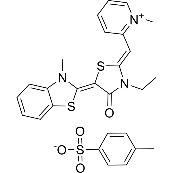
YM-1 tosylate is a stable MKT-077 (HY-15096) analog and an orally active Hsp70 inhibitor. YM-1 tosylate induces cell death of HeLa cells and up-regulates the level of p53 and p21 proteins.
For research use only. We do not sell to patients.
- CAS No.: 1427450-47-2
- Formula: C27H27N3O4S3
- Molecular Weight:553.72
-
Storage:
Please store the product under the recommended conditions in the Certificate of Analysis.

To place orders, for customer services and technical support, please contact: MedChemExpress USA
Tel: 609-228-6898 E-mail: [email protected] [email protected]


-
 Biological Activity
-
Chemical Information
- Protocols
- Purity & Documentation
- References
-
 Help & FAQs


Publications Citing Use of MedChemExpress (MCE) YM-1 tosylate
More

Biological Activity
Description
IC50 & Target
[1]|
HSP70 3.2 μM (IC50) |
Cellular Effect
|
Cell Line

|
Type | Value | Description | References |
|---|---|---|---|---|
| FM3A | EC50 |
1.1 x 10⁻⁵M
Compound: 8d
|
Cytotoxicity against mouse mammary tumor FM3A cells representing a model of host
Cytotoxicity against mouse mammary tumor FM3A cells representing a model of host
|
[PMID: 11855978] |
In Vitro
YM-1 tosylate promotes Hsp70-dependent steps in nNOS maturation and partially blocks formation of the ATP-bound form[1]. YM-1 (0-200 μM) tosylate activates the binding of Hsp70 to its unfolded substrate[1]. YM-1 (0.001-1000 μM) tosylate converts Hsp70 to its tight-affinity conformation and shows binding efficacy to Hsp70 with an IC50 value of 8.2 μM[1]. YM-1 (0, 0.1, 0.5 and 1 μM; 24 hours) tosylate promotes nNOS ubiquitination[1]. YM-1 (5 and 10 μM; 24 and 48 hours) tosylate induces cell death of HeLa cells and growth arrest of hTERT-RPE1 cells[2]. YM-1 (10 μM; 48 hours) tosylate up-regulates p53 and p21 proteins and down-regulates FoxM1 and survivin[2].
MedChemExpress (MCE) has not independently confirmed the accuracy of these methods. They are for reference only.
-
Cell Line:MCF7, MCF10A and MDA-MB-231
-
Concentration:0-30 μM
-
Incubation Time:72 h
-
Result:Inhibited viability with EC50s of 2.0 ± 0.2, 3.3 ± 0.3 and 5.2 ± 0.8 μM for MDA-MB-231, MCF10A and MCF7, respectively.
In Vivo
MedChemExpress (MCE) has not independently confirmed the accuracy of these methods. They are for reference only.
-
Animal Model:CD1 mice[1]
-
Dosage:20 mg/kg
-
Administration:Intravenous injection (Pharmacokinetic Analysis)
-
Result:Initial pharmacokinetics of HSP70-IN-4 (Compound YM-01; 20 mg/kg; i.v.)[1]
hour plasma (ng/mL) brain (ng/g) kidney (ng/g) 0.16 359 0 74378 1 324 0 63231
-
Animal Model:UAS-hAR52Q flies with polyQ AR-induced dihydrotestosterone (DHT) phenotype[1]
-
Dosage:1 mM
-
Administration:Oral administration; 1 mM, for 7 days
-
Result:Weakened the DHT-dependent eye degeneration phenotype and rescued DHT-dependent pupal toxicity of the polyQ AR.


Chemical Information
-
CAS No. 1427450-47-2
-
Molecular Weight 553.72
-
Formula C27H27N3O4S3
-
SMILES
CN(C1=CC=CC=C1S/2)C2=C3C(N(CC)/C(S\3)=C/C4=[N+](C)C=CC=C4)=O.[O-]S(=O)(C5=CC=C(C)C=C5)=O
-
Shipping
Room temperature in continental US; may vary elsewhere.
-
Storage
Please store the product under the recommended conditions in the Certificate of Analysis.


Publications (1)
-
Journal Impact Factor
-
Most Recent


Purity & Documentation
References

Calculators
Concentration (start) × Volume (start) = Concentration (final) × Volume (final)


YM-1 tosylate Related Classifications
HY-18406 Related Classifications
 Powered by Bioz