



Intetumumab

Based on 1 Customer Validation
Intetumumab (CNTO 95) is a human monoclonal antibody targeting αv integrin, with a Kd value of 1-24 nM. Through high-affinity binding to αv integrin, Intetumumab inhibits its interaction with extracellular matrix proteins (such as vitronectin and fibronectin), thereby blocking the downstream focal adhesion kinase signaling pathway. This further inhibits the adhesion, migration and invasion of tumor cells as well as the proliferation of vascular endothelial cells, promotes cell apoptosis, and exerts anti-tumor and anti-angiogenic effects. Intetumumab can be used in research related to head and neck cancer, non-small cell lung cancer and uterine serous papillary carcinoma.
商品は「研究用試薬」です。人や動物の医療用・臨床診断用・食品用の製品ではありません。
研究用途以外に使用した場合、当社は一切の責任を負いかねます。
- 純度 : 99.24%
- CAS 番号: 725735-28-4
- 分子量:145.14 kDa
-
保管条件:
Please store the product under the recommended conditions in the Certificate of Analysis.


To place orders, for customer services and technical support, please contact: MedChemExpress USA
Tel: 609-228-6898 E-mail: [email protected] [email protected]


-
 生物活性
生物活性
-
化学情報
- プロトコル
- 純度とドキュメンテーション
- 参考文献
-
 Help & FAQs


-

HY-P99183
AbituzumabHumanised Anti-integrin αV mAb -

HY-P99333
VolociximabIntegrin α5β1 mAb -

HY-P9911
Vedolizumab -

HY-P99291
Etaracizumabαvβ3 Integrin mAb -

HY-P9911A
Vedolizumab (anti-α4β7-integrin) -

HY-P99524
VatelizumabMonoclonal Antibody -

HY-P99184
AbrilumabAnti-Human α4β7 -

HY-P99946
ErlizumabITGB2/CD18 Antibody -

HY-P99560
TadocizumabAnti-αIIbβ3 mAb -
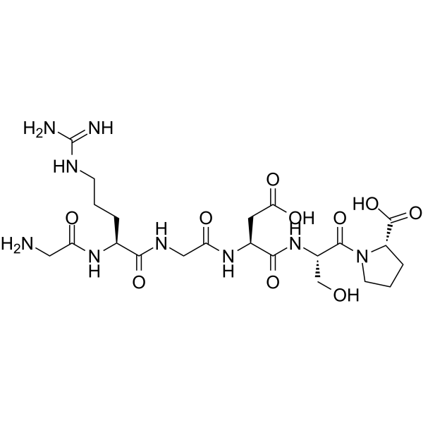
HY-P0290
GRGDSPIntegrin Inhibitor -
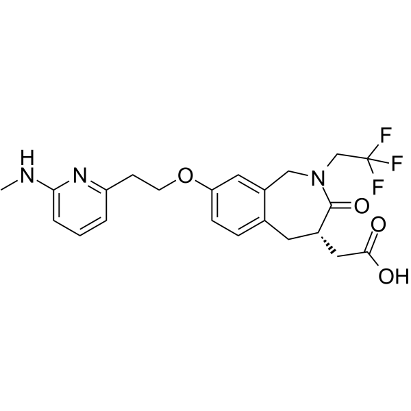
HY-19307
SB-273005αvβ3/αvβ5 Receptor Antagonist -
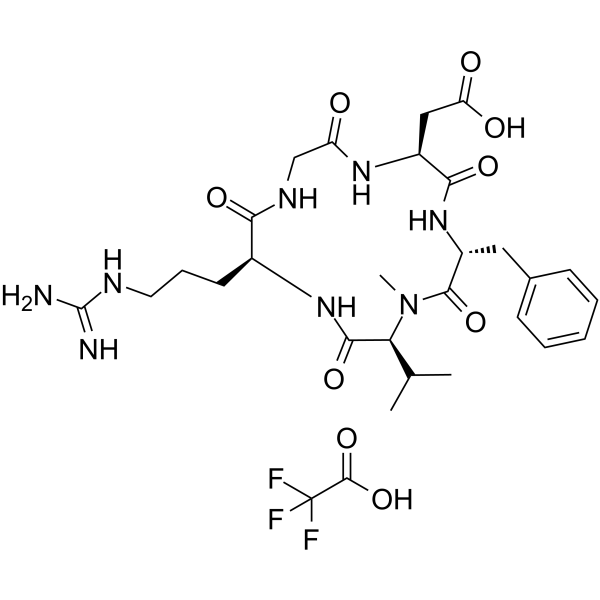
HY-16143
Cilengitide TFAIntegrin Inhibitor


生物活性
製品説明
Isotype
Human IgG1 kappa
Recommend Isotype Controls
Species Reactivity
Human
IC50 & Target
Integrin aV/ITGAV/CD51
体外実験
Intetumumab binds human αv integrin with a Kd of 1-24 nmol/L and rat αv integrin with a Kd of 220 nmol/L[1].
Intetumumab (0.15-5 μg/mL; 15 min pre-incubation with cells, 20 min incubation on coated plates) significantly inhibits adhesion of primary uterine serous papillary carcinoma cell lines to vitronectin, with 30% to 65% inhibition (P < 0.003)[2].
Intetumumab (1.25-20 μg/mL; 48 h incubation during migration assay) significantly inhibits migration of primary uterine serous papillary carcinoma cell lines through an 8.0-μm pore membrane, with 17% to 27% inhibition in USPC-ARK-2 cells (P < 0.03)[2].
MedChemExpress (MCE) has not independently confirmed the accuracy of these methods. They are for reference only.
-
Cell Line:primary uterine serous papillary carcinoma cell lines (USPC-ARK-2, USPC-ARK-4, USPC-ARK-6)
-
Concentration:1.25, 5, 20 μg/mL
-
Incubation Time:48 h (incubation during migration assay)
-
Result:Inhibited migration of USPC-ARK-2 cells by 17% to 27% relative to control cells without antibody.
Showed a statistically significant effect at all tested concentrations.
Induced similar significant inhibition in USPC-ARK-4 and USPC-ARK-6 cell lines.
体内実験
Intetumumab (10 mg/kg; intraperitoneal injection; once weekly for 60 days) significantly reduces the lung metastasis rate of intravenously injected A549 cells, with the number of affected rats and the average number of metastatic lesions per lung decreased by 56% and 77%, respectively[1].
Monotherapy with Intetumumab (10 mg/kg; intraperitoneal injection; once weekly for 60 days) reduces spontaneous lung metastasis of subcutaneously implanted A549 xenografts by 67% and enhances the anti-metastatic efficacy of radiotherapy[1].
Intetumumab (10 mg/kg; intraperitoneal injection; single administration) does not significantly enhance the radiosensitivity of intestinal crypt stem cells in nude mice[1].
Intetumumab enhances the efficacy of radiotherapy in a nude mouse xenograft model of human small cell lung cancer, delays tumor growth and reduces metastasis, without increasing the radiosensitivity of intestinal crypt stem cells[2].
Intetumumab is well tolerated, showing no toxicity, histopathological changes, or adverse effects on physiological angiogenesis in cynomolgus monkeys following long-term administration[2].
Intetumumab exerts anti-tumor and anti-angiogenic activities in nude mice bearing human melanoma xenografts[2].
Intetumumab inhibits spontaneous metastasis of human breast cancer xenografts in nude mice[2].
MedChemExpress (MCE) has not independently confirmed the accuracy of these methods. They are for reference only.
-
Animal Model:nude rats (male, 5-6 weeks old, 150-200 g, injected i.v. with A549 cells)[1]
-
Dosage:10 mg/kg
-
Administration:i.p.; once weekly for 60 days
-
Result:Reduced the proportion of rats with lung metastases from 9/10 in controls to 4/9.
Reduced the average number of metastatic lesions per lung from 56 in controls to 13.
-
Animal Model:nude rats (male, 5-6 weeks old, 150-200 g, inoculated s.c. with A549 cells)[1]
-
Dosage:10 mg/kg
-
Administration:i.p.; once weekly for 60 days
-
Result:Reduced the average number of spontaneous metastatic lung lesions per lung from 18 in controls to 6.
Reduced the proportion of rats with lung metastases from 6/6 in controls to 4/6.
When combined with fractionated radiation, reduced the average number of metastatic lesions per lung from 18 in controls to 4, and reduced the proportion of rats with lung metastases from 6/6 in controls to 5/6.
- Collagen: Sirius Red Staining
- Annexin V plus membrane-impermeant dye apoptosis staining
- Cell migration
- ECM-Embedded Organoid (Matrigel/Dome) Culture
- Matrigel Transwell/Boyden Chamber Invasion Assay
- Cell Viability Determination by MTT Colorimetric Assay
- Fibrosis/Collagen Morphometry
- Detection of Apoptosis
- TUNEL staining for apoptotic DNA fragmentation
- Endothelial Tube Formation Assay
- Apoptosis Solutions
- Matrigel/ECM Transwell Invasion Assay
- Cell invasion
臨床実験
| NCT Number | Sponsor | Condition | Start Date |
Phase

|
|---|---|---|---|---|
| NCT01329991 | 2011-05 | PHASE1 |
遺伝子ID
アクセッション番号
Conjugated
Unconjugated
Reconsititution
The product can be reconstituted/diluted with sterile PBS or saline.
Format
-
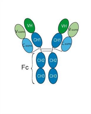
Human IgG1 kappa
アプリケーション
ELISA, FACS, Functional assay
Verified Bioactivity
-
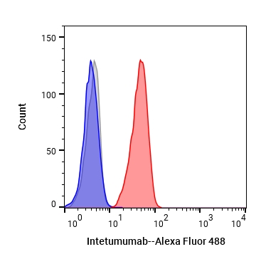 Flow cytometric analysis of 1X10⁶ MCF-7 cells with Intetumumab (HY-P99296, red). Cells were fixed with 4% paraformaldehyde. Then stained with the primary antibody at 1/200 dilution for an hour at 4℃. Alexa Fluor 488-conjugated AffiniPure Goat Anti-Human IgG H&L (AF488) (HY-P83776) was used as the secondary antibody at 1/1,000 dilution for 30 minutes at 4℃. Human IgG1 kappa (HY-P99001, blue) was used as the isotype control, cells without incubation with primary antibody were used as the unlabeled control (black).


化学情報
-
CAS 番号 725735-28-4
-
性状 Liquid
-
分子量 145.14 kDa
-
Color Colorless to light yellow
-
SMILES
[Intetumumab]
-
別名
CNTO 95; Anti-Human CD51 Recombinant Antibody
-
輸送条件
Shipping with dry ice.
-
Formulation
Please refer to the lot-specific COA for specific buffer information.
-
保管条件
Please store the product under the recommended conditions in the Certificate of Analysis.


純度とドキュメンテーション
-
データシート (264 KB)
-
SDS (252 KB)
- English - EN (252 KB)
- Français - FR (252 KB)
- Deutsch - DE (252 KB)
- Norwegian - NO (252 KB)
- Español - ES (252 KB)
- Swedish - SV (252 KB)
- Italian - IT (252 KB)
- Korean - KR (252 KB)
- Portuguese - PT (252 KB)
-
Inhibitory Antibodies User Guide (603 KB)
参考文献
[1]. Ning S, et al. Anti-alphav integrin monoclonal antibody intetumumab enhances the efficacy of radiation therapy and reduces metastasis of human cancer xenografts in nude rats. Cancer Res. 2010;70(19):7591-7599. [Content Brief]
[2]. Bellone M, et al. Expression of αV-integrins in uterine serous papillary carcinomas; implications for targeted therapy with intetumumab (CNTO 95), a fully human antagonist anti-αV-integrin antibody. Int J Gynecol Cancer. 2011;21(6):1084-1090. [Content Brief]

Calculators
濃度 (開始) × 体積 (開始) = 濃度 (終了) × 体積 (終了)


Intetumumab 関連分類
HY-P99296 関連分類
 Powered by Bioz
Keywords
- Intetumumab
- 725735-28-4
- CNTO 95
- Anti-Human CD51 Recombinant Antibody
- CNTO95
- CNTO-95
- Integrin
- Apoptosis
- radiosensitizing effects
- head and neck cancer
- human αv integrin
- xenograft tumors
- rat αv integrin
- antimetastatic effects
- non-small cell lung cancer
- antitumor effects
- αv integrin
- antiangiogenic effects
- Inhibitor
- inhibitor
- inhibit