From 11:00 pm to 12:00 pm EST ( 8:00 pm to 9:00 pm PST ) on January 6th, the website will be under maintenance. We are sorry for the inconvenience. Please arrange your schedule properly.
7rh (DDR1-IN-2) is a potent inhibitor of discoidin domain receptor 1(DDR1), with an IC50 of 13.1 nM, and also less potently inhibits DDR2, with an IC50 of 203 nM. 7rh is a click chemistry reagent, it contains an Alkyne group and can undergo copper-catalyzed azide-alkyne cycloaddition (CuAAc) with molecules containing Azide groups.
VU6015929 is a potent, selective and orally active dual discoidin domain receptor 1/2 (DDR1/2) inhibitor with IC50s of 4.67 nM and 7.39 nM, respectively. VU6015929 potently blocks collagen-induced DDR1 activation and collagen-IV production [1].
TA-02, an analog of SB 203580 (HY-10256), is a p38 MAPK inhibitor with an IC50 of 20 nM. TA-02 especially inhibits TGFBR-2. TA-02 exhibits similar cardiogenic properties as SB 203580 and SB 202190 (HY-10295) [1].
DDR1-IN-5 is a selective Discoidin Domain Receptor family, member 1(DDR1) inhibitor with an IC50 of 7.36 nM. DDR1-IN-5 inhibits auto-phosphorylation DDR1b (Y513) with an IC50 of 4.1 nM. DDR1-IN-5 has anti-cancer activity [1]. DDR1-IN-5 is a click chemistry reagent, it contains an Alkyne group and can undergo copper-catalyzed azide-alkyne cycloaddition (CuAAc) with molecules containing Azide groups.
DDR1-IN-6 is a selective Discoidin Domain Receptor family, member 1(DDR1) inhibitor with an IC50 of 9.72 nM. DDR1-IN-6 inhibits auto-phosphorylation DDR1b (Y513) with an IC50 of 9.7 nM. DDR1-IN-6 has anti-cancer activity [1]. DDR1-IN-6 is a click chemistry reagent, it contains an Alkyne group and can undergo copper-catalyzed azide-alkyne cycloaddition (CuAAc) with molecules containing Azide groups.
DDR1-IN-4 (Compound 2.45) is a selective and potent Discoidin Domain Receptor 1(DDR1) autophosphorylation inhibitor, with IC50 values of 29 nM and 1.9 μM for DDR1 and DDR2, respectively [1].
Collagen (bovine skin) is a three-dimensional cell culture matrix and morphoregulator extracted from bovine skin, which binds to integrins (such as α1β1, α2β1, α11β1) and discoidin domain receptors (DDR1 and DDR2). Collagen (bovine skin) can be reconstituted into a three-dimensional fibrous network to mimic the in vivo tissue environment. It can not only be modified through cross-linking or concentration adjustment, but also interact with fibronectin to enhance matrix-associated cellular activities. Collagen (bovine skin) mediates the proliferation, aggregation, durotactic migration and differentiation of fibroblasts, regulates the synthesis, remodeling and contraction of extracellular matrix, and modulates the expression, activation of MMP as well as cell apoptosis, etc. Collagen (bovine skin) can be used in studies related to the mechanisms of cancer occurrence and development [1] .
Ddr1 Mouse Pre-designed siRNA Set A contains three designed siRNAs for Ddr1 gene (Mouse), as well as a negative control, a positive control, and a FAM-labeled negative control.
DDR1 Human Pre-designed siRNA Set A contains three designed siRNAs for DDR1 gene (Human), as well as a negative control, a positive control, and a FAM-labeled negative control.
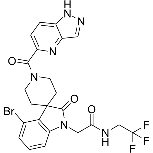
LLC355 is a discoidin domain receptor 1(DDR1) ATTEC degrader. LLC355 efficiently degrades DDR1 protein with a DC50 value of 150.8 nM in non-small cell lung cancer NCI-H23 cells. LLC355 induces DDR1 degradation via lysosome-mediated autophagy. LLC355 potently inhibits cancer cell tumorigenicity, migration, and invasion [1].
DDR1/2 inhibitor-2 (Example 31) is a DDR1/DDR2 inhibitor, with IC50 values less than 100 nM. DDR1/2 inhibitor-2 can be used for research of cancer and fibrotic diseases [1].
DDR1-IN-1 dihydrochloride is a potent and selective DDR1 receptor tyrosine kinase inhibitor with an IC50 of 105 nM; 4-fold less potent for DDR2 (IC50 = 413 nM) [1].
DDR-TRK-1, a chemical probe, is a selective Discoidin Domain Receptor 1(DDR1) inhibitor, with an IC50 value of 9.4 nM. DDR-TRK-1 also inhibits TRK family.
DDR1-IN-9 is a selective inhibitor of DDR1 with significant kinase activity suppression, exhibiting a Kd value of 4.7 nM and an IC50 value of 9.4 nM. DDR1-IN-9 demonstrates reduced potency against a diverse panel of 400 nonmutated kinases, indicating its specificity. Additionally, DDR1-IN-9 shows favorable pharmacokinetic properties and potential therapeutic effects in a model of pulmonary fibrosis.
SR-302, a chemical probe, is a potent and selectivity DDR/p38 inhibitor, with IC50 values of 0.125, 0.023 and 0.018 μM for p38α, DDR1 and DDR2, respectively. SR-302 can be used for the research of fibrotic disorders, such as renal and pulmonary fibrosis, atherosclerosis, and various forms of cancer [1].
DDR1 ligand 1 is a PROTAC target protein ligand (Ligands for Target Protein for PROTACs). DDR1 ligand 1 can be used for the synthesis of PROTAC DDR1degrader-1 (HY-176184) [1].
HG-7-85-01 is a type II ATP competitive inhibitor of wild-type and gatekeeper mutations forms of Bcr-Abl, PDGFRα, Kit, and Src kinases. HG-7-85-01 inhibits T315I mutant Bcr-Abl kinase, KDR and RET with IC50s of 3 nM, 20 nM and 30 nM, and is only weak or no inhibition of other kinases (IC50>2 μM). HG-7-85-01 inhibits the cell proliferation, which is mediated by the induction of apoptosis, and inhibition of cell-cycle progression [1].
DDR1 ligand 1-piperidine is a target protein ligand and linker conjugate, which can be used for the synthesis of PROTAC DDR1degrader-1 (HY-176184) [1].
DDR1-IN-11 (Compound 4) is an inhibitor of Discoidin domain receptor 1(DDR1) with an IC50 of 46.16 nM. DDR1-IN-11 can achieve an inhibition rate of 99.86% against Z-138 cells at a concentration of 10 μM, and it can be used in the research of acute myeloid leukemia (AML) [1].
Recombinant Humanized Type III Collagen 10.4kDa is a novel biomaterial that have anticancer effects. Recombinant Humanized Type III Collagen 10.4kDa activates discoidin domain receptor 1(DDR1), and thus inhibits autophagy, proliferation, and migration of cancer cells, and induces apoptosis[1].
M4K-2009 is an orally bioactive ALK2 inhibitor with an IC50 of 13 nM. M4K-2009 exerts comparable inhibitory potency against wild-type and mutant ALK2G328V, ALK2 R206H, and ALK2 R258G. M4K-2009 exhibits moderate off-target inhibitory activity against hERG potassium channels. M4K-2009 can be used in studies related to diffuse intrinsic pontine glioma [1] .
DDR1-IN-10 (compound 7q) is a DDR1 inhibitor. DDR1-IN-10 can be used in the study of pancreatic cancer, non-small cell lung cancer, and gastric carcinoma [1].
Ddr1 Rat Pre-designed siRNA Set A contains three designed siRNAs for Ddr1 gene (Rat), as well as a negative control, a positive control, and a FAM-labeled negative control.
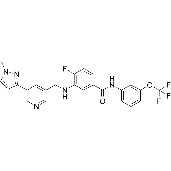
DDR1-IN-8 (compound 7s) is a potent inhibitor of DDR1/2, with the IC50 values of 0.045 μM and 0.126 μM, respectively. DDR1-IN-8 has anti-tumor activity [1].
SJ11646 is a Dasatinib (HY-10181)-based LCKPROTAC degrader with a DC50 of 0.00838 pM. SJ11646 has potent cytotoxicity against LCK-activated T-cell acute lymphoblastic leukemia (T-ALL) cells and primary leukemia samples with drastically prolonged suppression of LCK signaling, and induces T-ALL apoptosis. SJ11646 binds to 51 human kinases with a high affinity (particularly ABL1, KIT, and DDR1). SJ11646 has superior antileukemic efficacy in T-ALL mice model. [1] . Pink: LCK ligand (HY-107447); Blue: CRBN ligase ligand (HY-163169); Black: linker (HY-76667)
CHMFL-ABL/KIT-155 (CHMFL-ABL-KIT-155; compound 34) is a highly potent and orally active type II ABL/c-KIT dual kinase inhibitor (IC50s of 46 nM and 75 nM, respectively), and it also presents significant inhibitory activities to BLK (IC50=81 nM), CSF1R (IC50=227 nM), DDR1 (IC50=116 nM), DDR2 (IC50=325 nM), LCK (IC50=12 nM) and PDGFRβ (IC50=80 nM) kinases. CHMFL-ABL/KIT-155 (CHMFL-ABL-KIT-155) arrests cell cycle progression and induces apoptosis [1].
JNK3 inhibitor-2 is a potent and selective JNK3 inhibitor with IC50 values of >100, >100, 0.25 µM for JNK1, JNK2, JNK3, respectively. JNK3 inhibitor-2 shows DDR1 and EGFR (T790M, L858R) inhibition [1].
DDR1/2 inhibitor-3 (5n) is a DDR1/2 inhibitor, with IC50 valuesof 9.4 and 20.4 nM, respectively. DDR1/2 inhibitor-3 can be used in anti-inflammatory research [1].
Anti-DDR1/CD167a Antibody is a CHO-expressed antibody that targets DDR1/CD167a. The Anti-DDR1/CD167a Antibody has a mouse IgG1 type heavy chain and a mouse κ type light chain, with a predicted molecular weight (MW) of 143.32 kDa. The isotype control for Anti-DDR1/CD167a Antibody can refer to Mouse IgG1 kappa, Isotype Control (HY-P99977).
DDR1/2 IN-4 (Compound 37) is a selective dual DDR1 and DDR2 kinase inhibitor, with a pKi of 8.6 for DDR1 and a pKi of 8.2 for DDR2. DDR1/2 IN-4 functionally inhibits the kinase activities of DDR1 and DDR2. DDR1/2 IN-4 inhibits the release of MCP-1. DDR1/2 IN-4 can be used in studies related to idiopathic pulmonary fibrosis [1].
Lenalidomide-5-Br-amide-C2-Br is an E3 ligase ligand-linker conjugate. Lenalidomide-5-Br-amide-C2-Br can be used to synthesize PROTAC DDR1degrader-1 (HY-176184) [1].
TPKI-39 is a DDR1, DDR2, and FLT1 inhibitor, with a human DDR1IC50 of 380 nM, human DDR1Ka of 24 nM, human DDR2IC50 of 120 nM, human FLT1IC50 of 65 nM, and human FLT1Ka of 91 nM.TPKI-39 inhibits DDR1 enzymatic activity and autophosphorylation, DDR2 enzymatic activity, and FLT1 enzymatic activity in cells.TPKI-39 inhibits collagen-induced DDR1 autophosphorylation in cells [1].
AP4-43 is an orally active CLK1, CLK4, PI3K, DDR1, EGFR and NEK4 inhibitor. AP4-43 reduces growth of mammalian colorectal cancer organoids. AP4-43 improves survival in a transgenic Drosophila model of KRAS-mutant colorectal cancer. AP4-43 can be used for the research of KRAS-mutant colorectal cancer [1].
Anti-Mouse DDR2 Antibody (DAB0065) is a mAb that specifically targets mouse discoidin domain receptor DDR2 without cross-reacting with DDR1. Anti-Mouse DDR2 Antibody (DAB0065) binds to the extracellular domain of native mouse DDR2, induces endocytosis and lysosomal degradation of DDR2, and this process is independent of collagen binding. Anti-Mouse DDR2 Antibody (DAB0065) exhibits significant therapeutic effects in both the unilateral ureteral obstruction (UUO) mouse model of renal fibrosis and the bleomycin (HY-108345)-induced mouse model of pulmonary fibrosis, effectively downregulating the mRNA expression of type I collagen Col1a1 and fibronectin Fn1. Anti-Mouse DDR2 Antibody (DAB0065) can be humanized and has the potential to be developed as a targeted agent for diseases such as idiopathic pulmonary fibrosis and renal fibrosis [1] .
Axl-IN-21 is an orally active and selective AXL inhibitor (Kd = 2.7 nM, IC50 = 4.0 nM). Axl-IN-21 displays kinase selectivity and retains strong activity against cancer-related mul-kinases (Mer with Kd = 1.4 nM, DDR1 with IC50 = 22.2 nM, HIPK4 with Kd = 11.0 nM and LOK with Kd =10 nM). Axl-IN-21 overcomes tumor microenvironment-driven resistance by blocking CAF-derived GAS6-induced AXL/STAT3/ABCG1 signaling, restoring chemosensitivity and inhibiting drug efflux in gastric cancer (GC). Axl-IN-21 suppresses TGF-β1-induced epithelial-mesenchymal transition (EMT), migration, and invasion in MDA-MB-231 cells. Axl-IN-21 exhibits no significant cytotoxicity in non-cancerous cells. Axl-IN-21 can be research for triple negative breast cancer and gastric cancer [1] [2] .
TA-02 (Standard) is the analytical standard of TA-02 (HY-100115). This product is intended for research and analytical applications. TA-02, an analog of SB 203580 (HY-10256), is a p38 MAPK inhibitor with an IC50 of 20 nM. TA-02 especially inhibits TGFBR-2. TA-02 exhibits similar cardiogenic properties as SB 203580 and SB 202190 (HY-10295) [1].
Collagen (bovine skin) is a three-dimensional cell culture matrix and morphoregulator extracted from bovine skin, which binds to integrins (such as α1β1, α2β1, α11β1) and discoidin domain receptors (DDR1 and DDR2). Collagen (bovine skin) can be reconstituted into a three-dimensional fibrous network to mimic the in vivo tissue environment. It can not only be modified through cross-linking or concentration adjustment, but also interact with fibronectin to enhance matrix-associated cellular activities. Collagen (bovine skin) mediates the proliferation, aggregation, durotactic migration and differentiation of fibroblasts, regulates the synthesis, remodeling and contraction of extracellular matrix, and modulates the expression, activation of MMP as well as cell apoptosis, etc. Collagen (bovine skin) can be used in studies related to the mechanisms of cancer occurrence and development [1] .
Recombinant Humanized Type III Collagen 10.4kDa is a novel biomaterial that have anticancer effects. Recombinant Humanized Type III Collagen 10.4kDa activates discoidin domain receptor 1(DDR1), and thus inhibits autophagy, proliferation, and migration of cancer cells, and induces apoptosis[1].
Anti-DDR1/CD167a Antibody is a CHO-expressed antibody that targets DDR1/CD167a. The Anti-DDR1/CD167a Antibody has a mouse IgG1 type heavy chain and a mouse κ type light chain, with a predicted molecular weight (MW) of 143.32 kDa. The isotype control for Anti-DDR1/CD167a Antibody can refer to Mouse IgG1 kappa, Isotype Control (HY-P99977).
Anti-Mouse DDR2 Antibody (DAB0065) is a mAb that specifically targets mouse discoidin domain receptor DDR2 without cross-reacting with DDR1. Anti-Mouse DDR2 Antibody (DAB0065) binds to the extracellular domain of native mouse DDR2, induces endocytosis and lysosomal degradation of DDR2, and this process is independent of collagen binding. Anti-Mouse DDR2 Antibody (DAB0065) exhibits significant therapeutic effects in both the unilateral ureteral obstruction (UUO) mouse model of renal fibrosis and the bleomycin (HY-108345)-induced mouse model of pulmonary fibrosis, effectively downregulating the mRNA expression of type I collagen Col1a1 and fibronectin Fn1. Anti-Mouse DDR2 Antibody (DAB0065) can be humanized and has the potential to be developed as a targeted agent for diseases such as idiopathic pulmonary fibrosis and renal fibrosis [1] .
The DDR1 protein is a fibrillar collagen receptor that coordinates key cellular processes including matrix remodeling, migration, differentiation, survival, and proliferation. Upon collagen binding, DDR1 activates signaling cascades involving SRC and MAP kinases, promoting matrix remodeling through MMP upregulation, promoting cell migration, wound healing, and tumor invasion. DDR1 Protein, Mouse (HEK293, His) is the recombinant mouse-derived DDR1, expressed by HEK293, with C-6*His labeled tag.
The DDR1 protein is a fibrillar collagen receptor that coordinates key cellular processes including matrix remodeling, migration, differentiation, survival, and proliferation.Upon collagen binding, DDR1 activates signaling cascades involving SRC and MAP kinases, promoting matrix remodeling through MMP upregulation, promoting cell migration, wound healing, and tumor invasion.DDR1 Protein, Rat (HEK293, His) is the recombinant rat-derived DDR1 protein, expressed by HEK293 , with C-His labeled tag.
The DDR1 protein is a fibrillar collagen receptor that coordinates key cellular processes including matrix remodeling, migration, differentiation, survival, and proliferation.Upon collagen binding, DDR1 activates signaling cascades involving SRC and MAP kinases, promoting matrix remodeling through MMP upregulation, promoting cell migration, wound healing, and tumor invasion.DDR1 Protein, Rat (HEK293, hFc) is the recombinant rat-derived DDR1 protein, expressed by HEK293 , with C-hFc labeled tag.
The DDR1 protein is a tyrosine kinase and cell surface receptor for fibrillar collagen that tightly regulates cell attachment and affects extracellular matrix processes. Upon collagen binding, DDR1 initiates a signaling cascade involving SRC, activating MAP kinase. DDR1 Protein, Human (HEK293, His) is the recombinant human-derived DDR1 protein, expressed by HEK293 , with C-His labeled tag.
The DDR1 protein is a fibrillar collagen receptor that coordinates key cellular processes including matrix remodeling, migration, differentiation, survival, and proliferation. Upon collagen binding, DDR1 activates signaling cascades involving SRC and MAP kinases, promoting matrix remodeling through MMP upregulation, promoting cell migration, wound healing, and tumor invasion. DDR1 Protein, Mouse (HEK293, Fc) is the recombinant mouse-derived DDR1 protein, expressed by HEK293 , with C-hFc labeled tag.
The DDR1 protein is a fibrillar collagen receptor that coordinates key cellular processes including matrix remodeling, migration, differentiation, survival, and proliferation. Upon collagen binding, DDR1 activates signaling cascades involving SRC and MAP kinases, promoting matrix remodeling through MMP upregulation, promoting cell migration, wound healing, and tumor invasion. DDR1 Protein, Mouse (HEK293, His, solution) is the recombinant mouse-derived DDR1 protein, expressed by HEK293 , with C-His labeled tag.
The DDR1 protein is a fibrillar collagen receptor that coordinates key cellular processes including matrix remodeling, migration, differentiation, survival, and proliferation. Upon collagen binding, DDR1 activates signaling cascades involving SRC and MAP kinases, promoting matrix remodeling through MMP upregulation, promoting cell migration, wound healing, and tumor invasion. DDR1 Protein, Mouse (sf9, His-GST) is the recombinant mouse-derived DDR1 protein, expressed by Sf9 insect cells , with N-His, N-GST labeled tag.
The DDR1 protein is a tyrosine kinase and cell surface receptor for fibrillar collagen that tightly regulates cell attachment and affects extracellular matrix processes. Upon collagen binding, DDR1 initiates a signaling cascade involving SRC, activating MAP kinase. DDR1 Protein, Human (sf9, His-GST) is the recombinant human-derived DDR1 protein, expressed by Sf9 insect cells , with N-His, N-GST labeled tag.
The DDR1 protein is a tyrosine kinase and cell surface receptor for fibrillar collagen that tightly regulates cell attachment and affects extracellular matrix processes. Upon collagen binding, DDR1 initiates a signaling cascade involving SRC, activating MAP kinase. DDR1 Protein, Human (HEK293, Fc) is the recombinant human-derived DDR1 protein, expressed by HEK293, with C-hFc labeled tag.
Ddr1 Mouse Pre-designed siRNA Set A contains three designed siRNAs for Ddr1 gene (Mouse), as well as a negative control, a positive control, and a FAM-labeled negative control.
DDR1 Human Pre-designed siRNA Set A contains three designed siRNAs for DDR1 gene (Human), as well as a negative control, a positive control, and a FAM-labeled negative control.
Ddr1 Rat Pre-designed siRNA Set A contains three designed siRNAs for Ddr1 gene (Rat), as well as a negative control, a positive control, and a FAM-labeled negative control.
Inquiry Online
Your information is safe with us. * Required Fields.
Western blot analysis of extracts from THP-1(lane 2(20μg), Jurkat (lane 3(20μg) and NIH3T3(lane 4(20μg) using FOXO1A (HY-P80132) Rabbit mAb. Proteins were transferred
to a PVDF membrane and blocked with 5% non-fat milk in TBST for 2 hour at room temperature. The primary antibody (1/1000) and Loading control antibody (Beta Actin, HY-P80438, 1/10000) was
used in 5% non-fat milk in TBST at 4°C overnight. Goat Anti-Mouse/Rabbit IgG-HRP Secondary Antibody (1/10000) was used for 1 hour at room temperature.
Western blot analysis of extracts from THP-1(lane 2(20μg), Jurkat (lane 3(20μg) and NIH3T3(lane 4(20μg) using FOXO1A (HY-P80132) Rabbit mAb. Proteins were transferred
to a PVDF membrane and blocked with 5% non-fat milk in TBST for 2 hour at room temperature. The primary antibody (1/1000) and Loading control antibody (Beta Actin, HY-P80438, 1/10000) was
MedchemExpress Validation 03
Western blot analysis of extracts from THP-1(lane 2(20μg), Jurkat (lane 3(20μg) and NIH3T3(lane 4(20μg) using FOXO1A (HY-P80132) Rabbit mAb. Proteins were transferred
MedchemExpress Validation 04
Western blot analysis of extracts from THP-1(lane 2(20μg), Jurkat (lane 3(20μg) and NIH3T3(lane 4(20μg) using FOXO1A (HY-P80132) Rabbit mAb. Proteins were transferred
to a PVDF membrane and blocked with 5% non-fat milk in TBST for 2 hour at room temperature. The primary antibody (1/1000) and Loading control antibody (Beta Actin, HY-P80438, 1/10000) was
used in 5% non-fat milk in TBST at 4°C overnight. Goat Anti-Mouse/Rabbit IgG-HRP Secondary Antibody (1/10000) was used for 1 hour at room temperature.
MedchemExpress Validation
Western blot analysis of extracts from THP-1(lane 2(20μg), Jurkat (lane 3(20μg) and NIH3T3(lane 4(20μg) using FOXO1A (HY-P80132) Rabbit mAb. Proteins were transferred
to a PVDF membrane and blocked with 5% non-fat milk in TBST for 2 hour at room temperature. The primary antibody (1/1000) and Loading control antibody (Beta Actin, HY-P80438, 1/10000) was
used in 5% non-fat milk in TBST at 4°C overnight. Goat Anti-Mouse/Rabbit IgG-HRP Secondary Antibody (1/10000) was used for 1 hour at room temperature.
MedchemExpress Validation
Western blot analysis of extracts from THP-1(lane 2(20μg), Jurkat (lane 3(20μg) and NIH3T3(lane 4(20μg) using FOXO1A (HY-P80132) Rabbit mAb. Proteins were transferred
to a PVDF membrane and blocked with 5% non-fat milk in TBST for 2 hour at room temperature. The primary antibody (1/1000) and Loading control antibody (Beta Actin, HY-P80438, 1/10000) was
used in 5% non-fat milk in TBST at 4°C overnight. Goat Anti-Mouse/Rabbit IgG-HRP Secondary Antibody (1/10000) was used for 1 hour at room temperature.
MedChemExpress values your privacy and your trust is important to us. We use cookies to enhance your website experience. Some cookies are necessary to run the website.
Privacy and Cookie Policy