Search Result
Results for "
fluorescence microscopy
" in MedChemExpress (MCE) Product Catalog:
17
Biochemical Assay Reagents
| Cat. No. |
Product Name |
Target |
Research Areas |
Chemical Structure |
-
- HY-D0718
-
|
Nile Blue A oxazone; Phenoxazone 9
|
Fluorescent Dye
|
Others
|
|
Nile red (Nile blue oxazone) is a lipophilic stain. Nile red has environment-sensitive fluorescence. Nile red is intensely fluorescent in a lipid-rich environment while it has minimal fluorescence in aqueous media. Nile red is an excellent vital stain for the detection of intracellular lipid droplets by fluorescence microscopy and flow cytof uorometry. Nile red stains intracellular lipid droplets red. The fluorescence wavelength is 559/635 nm .
|
-
-
- HY-116215
-
|
|
Fluorescent Dye
|
Others
|
|
2-NBDG is a fluorescently-labeled deoxyglucose analog that is used primarily to directly monitor glucose uptake by living cells and tissues. It is also used as a topical contrast reagent for the detection of neoplasia. 2-NBDG can be used in real-time confocal, high-resolution, or wide-field fluorescence microscopy as well as in flow cytometry. The probe can be excited by the Argon laser at 488 nm to give the environment-sensitive fluorescence. It has lower photostability than the rhodamine-based fluorescent probes.
|
-
-
- HY-D0090
-
|
|
Fluorescent Dye
|
Others
|
|
MQAE is a chloride ion (Cl -) fluorescent probe that can be used to measure chloride concentrations. The fluorescence intensity of MQAE decreases proportionally as Cl - ions increase. MQAE has high cell permeability and is suitable for fluorescence detection such as confocal microscopy and flow cytometry (Ex/Em=350/460 nm) .
|
-
-
- HY-111330
-
|
HPF; 3'-p-(Hydroxyphenyl) fluorescein
|
Fluorescent Dye
Reactive Oxygen Species (ROS)
|
Others
|
|
Hydroxyphenyl Fluorescein (HPF) is a stable ROS fluorescent probe dye. Hydroxyphenyl Fluorescein has stronger specificity and stability than H2DCFDA (HY-D0940). Hydroxyphenyl Fluorescein can produce strong green fluorescence through hydroxyl radical reaction with intracellular peroxynitroso. Hydroxyphenyl Fluorescein can be applied for fluorescence microscopy, high-throughput imager, luciferase microplate reader or flow cytometry. Ex/Em=490/515 nm .
|
-

-
- HY-D0988
-
|
R-PE
|
Fluorescent Dye
Apoptosis
|
Cancer
|
|
R-Phycoerythrin is found in Heterosiphonia japonica. R-Phycoerythrin is an orange-red fluorescent probe with α, β, and γ subunits. R-Phycoerythrin can be used in photodynamic therapy (PDT) to induce apoptosis in tumor cells. R-Phycoerythrin can be used in fluorescence microscopy, flow cytometry, and immunofluorescence analysis (Ex: 495 nm).
|
-

-
- HY-D0722
-
|
5-(6)-Carboxyfluorescein diacetate; CFDA
|
Fluorescent Dye
|
Others
|
|
5(6)-CFDA is a common aliphatic luciferin-line organism. CFDA conducts free diffusion into cells, and then it is hydrolyzed into carboxyl fluorescein (CF) by intracellular non-specific lipase. CF containing portion contains an additional negative charge so that it is better retained in cells, compared to fluorescein dyes .
|
-
-
- HY-162543
-
|
18:1 Lissamine rhodamine PE
|
Fluorescent Dye
|
Others
|
|
18:1 Liss Rhod (18:1 Lissamine rhodamine) PE is a fluorescent phospholipid and fluorescent probe.18:1 Liss Rhod PE admixes into phospholipid inks for large-scale monitoring of dip-pen nanolithography-generated lithographic structures via fluorescence microscopy.18:1 Liss Rhod PE undergoes phase separation or self-quenching under certain conditions in thin lipid membrane stacks .
|
-

-
- HY-D2001
-
|
|
Fluorescent Dye
|
Others
|
|
ATTO 488 NHS ester is a new fluorescent marker based on the Rhodamine structure. It has strong absorption, high fluorescence quantum yield, high thermal stability and photochemical stability, and is suitable for single molecule detection and high-resolution microscopy. ATTO 488 NHS ester is an NHS ester derivative of ATTO 488 that can be used to label proteins or antibodies.
|
-
-
- HY-D0721
-
|
6-Carboxyfluorescein diacetate
|
Fluorescent Dye
|
Others
|
|
6-CFDA is a common aliphatic luciferin-line organism. CFDA conducts free diffusion into cells, and then it is hydrolyzed into carboxyl fluorescein (CF) by intracellular non-specific lipase. CF containing portion contains an additional negative charge so that it is better retained in cells, compared to fluorescein dyes .
|
-
-
- HY-DY1008
-
|
|
Fluorescent Dye
|
Others
|
Nile Red (solution) is a lipophilic stain. Nile red has environment-sensitive fluorescence. Nile red is intensely fluorescent in a lipid-rich environment while it has minimal fluorescence in aqueous media. Nile red is an excellent vital stain for the detection of intracellular lipid droplets by fluorescence microscopy and flow cytof uorometry. Nile red stains intracellular lipid droplets red. The fluorescence wavelength is 559/635 nm .
Solvent and concentration: DMSO: 1 mM
|
-

-
- HY-D1429
-
|
|
Fluorescent Dye
|
Others
|
|
ER-Tracker dye is a derivative of BODIPY series dyes coupled with Glibenclamide (HY-15206), highly selective binding to the endoplasmic reticulum, non-toxic to cells at low concentrations, this type of dye is an environmentally sensitive probe, and formaldehyde treatment can still retain part of the fluorescence, with high fluorescence life, good extinction coefficient and other characteristics. Glibenclamide is an atp-dependent K + channel blocker (Kir6, KATP) and CFTR Cl-channel blocker that binds in the endoplasmic reticulum. ER-Tracker is not suitable for staining fixed cells. Staining followed by fixation is possible, but cells fixed with aldehydes will only retain partial fluorescence (Ex/Em = 374/ 430-640 nm) .
|
-
-
- HY-DY1019
-
|
|
Fluorescent Dye
|
Others
|
2-NBDG (solution) is a fluorescently-labeled deoxyglucose analog that is used primarily to directly monitor glucose uptake by living cells and tissues. It is also used as a topical contrast reagent for the detection of neoplasia. 2-NBDG can be used in real-time confocal, high-resolution, or wide-field fluorescence microscopy as well as in flow cytometry. The probe can be excited by the Argon laser at 488 nm to give the environment-sensitive fluorescence. It has lower photostability than the rhodamine-based fluorescent probes.
Solvent and Concentration: Sterile PBS: 5 mM
|
-

-
- HY-NP163A
-
|
WGA-AF488
|
Fluorescent Dye
|
Cancer
|
Wheat germ agglutinin-AF488 (WGA-AF488) is a cell membrane-specific staining agent prepared by conjugating wheat germ agglutinin with the Alexa Fluor 488 (HY-D1304) fluorescent dye, and it binds to cell surface glycoproteins with high affinity. Wheat germ agglutinin-AF488 is applied in fluorescence microscopy and confocal imaging techniques, and it can clearly label the membrane structures of various cells including breast cancer cells, enabling high-resolution visual observation. Wheat germ agglutinin-AF488 is used in studies of breast cancer and triple-negative breast cancer to observe cell morphology and membrane dynamic changes .
|
-

-
- HY-DY1074
-
|
|
Fluorescent Dye
|
Others
|
ER-Tracker dye is a derivative of BODIPY series dyes coupled with Glibenclamide (HY-15206) , highly selective binding to the endoplasmic reticulum, non-toxic to cells at low concentrations, this type of dye is an environmentally sensitive probe, and formaldehyde treatment can still retain part of the fluorescence, with high fluorescence life, good extinction coefficient and other characteristics. Glibenclamide is an atp-dependent K + channel blocker (Kir6, KATP) and CFTR Cl-channel blocker that binds in the endoplasmic reticulum. ER-Tracker is not suitable for staining fixed cells. Staining followed by fixation is possible, but cells fixed with aldehydes will only retain partial fluorescence .
Solvent and concentration: DMSO: 1 mM
|
-

-
- HY-D2001A
-
|
|
Fluorescent Dye
|
Others
|
|
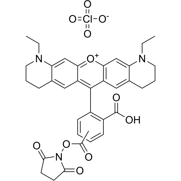
TTO 488 NHS ester TEA is a new fluorescent marker based on the Rhodamine structure. It has strong absorption, high fluorescence quantum yield, high thermal stability and photochemical stability, and is suitable for single molecule detection and high-resolution microscopy. TTO 488 NHS ester TEA is an NHS ester derivative of ATTO 488 that can be used to label proteins or antibodies.
|
-

-
- HY-131009
-
|
|
PARP
|
Others
|
|
Fluorescein-NAD+ is an alternative to radiolabeled NAD and a substrate for ADP-ribosylation. Fluorescein-NAD+ can be used in PARP assays by fluorescence microscopy. Extinction Coefficient: 262 nm.
|
-
-
- HY-D1950
-
|
|
Fluorescent Dye
|
Others
|
|
ATTO 633 is a fluorescent dye with an absorption peak at approximately 630 nm and a fluorescence emission peak at 651 nm. ATTO 633 can be used in nanomechanical photothermal microscopy studies .
|
-
-
- HY-D1305
-
|
|
Fluorescent Dye
|
Others
|
|
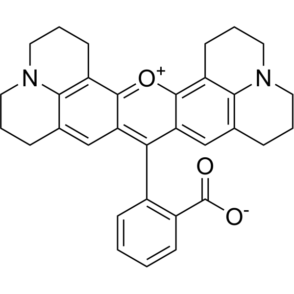
ATTO 488 carboxylic acid is a new fluorescent label based on the Rhodamine structure. It has strong absorption, high fluorescence quantum yield, high thermal stability and photochemical stability, and is suitable for single molecule detection and high-resolution microscopy. ATTO 488 carboxylic acid is a carboxylic acid derivative of ATTO 488, which can be used to label proteins or antibodies.
|
-
-
- HY-W440908
-
|
|
Liposome
|
Others
|
|
DSPE-PEG2000-Cy3 is a PEG lipid conjugated with a fluorophore. The Cy3 fluorophore is commonly used in applications such as immunolabeling, nucleic acid labeling, fluorescence microscopy and flow cytometry. The absorption wavelength of the Cy3 fluorophore peaks at 548-552 nm, while its emission wavelength reaches a maximum at 562-570 nm .
|
-
-
- HY-110334
-
|
|
Fluorescent Dye
|
Others
|
|
FFN 206 dihydrochloride, a fluorescent probe, is used as an excellent Vesicular Monoamine Transporter 2 (VMAT2) substrate with an apparent Km of 1.16 μM. FFN 206 dihydrochloride is capable of detecting VMAT2 activity in intact cells using fluorescence microscopy, with subcellular localization to VMAT2-expressing acidic compartments without apparent labeling of other organelles .
|
-
-
- HY-D2438
-
|
|
Fluorescent Dye
|
Cancer
|
|
CDDP-PEG-Cy3 is a CDDP-PEG conjugate labeled with Cy3 (HY-D0822). The Cy3 fluorophore is commonly used in applications such as immunolabeling, nucleic acid labeling, fluorescence microscopy, and flow cytometry. The maximum emission wavelength of Cy3 is approximately 562-570 nm. Cisplatin (CDDP) (HY-17394) is an antineoplastic chemotherapy agent by cross-linking with DNA and causing DNA damage in cancer cells. Cisplatin activates ferroptosis and induces autophagy .
|
-

-
- HY-D2002
-
|
|
Fluorescent Dye
|
Others
|
|
ATTO 488 maleimide is a new fluorescent marker based on the Rhodamine structure. It has strong absorption, high fluorescence quantum yield, high thermal stability and photochemical stability, and is suitable for single molecule detection and high-resolution microscopy. ATTO 488 maleimide is a maleimide derivative of ATTO 488, which can be used to label proteins or antibodies.
|
-
-
- HY-D1959
-
|
|
Fluorescent Dye
|
Others
|
|
ATTO 565 NHS ester is a new fluorescent marker based on the Rhodamine structure. It has strong absorption, high fluorescence quantum yield, high thermal stability and photochemical stability, and is suitable for single molecule detection and high-resolution microscopy. ATTO 565 NHS ester is an NHS ester derivative of ATTO 565 that can be used to label proteins or antibodies.
|
-
-
- HY-D1240
-
|
|
Biochemical Assay Reagents
|
Others
|
|
Rhodamine 101 inner salt is a bright fluorescent dye with excitation and emission maxima at 565 and 595 nm, respectively. It can be used in various biological applications such as fluorescence microscopy, flow cytometry, fluorescence correlation spectroscopy, and ELISA.
|
-
-
- HY-105552
-
|
|
Biochemical Assay Reagents
|
Others
|
|
Prolonium (iodide) is membrane impermeant. Prolonium (iodide) can easily penetrate dead or damaged cells. Prolonium (iodide) is a probe that can be used for fluorescence microscopy and flow cytometry .
|
-

-
- HY-DN0194F
-
|
|
Fluorescent Dye
|
Others
|
|
Cy3-Asiatic acid is a fluorescent labeling reagent that combines Cy3 (HY-D0822) and Asiatic acid (HY-N0194). Cy3-Asiatic acid can be used to observe and track Asiatic acid under fluorescence microscopy (Ex/Em = 550/570 nm).
|
-

-
- HY-W440910
-
|
|
Liposome
|
Others
|
|
DSPE-PEG5000-Cy3 is a phospholipid PEG polymer with Cy3 dye used in labeling and fluorescence microscopy. The polymer can self-assemble in aqueous solution to form micelles/lipid bilayer and used to prepare liposomes or nanoparticles for nutrients delivery such as mRNA or DNA vaccine.
|
-

-
- HY-DY1035
-
|
|
Reactive Oxygen Species (ROS)
Fluorescent Dye
|
Others
|
Hydroxyphenyl Fluorescein (HPF) (solution) is a stable ROS fluorescent probe dye. Hydroxyphenyl Fluorescein has stronger specificity and stability than H2DCFDA (HY-D0940). Hydroxyphenyl Fluorescein can produce strong green fluorescence through hydroxyl radical reaction with intracellular peroxynitroso. Hydroxyphenyl Fluorescein can be applied for fluorescence microscopy, high-throughput imager, luciferase microplate reader or flow cytometry. Ex/Em=490/515 nm .
Solvent and concentration: DMSO: 10 mM
|
-

-
- HY-152901
-
|
|
Fluorescent Dye
|
Others
|
|
Chol-N3 is a bioorthogonal-based chol probe. Chol-N3 can combine with super-resolution fluorescence microscopy (SRM), providing direct visualization of nanoscale lipid heterogeneity in the cell surface of resting living cells . Chol-N3 is a click chemistry reagent, it contains an Azide group and can undergo copper-catalyzed azide-alkyne cycloaddition reaction (CuAAc) with molecules containing Alkyne groups. It can also undergo strain-promoted alkyne-azide cycloaddition (SPAAC) reactions with molecules containing DBCO or BCN groups.
|
-
-
- HY-153524
-
|
|
Fluorescent Dye
|
Others
|
|
ATTO 425 NHS ester is a new fluorescent marker based on the Rhodamine structure. It has strong absorption, high fluorescence quantum yield, high thermal stability and photochemical stability, and is suitable for single molecule detection and high-resolution microscopy. ATTO 425 NHS ester is an NHS ester derivative of ATTO 425 that can be used to label proteins or antibodies.
|
-
-
- HY-D1993
-
|
|
Fluorescent Dye
HIV
|
Infection
|
|
ATTO 647 NHS ester is a fluorescent label targeting free amino groups. ATTO 647 NHS ester can undergo a nucleophilic reaction with the free amino groups of EF-C peptide via its activated carboxylic acid group to form a stable covalent conjugate. ATTO 647 NHS ester-labeled nanofibers not only retain retroviral transduction-enhancing activity but also maintain stable fluorescent signals in both buffer and cell culture systems. ATTO 647 NHS ester is suitable for detection applications including fluorescence spectroscopy, microscopy and flow cytometry. ATTO 647 NHS ester has been used in studies related to HIV-1 infection .
|
-

-
- HY-N16309
-
|
|
Fluorescent Dye
|
Others
|
|
Cyanine 7 free acid bromide is a near-infrared fluorescent probe for labelling amine groups such as those on antibodies, nucleic acids, and proteins and can be detected using a variety of fluorescence detection techniques such as microscopy and flow cytometry.
|
-

-
- HY-D2506
-
|
|
Fluorescent Dye
|
Others
|
|
Cy3-PEG3400-NH2 is a polyethylene glycol derivative containing Cy3 (HY-D0822) fluorescent dye and polyethylene glycol (PEG) and an amino group. The Cy3 fluorophore is commonly used in applications such as immunolabeling, nucleic acid labeling, fluorescence microscopy, and flow cytometry. Cy3 has an emission maximum around 562-570 nm. Cy3-PEG3400-NH2 can be used for fluorescence imaging, fluorescence tracing and fluorescence labeling .
|
-

-
- HY-W440912
-
|
|
Liposome
|
Others
|
|
DSPE-PEG3400-Cy5 is a PEG phospholipid with Cy5 dye used in protein/nucelic acid labeling and fluorescence microscopy. The polymer can self-assemble in aqueous solution to form micelles/lipid bilayer and used to prepare liposomes or nanoparticles for nutrients delivery such as mRNA or DNA vaccine.
|
-
-
- HY-D2440
-
|
|
Fluorescent Dye
|
Others
|
|
cRGD-PEG-Cy3 is a Cy3 (HY-D0822) labeled cRGD-PEG conjugate. The Cy3 fluorophore is commonly used in applications such as immunolabeling, nucleic acid labeling, fluorescence microscopy, and flow cytometry. Cy3 has an emission maximum around 562-570 nm. cRGD can be used to modify lipid for improved stability .
|
-

-
- HY-D1915
-
|
|
Fluorescent Dye
|
Others
|
|
ATTO 390 is a new fluorescent marker based on the Rhodamine structure. It has strong absorption, high fluorescence quantum yield, high thermal stability and photochemical stability, and is suitable for single molecule detection and high-resolution microscopy.
|
-
-
- HY-D2046
-
|
|
Fluorescent Dye
|
Others
|
|
ATTO 532 NHS ester is a new fluorescent marker based on the Rhodamine structure. It has strong absorption, high fluorescence quantum yield, high thermal stability and photochemical stability, and is suitable for single molecule detection and high-resolution microscopy. ATTO 532 NHS ester is an NHS ester derivative of ATTO 532 that can be used to label proteins or antibodies.
|
-
-
- HY-D1957
-
|
|
Fluorescent Dye
|
Others
|
|
ATTO 633 NHS ester is a new fluorescent marker based on the Rhodamine structure. It has strong absorption, high fluorescence quantum yield, high thermal stability and photochemical stability, and is suitable for single molecule detection and high-resolution microscopy. ATTO 633 NHS ester is an NHS ester derivative of ATTO 633 that can be used to label proteins or antibodies.
|
-
-
- HY-D2019
-
|
|
Fluorescent Dye
|
Others
|
|
ATTO 550 NHS ester is a new fluorescent marker based on the Rhodamine structure. It has strong absorption, high fluorescence quantum yield, high thermal stability and photochemical stability, and is suitable for single molecule detection and high-resolution microscopy. ATTO 550 NHS ester is an NHS ester derivative of ATTO 550 that can be used to label proteins or antibodies.
|
-

-
- HY-D2016
-
|
|
Fluorescent Dye
|
Others
|
|
ATTO 565 cadaverine is a new fluorescent marker based on the Rhodamine structure. It has strong absorption, high fluorescence quantum yield, high thermal stability and photochemical stability, and is suitable for single molecule detection and high-resolution microscopy. ATTO 565 cadaverine is a cadaverine derivative of ATTO 565, which can be used to label proteins or antibodies.
|
-
-
- HY-D2058
-
|
|
Fluorescent Dye
|
Others
|
|
ATTO 700 NHS ester is a new fluorescent marker based on the Rhodamine structure. It has strong absorption, high fluorescence quantum yield, high thermal stability and photochemical stability, and is suitable for single molecule detection and high-resolution microscopy. ATTO 700 NHS ester is an NHS ester derivative of ATTO 700 that can be used to label proteins or antibodies.
|
-
-
- HY-D2073
-
|
|
Fluorescent Dye
|
Others
|
|
ATTO 680 NHS ester is a new fluorescent marker based on the Rhodamine structure. It has strong absorption, high fluorescence quantum yield, high thermal stability and photochemical stability, and is suitable for single molecule detection and high-resolution microscopy. ATTO 680 NHS ester is an NHS ester derivative of ATTO 680 that can be used to label proteins or antibodies.
|
-
-
- HY-D2021
-
|
|
Fluorescent Dye
|
Others
|
|
ATTO 550 maleimide is a new fluorescent marker based on the Rhodamine structure. It has strong absorption, high fluorescence quantum yield, high thermal stability and photochemical stability, and is suitable for single molecule detection and high-resolution microscopy. ATTO 550 maleimide is a maleimide derivative of ATTO 550, which can be used to label proteins or antibodies.
|
-

-
- HY-D1935
-
|
|
Fluorescent Dye
|
Others
|
|
ATTO 465 NHS ester is a new fluorescent marker based on the Rhodamine structure. It has strong absorption, high fluorescence quantum yield, high thermal stability and photochemical stability, and is suitable for single molecule detection and high-resolution microscopy. ATTO 465 NHS ester is an NHS ester derivative of ATTO 465 that can be used to label proteins or antibodies.
|
-
-
- HY-131009A
-
|
|
PARP
|
Others
|
|
Fluorescein-NAD+ sodium is an alternative to radiolabeled NAD and a substrate for ADP-ribosylation. Fluorescein-NAD+ sodium can be used in PARP assays by fluorescence microscopy. Extinction Coefficient: 262 nm.
|
-

-
- HY-D2602
-
|
|
Fluorescent Dye
|
Others
|
|
ICG PEG5000 NH2 is a polyethylene glycol derivative containing Indocyanine green (ICG) (HY-D0711) fluorescent dye and polyethylene glycol (PEG) and an amino group. The Indocyanine green fluorophore is commonly used in applications such as immunolabeling, nucleic acid labeling, fluorescence microscopy, and flow cytometry. ICG PEG5000 NH2 can be used for fluorescence imaging, fluorescence tracing and fluorescence labeling (Ex/Em=785/813 nm).
|
-

-
- HY-D2599
-
|
|
Fluorescent Dye
|
Others
|
|
ICG PEG1000 NH2 is a polyethylene glycol derivative containing Indocyanine green (ICG) (HY-D0711) fluorescent dye and polyethylene glycol (PEG) and an amino group. The Indocyanine green fluorophore is commonly used in applications such as immunolabeling, nucleic acid labeling, fluorescence microscopy, and flow cytometry. ICG PEG1000 NH2 can be used for fluorescence imaging, fluorescence tracing and fluorescence labeling (Ex/Em=785/813 nm).
|
-

-
- HY-D2554
-
|
|
Fluorescent Dye
|
Others
|
|
Cy5.5-PEG5000-NH2 is a polyethylene glycol derivative containing Cy5.5 (HY-D0924) fluorescent dye and polyethylene glycol (PEG) and an amino group. The Cy5.5 fluorophore is commonly used in applications such as immunolabeling, nucleic acid labeling, fluorescence microscopy, and flow cytometry. Cy5.5 has an emission maximum around 710 nm. Cy5.5-PEG5000-NH2 can be used for fluorescence imaging, fluorescence tracing and fluorescence labeling.
|
-

-
- HY-D2553
-
|
|
Fluorescent Dye
|
Others
|
|
Cy5.5-PEG3400-NH2 is a polyethylene glycol derivative containing Cy5.5 (HY-D0924) fluorescent dye and polyethylene glycol (PEG) and an amino group. The Cy5.5 fluorophore is commonly used in applications such as immunolabeling, nucleic acid labeling, fluorescence microscopy, and flow cytometry. Cy5.5 has an emission maximum around 710 nm. Cy5.5-PEG3400-NH2 can be used for fluorescence imaging, fluorescence tracing and fluorescence labeling.
|
-

-
- HY-D2552
-
|
|
Fluorescent Dye
|
Others
|
|
Cy5.5-PEG1000-NH2 is a polyethylene glycol derivative containing Cy5.5 (HY-D0924) fluorescent dye and polyethylene glycol (PEG) and an amino group. The Cy5.5 fluorophore is commonly used in applications such as immunolabeling, nucleic acid labeling, fluorescence microscopy, and flow cytometry. Cy5.5 has an emission maximum around 710 nm. Cy5.5-PEG1000-NH2 can be used for fluorescence imaging, fluorescence tracing and fluorescence labeling.
|
-

- HY-D2555
-
|
|
Fluorescent Dye
|
Others
|
|
Cy5.5-PEG10000-NH2 is a polyethylene glycol derivative containing Cy5.5 (HY-D0924) fluorescent dye and polyethylene glycol (PEG) and an amino group. The Cy5.5 fluorophore is commonly used in applications such as immunolabeling, nucleic acid labeling, fluorescence microscopy, and flow cytometry. Cy5.5 has an emission maximum around 710 nm. Cy5.5-PEG10000-NH2 can be used for fluorescence imaging, fluorescence tracing and fluorescence labeling.
|
-

- HY-D1396
-
Br-DAPI
3 Publications Verification
|
Fluorescent Dye
DNA Stain
|
Others
|
|
Br-DAPI is a marker dye in DAPI series. DAPI is a fluorescent dye that binds strongly to DNA. It binds to the AT base pair of the double-stranded DNA minor groove, and one DAPI molecule can occupy three base pair positions. The fluorescence intensity of DAPI molecules bound to double-stranded DNA is increased by about 20 times, and it is commonly observed with fluorescence microscopy, and the amount of DNA can be determined based on the intensity of fluorescence. In addition, because DAPI can pass through intact cell membranes, it can be used to stain both live and fixed cells . Storage: Keep away from light.
|
-
- HY-160270
-
|
|
Liposome
|
Others
|
|
DSPE-PEG5000-Fluor 488 is a PEG-dye-lipid conjugate consists of a DSPE phospholipid which is an unsaturated phospholipid, a Fluor 488 dye which is a cyanine dye that is prominently used in fluorescence microscopy with excitation and emission maxima at 499 nm and 520 nm and a large PEG spacer which links the former substance together.
|
-
- HY-160276
-
|
|
Liposome
|
Others
|
|
DOPE-PEG5000-Fluor 555 is consist of a DOPE phospholipid which is an unsaturated phospholipid and a Fluor 555 dye which is a bright orange cyanine dye that can be used in fluorescence microscopy, FRET and other in vivo imaging techniques.
|
-
- HY-D2435
-
|
|
Fluorescent Dye
|
Inflammation/Immunology
Cancer
|
|
CDDP-PEG-Cy3 is a MTX-PEG conjugate labeled with Cy3 (HY-D0822). The Cy3 fluorophore is commonly used in applications such as immunolabeling, nucleic acid labeling, fluorescence microscopy, and flow cytometry. The maximum emission wavelength of Cy3 is approximately 562-570 nm. Methotrexate (Amethopterin; MTX) (HY-14519), an antimetabolite and antifolate agent, inhibits the enzyme dihydrofolate reductase, thereby preventing the conversion of folic acid into tetrahydrofolate, and inhibiting DNA synthesis. Methotrexate, also an immunosuppressant and antineoplastic agent, is used for the research of rheumatoid arthritis and a number of different cancers (such as acute lymphoblastic leukemia) .
|
-

- HY-D1917
-
|
|
Fluorescent Dye
|
Others
|
|
ATTO 390 NHS ester is a new fluorescent marker based on the Rhodamine structure. It has strong absorption, high fluorescence quantum yield, high thermal stability and photochemical stability, and is suitable for single molecule detection and high-resolution microscopy. ATTO 390 NHS ester is an NHS ester derivative of ATTO 390 that can be used to label proteins or antibodies.
|
-
- HY-D2038
-
|
|
Fluorescent Dye
|
Others
|
|
ATTO 550 alkyne is a new fluorescent marker based on the Rhodamine structure. It has strong absorption, high fluorescence quantum yield, high thermal stability and photochemical stability, and is suitable for single molecule detection and high-resolution microscopy. ATTO 550 alkyne is an alkyne derivative of ATTO 550 and can be used to label proteins or antibodies.
|
-
- HY-D1961
-
|
|
Fluorescent Dye
|
Others
|
|
ATTO 565 maleimide is a new fluorescent marker based on the Rhodamine structure. It has strong absorption, high fluorescence quantum yield, high thermal stability and photochemical stability, and is suitable for single molecule detection and high-resolution microscopy. ATTO 565 maleimide is a maleimide derivative of ATTO 565, which can be used to label proteins or antibodies.
|
-
- HY-D1929
-
|
|
Fluorescent Dye
|
Others
|
|
ATTO 594 NHS ester is a new fluorescent marker based on the Rhodamine structure. It has strong absorption, high fluorescence quantum yield, high thermal stability and photochemical stability, and is suitable for single molecule detection and high-resolution microscopy. ATTO 594 NHS ester is an NHS ester derivative of ATTO 594 that can be used to label proteins or antibodies.
|
-

- HY-D2014
-
|
|
Fluorescent Dye
|
Others
|
|
ATTO 565 alkyne is a new fluorescent marker based on the Rhodamine structure. It has strong absorption, high fluorescence quantum yield, high thermal stability and photochemical stability, and is suitable for single molecule detection and high-resolution microscopy. ATTO 565 alkyne is an alkyne derivative of ATTO 565 and can be used to label proteins or antibodies.
|
-
- HY-D2035
-
|
|
Fluorescent Dye
|
Others
|
|
ATTO 514 alkyne is a new fluorescent marker based on the Rhodamine structure. It has strong absorption, high fluorescence quantum yield, high thermal stability and photochemical stability, and is suitable for single molecule detection and high-resolution microscopy. ATTO 514 alkyne is an alkyne derivative of ATTO 514 and can be used to label proteins or antibodies.
|
-

- HY-D2047
-
|
|
Fluorescent Dye
|
Others
|
|
ATTO 532 maleimide is a new fluorescent marker based on the Rhodamine structure. It has strong absorption, high fluorescence quantum yield, high thermal stability and photochemical stability, and is suitable for single molecule detection and high-resolution microscopy. ATTO 532 maleimide is a maleimide derivative of ATTO 532, which can be used to label proteins or antibodies.
|
-
- HY-D1939
-
|
|
Fluorescent Dye
|
Others
|
|
ATTO 465 amine is a new fluorescent marker based on the Rhodamine structure. It has strong absorption, high fluorescence quantum yield, high thermal stability and photochemical stability, and is suitable for single molecule detection and high-resolution microscopy. ATTO 465 amine is an amine derivative of ATTO 465 and can be used to label proteins or antibodies.
|
-
- HY-D2063
-
|
|
Fluorescent Dye
|
Others
|
|
ATTO 740 maleimide is a new fluorescent marker based on the Rhodamine structure. It has strong absorption, high fluorescence quantum yield, high thermal stability and photochemical stability, and is suitable for single molecule detection and high-resolution microscopy. ATTO 740 maleimide is a maleimide derivative of ATTO 740, which can be used to label proteins or antibodies.
|
-
- HY-D2015
-
|
|
Fluorescent Dye
|
Others
|
|
ATTO 488 alkyne is a new fluorescent marker based on the Rhodamine structure. It has strong absorption, high fluorescence quantum yield, high thermal stability and photochemical stability, and is suitable for single molecule detection and high-resolution microscopy. ATTO 488 alkyne is an alkyne derivative of ATTO 488 and can be used to label proteins or antibodies.
|
-
- HY-D1933
-
|
|
Fluorescent Dye
|
Others
|
|
ATTO 590 maleimide is a new fluorescent marker based on the Rhodamine structure. It has strong absorption, high fluorescence quantum yield, high thermal stability and photochemical stability, and is suitable for single molecule detection and high-resolution microscopy. ATTO 590 maleimide is a maleimide derivative of ATTO 590, which can be used to label proteins or antibodies.
|
-
- HY-D2059
-
|
|
Fluorescent Dye
|
Others
|
|
ATTO 700 maleimide is a new fluorescent marker based on the Rhodamine structure. It has strong absorption, high fluorescence quantum yield, high thermal stability and photochemical stability, and is suitable for single molecule detection and high-resolution microscopy. ATTO 700 maleimide is a maleimide derivative of ATTO 700, which can be used to label proteins or antibodies.
|
-

- HY-D2026
-
|
|
Fluorescent Dye
|
Others
|
|
ATTO 514 NHS ester is a new fluorescent marker based on the Rhodamine structure. It has strong absorption, high fluorescence quantum yield, high thermal stability and photochemical stability, and is suitable for single molecule detection and high-resolution microscopy. ATTO 514 NHS ester is an NHS ester derivative of ATTO 514 that can be used to label proteins or antibodies.
|
-

- HY-D1999
-
|
|
Fluorescent Dye
|
Others
|
|
ATTO 665 NHS ester is a new fluorescent marker based on the Rhodamine structure. It has strong absorption, high fluorescence quantum yield, high thermal stability and photochemical stability, and is suitable for single molecule detection and high-resolution microscopy. ATTO 665 NHS ester is an NHS ester derivative of ATTO 665 that can be used to label proteins or antibodies.
|
-

- HY-D1932
-
|
|
Fluorescent Dye
|
Others
|
|
ATTO 590 NHS ester is a new fluorescent marker based on the Rhodamine structure. It has strong absorption, high fluorescence quantum yield, high thermal stability and photochemical stability, and is suitable for single molecule detection and high-resolution microscopy. ATTO 590 NHS ester is an NHS ester derivative of ATTO 590 that can be used to label proteins or antibodies.
|
-
- HY-D2074
-
|
|
Fluorescent Dye
|
Others
|
|
ATTO 680 maleimide is a new fluorescent marker based on the Rhodamine structure. It has strong absorption, high fluorescence quantum yield, high thermal stability and photochemical stability, and is suitable for single molecule detection and high-resolution microscopy. ATTO 680 maleimide is a maleimide derivative of ATTO 680, which can be used to label proteins or antibodies.
|
-

- HY-D2052
-
|
|
Fluorescent Dye
|
Others
|
|
ATTO 532 iodacetamid is a new fluorescent marker based on the Rhodamine structure. It has strong absorption, high fluorescence quantum yield, high thermal stability and photochemical stability, and is suitable for single molecule detection and high-resolution microscopy. ATTO 532 iodacetamid is an iodoacetamide derivative of ATTO 532, which can be used to label proteins or antibodies.
|
-
- HY-D1946
-
|
|
Fluorescent Dye
|
Others
|
|
ATTO 590 alkyne is a new fluorescent marker based on the Rhodamine structure. It has strong absorption, high fluorescence quantum yield, high thermal stability and photochemical stability, and is suitable for single molecule detection and high-resolution microscopy. ATTO 590 alkyne is an alkyne derivative of ATTO 590 and can be used to label proteins or antibodies.
|
-

- HY-121364
-
|
|
Histamine Receptor
Fluorescent Dye
|
Others
|
|
Bodilisant is a histamine H3 receptor (hH3R) ligand and imaging/labeling agent, with a Ki value of 6.51 nM for hH3R. Bodilisant binds to hH3R to produce strong green fluorescence, localizes to the extracellular membrane without internalization, and generates clear, displaceable fluorescent labeling of hH3R in native human brain tissues. Bodilisant serves as a pharmacological tool to visualize the distribution of hH3R via fluorescence confocal laser scanning microscopy .
|
-

- HY-D2601
-
|
|
Fluorescent Dye
|
Others
|
|
ICG PEG3400 NH2 is a polyethylene glycol derivative containing Indocyanine green (ICG) (HY-D0711) fluorescent dye and polyethylene glycol (PEG) and an amino group. The Indocyanine green fluorophore is commonly used in applications such as immunolabeling, nucleic acid labeling, fluorescence microscopy, and flow cytometry. ICG PEG3400 NH2 can be used for fluorescence imaging, fluorescence tracing and fluorescence labeling (Ex/Em=785/813 nm).
|
-

- HY-D2600
-
|
|
Fluorescent Dye
|
Others
|
|
ICG PEG2000 NH2 is a polyethylene glycol derivative containing Indocyanine green (ICG) (HY-D0711) fluorescent dye and polyethylene glycol (PEG) and an amino group. The Indocyanine green fluorophore is commonly used in applications such as immunolabeling, nucleic acid labeling, fluorescence microscopy, and flow cytometry. ICG PEG2000 NH2 can be used for fluorescence imaging, fluorescence tracing and fluorescence labeling (Ex/Em=785/813 nm).
|
-

- HY-D2603
-
|
|
Fluorescent Dye
|
Others
|
|
ICG PEG10000 NH2 is a polyethylene glycol derivative containing Indocyanine green (ICG) (HY-D0711) fluorescent dye and polyethylene glycol (PEG) and an amino group. The Indocyanine green fluorophore is commonly used in applications such as immunolabeling, nucleic acid labeling, fluorescence microscopy, and flow cytometry. ICG PEG10000 NH2 can be used for fluorescence imaging, fluorescence tracing and fluorescence labeling (Ex/Em=785/813 nm).
|
-

- HY-D2588
-
|
|
Fluorescent Dye
|
Others
|
|
Cy7.5 PEG-NH2 is a polyethylene glycol derivative containing CY7.5 (HY-D0926) fluorescent dye and polyethylene glycol (PEG) and an amino group. The CY7.5 fluorophore is commonly used in applications such as immunolabeling, nucleic acid labeling, fluorescence microscopy, and flow cytometry. Cy7.5 PEG-NH2 can be used for fluorescence imaging, fluorescence tracing and fluorescence labeling.
|
-

- HY-P4900
-
|
|
Caspase
|
Others
|
|
Fluorescein-6-carbonyl-Asp(OMe)-Glu(OMe)-Val-DL-Asp(OMe)-fluoromethylketone is a cell-permeable, non-toxic inhibitor that binds irreversibly to activated caspase-3 in apoptotic cells. The fluorescence intensity can be measured by flow cytometry, microwell plate reader, or fluorescence microscopy .
|
-
- HY-D1349
-
|
|
Fluorescent Dye
|
|
|
Bodipy TR alkyneis one of a boron dipyrromethene fluorophore for the ROX (Texas Red) channel. This is a versatile fluorophore that can be used in microscopy, fluorescence polarization measurements, and other applications. This derivative is a terminal alkyne of copper-catalyzed click chemistry.
|
-
- HY-D2505
-
|
|
Fluorescent Dye
|
Others
|
|
Cy3-PEG1000-NH2 is a polyethylene glycol derivative containing Cy3 (HY-D0822) fluorescent dye and polyethylene glycol (PEG) and an amino group. The Cy3 fluorophore is commonly used in applications such as immunolabeling, nucleic acid labeling, fluorescence microscopy, and flow cytometry. Cy3 has an emission maximum around 562-570 nm. Cy3-PEG1000-NH2 can be used for fluorescence imaging, fluorescence tracing and fluorescence labeling .
|
-

- HY-D2508
-
|
|
Fluorescent Dye
|
Others
|
|
Cy3-PEG10000-NH2 is a polyethylene glycol derivative containing Cy3 (HY-D0822) fluorescent dye and polyethylene glycol (PEG) and an amino group. The Cy3 fluorophore is commonly used in applications such as immunolabeling, nucleic acid labeling, fluorescence microscopy, and flow cytometry. Cy3 has an emission maximum around 562-570 nm. Cy3-PEG10000-NH2 can be used for fluorescence imaging, fluorescence tracing and fluorescence labeling .
|
-

- HY-D2507
-
|
|
Fluorescent Dye
|
Others
|
|
Cy3-PEG5000-NH2 is a polyethylene glycol derivative containing Cy3 (HY-D0822) fluorescent dye and polyethylene glycol (PEG) and an amino group. The Cy3 fluorophore is commonly used in applications such as immunolabeling, nucleic acid labeling, fluorescence microscopy, and flow cytometry. Cy3 has an emission maximum around 562-570 nm. Cy3-PEG5000-NH2 can be used for fluorescence imaging, fluorescence tracing and fluorescence labeling .
|
-

- HY-W440934
-
|
|
Liposome
|
Others
|
|
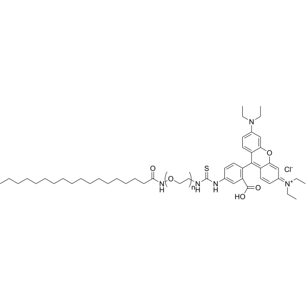
Stearic acid-PEG2000-Rhodamine is an amphiphilic PEG polymer which can form micelles in water. The rhodamine can be used for staining sample and easily traced by fluorescence microscopy. Rhodamine has maximum absorption at 570 nm and emission around 595 nm.
|
-

- HY-D2434
-
|
|
Fluorescent Dye
|
Others
|
|
HRP-PEG-Cy3 is a HRP and Cy3 (HY-D0822) labeled PEG. The Cy3 fluorophore is commonly used in applications such as immunolabeling, nucleic acid labeling, fluorescence microscopy, and flow cytometry. Cy3 has an emission maximum around 562-570 nm.
|
-

- HY-W440909
-
|
|
Liposome
|
Others
|
|
DSPE-PEG3400-Cy3 is a phospholipid PEG polymer with Cy3 dye used in labeling and fluorescence microscopy. The polymer can self-assemble in aqueous solution to form micelles/lipid bilayer and used to prepare liposomes or nanoparticles for nutrients delivery such as mRNA or DNA vaccine.
|
-

- HY-D2012
-
|
|
Fluorescent Dye
|
Others
|
|
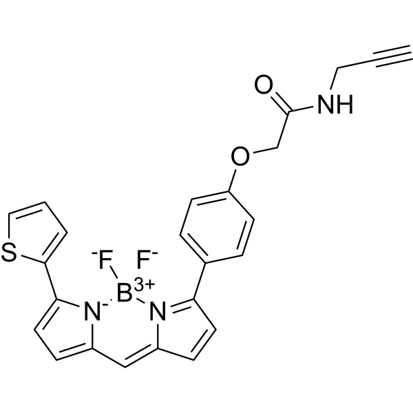
ATTO 488 iodacetamid is a fluorescent dye suitable for single molecule detection applications and high-resolution microscopy for use in flow cytometry (FACS), fluorescence in situ hybridization (FISH) experiments. ATTO 488 iodacetamid has an effective excitation wavelength of 480-515 nm. When using an argon ion laser, the excitation wavelength is recommended to be 488 nm.
|
-
- HY-D2761
-
|
|
Fluorescent Dye
|
Others
|
|
DY-680-NHS ester is an amine reactive hydrophilic fluorochrome. It can be conjugated to an antibody with higher D/P ratio without causing fluorescence quenching and conjugate precipitation, and it is widely used in applications including western blotting, microscopy, flow cytometry, and cell-based assays. The spectrum has an excitation of 690nm and emission at 709nm.
|
-

- HY-N16316
-
|
|
Fluorescent Dye
|
Others
|
|
Meso-Cl cyanine 7 free acid chloride is a Meso-Cl cyanine fluorescent dye. Meso-Cl cyanine 7 free acid chloride can be used for labelling amine groups such as those on antibodies, nucleic acids, and proteins and can be detected using a variety of fluorescence detection techniques such as microscopy and flow cytometry (Ex/Em = 778/805 nm) .
|
-

- HY-D0150A
-
|
|
Fluorescent Dye
|
Others
|
|
(Z)-Thiazole Orange iodide is an asymmetric cyanine dye whose fluorescence highly depends on the local environment.(Z)-Thiazole Orange iodide is essentially dark in solution; however, its fluorescence increases a thousandfold when (Z)-Thiazole Orange iodide is introduced into double-stranded DNA and RNA (dsDNA or dsRNA). The maximum absorption of Thiazole Orange in complex with DNA is 509 nm, and the maximum emission is 532 nm. Thiazole Orange solution is widely used for determining the percentage of reticulocytes in human peripheral blood with microscopy and flow cytometry.
|
-

- HY-D2526
-
|
|
Fluorescent Dye
Liposome
|
Others
|
|
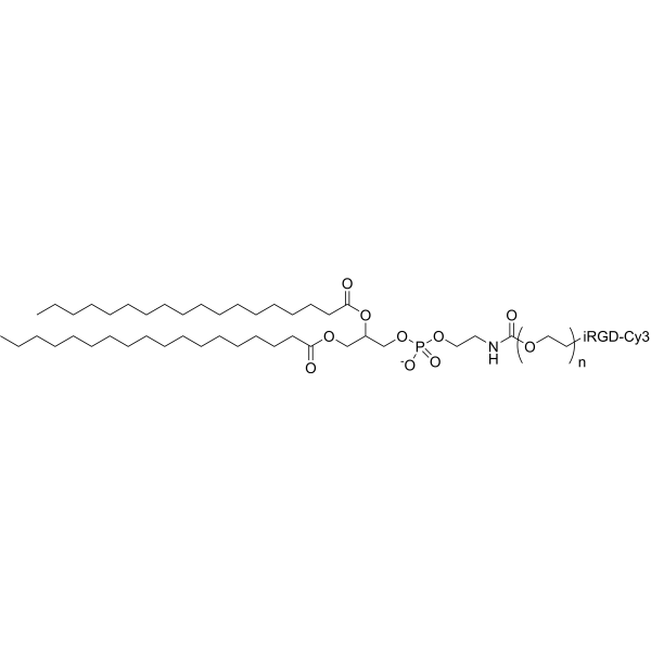
Cy3 IRGD-PEG-DSPE is a PEG phospholipid with Cy3 (HY-D0822) dye used in protein/nucelic acid labeling and fluorescence microscopy. Cy3 IRGD-PEG-DSPE can self-assemble in aqueous solution to form micelles/lipid bilayer and used to prepare liposomes or nanoparticles for nutrients delivery such as mRNA or DNA vaccine.
|
-
- HY-W440938
-
|
|
Liposome
|
Others
|
|
Stearic acid-PEG2000-FITC is an amphiphatic polyPEG which can self assemble to form micelles in water. The polymer can be used to encapsulate therapeutic agent. FITC is a green dye with peak absorption at 494 nm and maximum emission at 520 nm and can be used for staining biological samples or nanoparticles. FITC can be easily traced by fluorescence microscopy.
|
-

- HY-W440936
-
|
|
Liposome
|
Others
|
|
Stearic acid-PEG5000-Rhodamine is a fatty acid containing PEG polymer which can self assemble in an aqueous solution to form micelles. The polymer can be used to prepare nanoparticles for drug encapsulation. The red dye rhodamine can be easily traced by fluorescence microscopy. Rhodamine has maximum absorption at 570 nm and emission around 595 nm.
|
-

- HY-W440935
-
|
|
Liposome
|
Others
|
|
Stearic acid-PEG3400-Rhodamine is a fatty acid containing PEG polymer which can self assemble in an aqueous solution to form micelles. The polymer can be used to prepare nanoparticles for drug encapsulation. The red dye rhodamine can be easily traced by fluorescence microscopy. Rhodamine has maximum absorption at 570 nm and emission around 595 nm.
|
-
- HY-203233
-
|
|
Fluorescent Dye
|
Others
|
|
Rhodamine-DHPE is a fluorescently labeled phosphatidylethanolamine lipid that labels phospholipid bilayers. Rhodamine-DHPE serves as a fluorescence quenching substrate and membrane stain. The fluorescence lifetime of Rhodamine-DHPE decreases significantly in the presence of Cu 2+-PS complexes. Rhodamine-DHPE effectively stains the membranes of human red blood cells and mouse fibroblasts, and supports lifetime-resolved imaging via pump-probe fluorescence microscopy .
|
-

- HY-D2428
-
|
|
Fluorescent Dye
|
Cardiovascular Disease
Infection
Inflammation/Immunology
Cancer
|
|
OVA-PEG-Cy3 is a Cy3 (HY-D0822)-labeled OVA-PEG conjugate. The Cy3 fluorophore is commonly used in applications such as immunolabeling, nucleic acid labeling, fluorescence microscopy, and flow cytometry. The maximum emission wavelength of Cy3 is approximately 562-570 nm. Ovalbumins (OVA), the main protein found in egg whites, have various biological activities such as anticancer, antihypertensive, antibacterial, antioxidant and immunomodulatory activities. Ovalbumins are the most abundant proteins synthesized in progesterone- or estrogen-treated fallopian tubes and are commonly used as markers to study hormone regulation of gene expression in tissues .
|
-

- HY-D3252
-
|
|
Fluorescent Dye
|
Others
|
|
IR 650 maleimide is a near-infrared fluorescent dye. It should be noted that IR 650 maleimide‘s fluorescence stability is poor, with less than 16% of the fluorescence signal retained after the expansion microscopy experimental procedure .
|
-

- HY-P0281F
-
|
Cy5-TAT(47-57) acetate; Cy5-HIV-1 TAT protein (47-57) acetate
|
Fluorescent Dye
|
Others
|
|
Cy5-TAT acetate is a TAT peptide labeled with CY5 (HY-D0821) (Ex= 600-620 nm, Em= 670 nm). Cy5-TAT acetate can be used for fluorescence microscopy especially single-molecule fluorescence imaging .
|
-

- HY-D3209
-
|
|
Fluorescent Dye
Quinone Reductase
|
Cancer
|
|
NIR-ASM is a near-infrared fluorescent probe that can cross cell membranes and be activated by NQO1. NIR-ASM can distinguish NQO1-expressing cancer cells from normal cells via fluorescence microscopy and flow cytometry. NIR-ASM generates near-infrared fluorescence with a high signal-to-noise ratio in tumor models with NQO1 activity, enabling the detection of endogenous NQO1 activity in vivo. NIR-ASM is applicable to the research of lung cancer and breast cancer .
|
-

- HY-N16307
-
|
|
Fluorescent Dye
|
Others
|
|
Meso-Cl cyanine 7.5 free acid chloride is a Meso-Cl cyanine fluorescent dye. Meso-Cl cyanine 7.5 free acid chloride can be used for labelling amine groups such as those on antibodies, nucleic acids, and proteins and can be detected using a variety of fluorescence detection techniques such as microscopy and flow cytometry (Ex/Em = 815/825 nm) .
|
-

- HY-W440939
-
|
|
Liposome
|
Others
|
|
Stearic acid-PEG3400-FITC is a PEG lipid which forms micelles in water and can be used for drug delivery applications. The FITC fluorescent can be easily traced by miscroscopy. FITC is a green dye with peak absorption at 494 nm and maximum emission at 520 nm and can be used for staining biological samples or nanoparticles. FITC can be easily traced by fluorescence microscopy.
|
-
- HY-W440940
-
|
|
Liposome
|
Others
|
|
Stearic acid-PEG5000-FITC is a PEG lipid which forms micelles in water and can be used for drug delivery applications. The FITC fluorescent can be easily traced by miscroscopy. FITC is a green dye with peak absorption at 494 nm and maximum emission at 520 nm and can be used for staining biological samples or nanoparticles. FITC can be easily traced by fluorescence microscopy.
|
-
- HY-D2609
-
|
|
Fluorescent Dye
|
Others
|
|
ICG PEG1000 DSPE is a PEG phospholipid with Indocyanine green (ICG) (HY-D0711) dye used in protein/nucelic acid labeling and fluorescence microscopy. ICG PEG1000 DSPE can self-assemble in aqueous solution to form micelles/lipid bilayer and used to prepare liposomes or nanoparticles for nutrients delivery such as mRNA or DNA vaccine (Ex/Em=785/813 nm).
|
-

- HY-D2611
-
|
|
Fluorescent Dye
|
Others
|
|
ICG PEG3400 DSPE is a PEG phospholipid with Indocyanine green (ICG) (HY-D0711) dye used in protein/nucelic acid labeling and fluorescence microscopy. ICG PEG3400 DSPE can self-assemble in aqueous solution to form micelles/lipid bilayer and used to prepare liposomes or nanoparticles for nutrients delivery such as mRNA or DNA vaccine (Ex/Em=785/813 nm).
|
-

- HY-D2613
-
|
|
Fluorescent Dye
|
Others
|
|
ICG PEG10000 DSPE is a PEG phospholipid with Indocyanine green (ICG) (HY-D0711) dye used in protein/nucelic acid labeling and fluorescence microscopy. ICG PEG10000 DSPE can self-assemble in aqueous solution to form micelles/lipid bilayer and used to prepare liposomes or nanoparticles for nutrients delivery such as mRNA or DNA vaccine (Ex/Em=785/813 nm).
|
-

- HY-D2612
-
|
|
Fluorescent Dye
|
Others
|
|
ICG PEG5000 DSPE is a PEG phospholipid with Indocyanine green (ICG) (HY-D0711) dye used in protein/nucelic acid labeling and fluorescence microscopy. ICG PEG5000 DSPE can self-assemble in aqueous solution to form micelles/lipid bilayer and used to prepare liposomes or nanoparticles for nutrients delivery such as mRNA or DNA vaccine (Ex/Em=785/813 nm).
|
-

- HY-D2610
-
|
|
Fluorescent Dye
|
Others
|
|
ICG PEG2000 DSPE is a PEG phospholipid with Indocyanine green (ICG) (HY-D0711) dye used in protein/nucelic acid labeling and fluorescence microscopy. ICG PEG2000 DSPE can self-assemble in aqueous solution to form micelles/lipid bilayer and used to prepare liposomes or nanoparticles for nutrients delivery such as mRNA or DNA vaccine (Ex/Em=785/813 nm).
|
-

- HY-160269
-
|
|
Fluorescent Dye
|
Others
|
|
DSPE-PEG2000-Fluor 488 is a PEG-dye-lipid conjugate consisting of a DSPE phospholipid and a Fluor 488 dye. DSPE is a phospholipid that spontaneously forms micelles in a water medium, and Fluor 488 is a cyanine dye that is widely used in fluorescence microscopy. Fluor 488 has excitation and emission maxima at 499 nm and 520 nm. Polyethylene glycol lipids are commonly used for the stabilization of lipid nanoparticles .
|
-

- HY-D2437
-
|
|
Fluorescent Dye
Antibiotic
|
Cardiovascular Disease
Cancer
|
|
DOX-PEG-Cy3 (Doxorubicin-PEG-Cy3) is a Cy3 (HY-D0822) labeled DOX-PEG conjugate. The Cy3 fluorophore is commonly used in applications such as immunolabeling, nucleic acid labeling, fluorescence microscopy, and flow cytometry. Cy3 has an emission maximum around 562-570 nm. DOX is a broad-spectrum anthracycline antibiotic with cytotoxic properties .
|
-

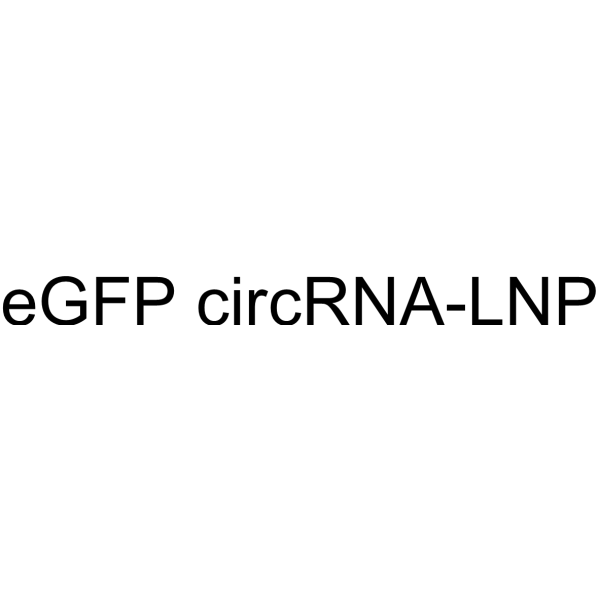
- HY-153232
-
|
|
Fluorescent Dye
Liposome
|
Others
|
|
eGFP circRNA-LNP is a lipid nanoparticle (LNP) containing eGFP circRNA, suitable for assays of RNA delivery, translation efficiency, cell viability, etc. eGFP circRNA carries Enhanced Green Fluorescent Protein (Enhanced Green Fluorescent Protein) eGFP, which will express green fluorescent protein after entering the cell. eGFP is commonly used as a reporter gene detectable by fluorescence microscopy or flow cytometry .
|
-
- HY-D2706
-
|
|
Fluorescent Dye
|
Others
|
|
Cy3 Dextran (MW 3000) is a fluorescent labeling reagent that combines Cy3 (HY-D0822) fluorescent dye and Dextran (HY-112624). The Cy3 fluorophore is commonly used in applications such as immunolabeling, nucleic acid labeling, fluorescence microscopy, and flow cytometry. Dextran has an inhibitory effect on thrombocyte aggregation and coagulation factors and is used as a plasma volume expander (Ex/Em = 550/570 nm) .
|
-

- HY-D2836I
-
|
FITC-Hyaluronate (MW 5000)
|
Fluorescent Dye
|
Others
|
|
FITC-HA (MW 5000) is hyaluronic acid (HA) (HY-B0633A) labeled with FITC (HY-66019). FITC-HA retains the ability of HA to bind to receptors (such as CD44) and form extracellular matrices, while it can be detected by fluorescence microscopy or flow cytometry for tracing the localization, binding, internalization and metabolic pathways of HA in cells, tissues or living organisms (Ex/Em ≈ 490/520 nM) .
|
-

- HY-D2544
-
|
|
Fluorescent Dye
|
Others
|
|
Cy5-PEG5000-DSPE is a PEG phospholipid with Cy5 (HY-D0821) dye used in protein/nucelic acid labeling and fluorescence microscopy. Cy5-PEG5000-DSPE can self-assemble in aqueous solution to form micelles/lipid bilayer and used to prepare liposomes or nanoparticles for nutrients delivery such as mRNA or DNA vaccine.
|
-

- HY-D2542
-
|
|
Fluorescent Dye
|
Others
|
|
Cy5-PEG2000-DSPE is a PEG phospholipid with Cy5 (HY-D0821) dye used in protein/nucelic acid labeling and fluorescence microscopy. Cy5-PEG2000-DSPE can self-assemble in aqueous solution to form micelles/lipid bilayer and used to prepare liposomes or nanoparticles for nutrients delivery such as mRNA or DNA vaccine.
|
-

- HY-D2712
-
|
|
Fluorescent Dye
|
Others
|
|
Cy3 Dextran (MW 500000) is a fluorescent labeling reagent that combines Cy3 (HY-D0822) fluorescent dye and Dextran (HY-112624). The Cy3 fluorophore is commonly used in applications such as immunolabeling, nucleic acid labeling, fluorescence microscopy, and flow cytometry. Dextran has an inhibitory effect on thrombocyte aggregation and coagulation factors and is used as a plasma volume expander (Ex/Em = 550/570 nm) .
|
-

- HY-D2708
-
|
|
Fluorescent Dye
|
Others
|
|
Cy3 Dextran (MW 10000) is a fluorescent labeling reagent that combines Cy3 (HY-D0822) fluorescent dye and Dextran (HY-112624). The Cy3 fluorophore is commonly used in applications such as immunolabeling, nucleic acid labeling, fluorescence microscopy, and flow cytometry. Dextran has an inhibitory effect on thrombocyte aggregation and coagulation factors and is used as a plasma volume expander (Ex/Em = 550/570 nm) .
|
-

- HY-D2710
-
|
|
Fluorescent Dye
|
Others
|
|
Cy3 Dextran (MW 40000) is a fluorescent labeling reagent that combines Cy3 (HY-D0822) fluorescent dye and Dextran (HY-112624). The Cy3 fluorophore is commonly used in applications such as immunolabeling, nucleic acid labeling, fluorescence microscopy, and flow cytometry. Dextran has an inhibitory effect on thrombocyte aggregation and coagulation factors and is used as a plasma volume expander (Ex/Em = 550/570 nm) .
|
-

- HY-D2836B
-
|
FITC-Hyaluronate (MW 200000)
|
Fluorescent Dye
|
Others
|
|
FITC-HA (MW 200000) is hyaluronic acid (HA) (HY-B0633A) labeled with FITC (HY-66019). FITC-HA retains the ability of HA to bind to receptors (such as CD44) and form extracellular matrices, while it can be detected by fluorescence microscopy or flow cytometry for tracing the localization, binding, internalization and metabolic pathways of HA in cells, tissues or living organisms (Ex/Em ≈ 490/520 nM) .
|
-

- HY-D2541
-
|
|
Fluorescent Dye
|
Others
|
|
Cy5-PEG1000-DSPE is a PEG phospholipid with Cy5 (HY-D0821) dye used in protein/nucelic acid labeling and fluorescence microscopy. Cy5-PEG1000-DSPE can self-assemble in aqueous solution to form micelles/lipid bilayer and used to prepare liposomes or nanoparticles for nutrients delivery such as mRNA or DNA vaccine.
|
-

- HY-D2516
-
|
|
Fluorescent Dye
Liposome
|
Others
|
|
Cy3-PEG3400-DSPE is a PEG phospholipid with Cy3 (HY-D0822) dye used in protein/nucelic acid labeling and fluorescence microscopy. Cy3-PEG3400-DSPE can self-assemble in aqueous solution to form micelles/lipid bilayer and used to prepare liposomes or nanoparticles for nutrients delivery such as mRNA or DNA vaccine.
|
-

- HY-D2707
-
|
|
Fluorescent Dye
|
Others
|
|
Cy3 Dextran (MW 5000) is a fluorescent labeling reagent that combines Cy3 (HY-D0822) fluorescent dye and Dextran (HY-112624). The Cy3 fluorophore is commonly used in applications such as immunolabeling, nucleic acid labeling, fluorescence microscopy, and flow cytometry. Dextran has an inhibitory effect on thrombocyte aggregation and coagulation factors and is used as a plasma volume expander (Ex/Em = 550/570 nm) .
|
-

- HY-D2705
-
|
|
Fluorescent Dye
|
Others
|
|
Cy3 Dextran (MW 2000) is a fluorescent labeling reagent that combines Cy3 (HY-D0822) fluorescent dye and Dextran (HY-112624). The Cy3 fluorophore is commonly used in applications such as immunolabeling, nucleic acid labeling, fluorescence microscopy, and flow cytometry. Dextran has an inhibitory effect on thrombocyte aggregation and coagulation factors and is used as a plasma volume expander (Ex/Em = 550/570 nm) .
|
-

- HY-D2836E
-
|
FITC-Hyaluronate (MW 10000)
|
Fluorescent Dye
|
Others
|
|
FITC-HA (MW 10000) is hyaluronic acid (HA) (HY-B0633A) labeled with FITC (HY-66019). FITC-HA retains the ability of HA to bind to receptors (such as CD44) and form extracellular matrices, while it can be detected by fluorescence microscopy or flow cytometry for tracing the localization, binding, internalization and metabolic pathways of HA in cells, tissues or living organisms (Ex/Em ≈ 490/520 nM) .
|
-

- HY-D2836J
-
|
FITC-Hyaluronate (MW 3000)
|
Fluorescent Dye
|
Others
|
|
FITC-HA (MW 3000) is hyaluronic acid (HA) (HY-B0633A) labeled with FITC (HY-66019). FITC-HA retains the ability of HA to bind to receptors (such as CD44) and form extracellular matrices, while it can be detected by fluorescence microscopy or flow cytometry for tracing the localization, binding, internalization and metabolic pathways of HA in cells, tissues or living organisms (Ex/Em ≈ 490/520 nM) .
|
-

- HY-D2711
-
|
|
Fluorescent Dye
|
Others
|
|
Cy3 Dextran (MW 100000) is a fluorescent labeling reagent that combines Cy3 (HY-D0822) fluorescent dye and Dextran (HY-112624). The Cy3 fluorophore is commonly used in applications such as immunolabeling, nucleic acid labeling, fluorescence microscopy, and flow cytometry. Dextran has an inhibitory effect on thrombocyte aggregation and coagulation factors and is used as a plasma volume expander (Ex/Em = 550/570 nm) .
|
-

- HY-D2431
-
|
|
Fluorescent Dye
|
Others
|
|
Galactose-PEG-Cy3 is a Cy3 (HY-D0822) labeled Galactose-PEG conjugate. The Cy3 fluorophore is commonly used in applications such as immunolabeling, nucleic acid labeling, fluorescence microscopy, and flow cytometry. Cy3 has an emission maximum around 562-570 nm. Galactose-PEG improves drug cellular uptake and reduces endosomal degradation, and can be used in drug delivery .
|
-

- HY-D2543
-
|
|
Fluorescent Dye
|
Others
|
|
Cy5-PEG3400-DSPE is a PEG phospholipid with Cy5 (HY-D0821) dye used in protein/nucelic acid labeling and fluorescence microscopy. Cy5-PEG3400-DSPE can self-assemble in aqueous solution to form micelles/lipid bilayer and used to prepare liposomes or nanoparticles for nutrients delivery such as mRNA or DNA vaccine.
|
-

- HY-D2836D
-
|
FITC-Hyaluronate (MW 50000)
|
Fluorescent Dye
|
Others
|
|
FITC-HA (MW 50000) is hyaluronic acid (HA) (HY-B0633A) labeled with FITC (HY-66019). FITC-HA retains the ability of HA to bind to receptors (such as CD44) and form extracellular matrices, while it can be detected by fluorescence microscopy or flow cytometry for tracing the localization, binding, internalization and metabolic pathways of HA in cells, tissues or living organisms (Ex/Em ≈ 490/520 nM) .
|
-

- HY-D2836C
-
|
FITC-Hyaluronate (MW 100000)
|
Fluorescent Dye
|
Others
|
|
FITC-HA (MW 100000) is hyaluronic acid (HA) (HY-B0633A) labeled with FITC (HY-66019). FITC-HA retains the ability of HA to bind to receptors (such as CD44) and form extracellular matrices, while it can be detected by fluorescence microscopy or flow cytometry for tracing the localization, binding, internalization and metabolic pathways of HA in cells, tissues or living organisms (Ex/Em ≈ 490/520 nM) .
|
-

- HY-D2836A
-
|
FITC-Hyaluronate (MW 500000)
|
Fluorescent Dye
|
Others
|
|
FITC-HA (MW 500000) is hyaluronic acid (HA) (HY-B0633A) labeled with FITC (HY-66019). FITC-HA retains the ability of HA to bind to receptors (such as CD44) and form extracellular matrices, while it can be detected by fluorescence microscopy or flow cytometry for tracing the localization, binding, internalization and metabolic pathways of HA in cells, tissues or living organisms (Ex/Em ≈ 490/520 nM) .
|
-

- HY-D2430
-
|
|
Fluorescent Dye
Bacterial
Fungal
|
Infection
Cancer
|
|
Chitosan-PEG-Cy3 is a fluorescent labeling reagent that combines Cy3 (HY-D0822) fluorescent dye, Chitosan and polyethylene glycol (PEG). The Cy3 fluorophore is commonly used in applications such as immunolabeling, nucleic acid labeling, fluorescence microscopy, and flow cytometry. Cy3 has an emission maximum around 562-570 nm. Chitosan exhibits antimicrobial activity against various bacteria and fungi .
|
-

- HY-D2836H
-
|
FITC-Hyaluronate (MW 7000)
|
Fluorescent Dye
|
Others
|
|
FITC-HA (MW 7000) is hyaluronic acid (HA) (HY-B0633A) labeled with FITC (HY-66019). FITC-HA retains the ability of HA to bind to receptors (such as CD44) and form extracellular matrices, while it can be detected by fluorescence microscopy or flow cytometry for tracing the localization, binding, internalization and metabolic pathways of HA in cells, tissues or living organisms (Ex/Em ≈ 490/520 nM) .
|
-

- HY-D2836
-
|
FITC-Hyaluronate (MW 1000000)
|
Fluorescent Dye
|
Others
|
|
FITC-HA (MW 1000000) is hyaluronic acid (HA) (HY-B0633A) labeled with FITC (HY-66019). FITC-HA retains the ability of HA to bind to receptors (such as CD44) and form extracellular matrices, while it can be detected by fluorescence microscopy or flow cytometry for tracing the localization, binding, internalization and metabolic pathways of HA in cells, tissues or living organisms (Ex/Em ≈ 490/520 nM) .
|
-

- HY-D2545
-
|
|
Fluorescent Dye
|
Others
|
|
Cy5-PEG10000-DSPE is a PEG phospholipid with Cy5 (HY-D0821) dye used in protein/nucelic acid labeling and fluorescence microscopy. Cy5-PEG10000-DSPE can self-assemble in aqueous solution to form micelles/lipid bilayer and used to prepare liposomes or nanoparticles for nutrients delivery such as mRNA or DNA vaccine.
|
-

- HY-137018
-
|
|
Biochemical Assay Reagents
|
Others
|
|
27-Alkyne cholesterol is a modified lipid containing an omega-terminal alkyne. The terminal alkyne group can be used in a highly specific linking reaction with azide-containing reagents, known as click chemistry, in the presence of a copper-containing catalyst.
Alkyne cholesterol represents a versatile, sensitive, and easy-to-use tool for tracking cellular cholesterol metabolism and localization as it allows for manifold detection methods including mass spectrometry, and fluorescence microscopy.
|
-

- HY-D0988A
-
|
R-PE (concentrated solution)
|
Fluorescent Dye
Apoptosis
|
Others
|
|
R-Phycoerythrin (R-PE) (concentrated solution) is found in Heterosiphonia japonica. R-Phycoerythrin (concentrated solution) is an orange-red fluorescent probe with α, β, and γ subunits. R-Phycoerythrin (concentrated solution) can be used in photodynamic therapy (PDT) to induce apoptosis in tumor cells. R-Phycoerythrin (concentrated solution) can be used in fluorescence microscopy, flow cytometry, and immunofluorescence analysis (Ex/Em = 496/578 nm) .
|
-

- HY-D2433
-
|
|
Fluorescent Dye
|
Others
|
|
Glucose-PEG2000-Cy3 is a Cy3 (HY-D0822) labeled Glucose-PEG conjugate. The Cy3 fluorophore is commonly used in applications such as immunolabeling, nucleic acid labeling, fluorescence microscopy, and flow cytometry. Cy3 has an emission maximum around 562-570 nm. Glucose-PEG improves drug cellular uptake and reduces endosomal degradation, and can be used in drug delivery .
|
-

- HY-D1540
-
|
Cy 5.5 amine; Lumiprobe Cy 5.5 amine
|
Fluorescent Dye
|
Others
|
|
Cyanine5.5 amine (Cy 5.5 amine), a Cy5.5 Analogue, is a near-infrared (NIR) fluorescent dye (Ex=648 nm, Em=710 nm). Cyanine5.5 amine can be used in the preparation of Cy5.5-labeled nanoparticles, which can be tracked and imaged with low fluorescence background using confocal microscopy .
|
-
- HY-D2466
-
|
|
Fluorescent Dye
|
Cardiovascular Disease
|
|
Cy3 Dextran (MW 70000) is a fluorescent labeling reagent that conjugates the Cy3 (HY-D0822) fluorescent dye with Dextran (HY-112624). The Cy3 fluorophore is commonly used in applications such as immunolabeling, nucleic acid labeling, fluorescence microscopy, and flow cytometry. The maximum emission wavelength of Cy3 is approximately 562-570 nm. Dextran inhibits platelet aggregation and coagulation factors, and serves as a plasma volume expander .
|
-

- HY-D2709
-
|
|
Fluorescent Dye
|
Others
|
|
Cy3 Dextran (MW 20000) is a fluorescent labeling reagent that combines Cy3 (HY-D0822) fluorescent dye and Dextran (HY-112624). The Cy3 fluorophore is commonly used in applications such as immunolabeling, nucleic acid labeling, fluorescence microscopy, and flow cytometry. Dextran has an inhibitory effect on thrombocyte aggregation and coagulation factors and is used as a plasma volume expander (Ex/Em = 550/570 nm) .
|
-

- HY-D1738
-
|
4',6-Diamidino-2-phenylindole dilactate
|
Fluorescent Dye
|
Others
|
|
DAPI (4',6-Diamidino-2-phenylindole) dilactate is a DAPI dye. DAPI is a fluorescent dye that binds strongly to DNA. It binds to the AT base pair of the double-stranded DNA minor groove, and one DAPI molecule can occupy three base pair positions. The fluorescence intensity of DAPI molecules bound to double-stranded DNA is increased by about 20 times, and it is commonly observed with fluorescence microscopy, and the amount of DNA can be determined based on the intensity of fluorescence. DAPI cannot penetrate intact cell membranes and is commonly used for staining both live and fixed cells . DAPI (Compound 3) is an acid-sensing ion channel 3 (ASIC3) inhibitor. DAPI binds to ASIC3 and blocks the channel function. DAPI can be used in the study of chronic pain treatment (Ex/Em = 356/451 nm) .
|
-
- HY-D2567
-
|
|
Fluorescent Dye
|
Others
|
|
Cy5.5-PEG5000-DSPE is a PEG phospholipid with Cy5.5 (HY-D0924) dye used in protein/nucelic acid labeling and fluorescence microscopy. Cy5.5-PEG5000-DSPE can self-assemble in aqueous solution to form micelles/lipid bilayer and used to prepare liposomes or nanoparticles for nutrients delivery such as mRNA or DNA vaccine.
|
-

- HY-D2566
-
|
|
Fluorescent Dye
|
Others
|
|
Cy5.5-PEG3400-DSPE is a PEG phospholipid with Cy5.5 (HY-D0924) dye used in protein/nucelic acid labeling and fluorescence microscopy. Cy5.5-PEG3400-DSPE can self-assemble in aqueous solution to form micelles/lipid bilayer and used to prepare liposomes or nanoparticles for nutrients delivery such as mRNA or DNA vaccine.
|
-

- HY-D2565
-
|
|
Fluorescent Dye
|
Others
|
|
Cy5.5-PEG2000-DSPE is a PEG phospholipid with Cy5.5 (HY-D0924) dye used in protein/nucelic acid labeling and fluorescence microscopy. Cy5.5-PEG2000-DSPE can self-assemble in aqueous solution to form micelles/lipid bilayer and used to prepare liposomes or nanoparticles for nutrients delivery such as mRNA or DNA vaccine.
|
-
- HY-D2564
-
|
|
Fluorescent Dye
|
Others
|
|
Cy5.5-PEG1000-DSPE is a PEG phospholipid with Cy5.5 (HY-D0924) dye used in protein/nucelic acid labeling and fluorescence microscopy. Cy5.5-PEG1000-DSPE can self-assemble in aqueous solution to form micelles/lipid bilayer and used to prepare liposomes or nanoparticles for nutrients delivery such as mRNA or DNA vaccine.
|
-

- HY-D2568
-
|
|
Fluorescent Dye
|
Others
|
|
Cy5.5-PEG10000-DSPE is a PEG phospholipid with Cy5.5 (HY-D0924) dye used in protein/nucelic acid labeling and fluorescence microscopy. Cy5.5-PEG10000-DSPE can self-assemble in aqueous solution to form micelles/lipid bilayer and used to prepare liposomes or nanoparticles for nutrients delivery such as mRNA or DNA vaccine.
|
-

- HY-D0988B
-
|
R-PE ammonium sulfate precipitate
|
Fluorescent Dye
Apoptosis
|
Others
|
|
R-Phycoerythrin (R-PE) (ammonium sulfate precipitate) is found in Heterosiphonia japonica. R-Phycoerythrin (ammonium sulfate precipitate) is an orange-red fluorescent probe with α, β, and γ subunits. R-Phycoerythrin (concentrated solution) can be used in photodynamic therapy (PDT) to induce apoptosis in tumor cells. R-Phycoerythrin (ammonium sulfate precipitate) can be used in fluorescence microscopy, flow cytometry, and immunofluorescence analysis (Ex/Em = 496/578 nm) .
|
-

- HY-D2441
-
|
|
Fluorescent Dye
|
Cancer
|
|
TAT-PEG-Cy3 is a fluorescent labeling reagent that combines Cy3 fluorescent dye, Cell membrane penetrating peptide (TAT) and polyethylene glycol (PEG). The Cy3 fluorophore is commonly used in applications such as immunolabeling, nucleic acid labeling, fluorescence microscopy, and flow cytometry. Cy3 has an emission maximum around 562-570 nm. TAT-PEG-Cy3 can be used for cell targeted delivery and biological imaging .
|
-

- HY-P10322
-
|
|
Fluorescent Dye
|
Cancer
|
|
Z-IETD-R110 is a fluorescent substrate of caspases. Z-IETD-R110 acts as a substrate for caspase-8. When caspase-8 is activated, it can recognize and cut Z-IETD-R110, releasing fluorophore, which can be detected by fluorescence microscopy. Z-IETD-R110 can be used to study oxidative stress-induced apoptosis, particularly in pancreatic acinar cells .
|
-

- HY-D2940
-
|
SiR650-BG
|
Fluorescent Dye
|
Others
|
SiR-SNAP (SiR650-BG) is a SiR-labeled SNAP tag near-infrared fluorescent probe (Ex/Em: 645 nm/661 nm). SiR-SNAP combines excellent optical properties, good cell membrane permeability, and environmentally sensitive fluorescence characteristics, providing a powerful tool for the dynamic study of proteins in living cells .
|
-

- HY-137855
-
|
|
Biochemical Assay Reagents
|
Others
|
|
4-Methylumbelliferyl sulfate (potassium), a fluorescent substrate, is commonly used to detect sulfatase activity in biochemical and biomedical research. It consists of a sulfate group attached to a fluorescent molecule, which can be cleaved by sulfatase enzymes. Upon cleavage, 4-Methylumbelliferyl sulfate releases a highly fluorescent product that can be detected using fluorescence microscopy or spectroscopy. The use of 4-Methylumbelliferyl sulfate as a substrate for sulfatase enzymes allows accurate detection and quantification of these enzymes in a variety of biological samples.
|
-
- HY-153231A
-
|
|
Fluorescent Dye
Liposome
|
Others
|
|
eGFP mRNA-LNP (500 ng/μL) is a lipid nanoparticle (LNP) containing eGFP mRNA. eGFP mRNA-LNP carries Enhanced Green Fluorescent Protein (eGFP), which will express green fluorescent protein after entering the cell. eGFP is commonly used as a reporter gene detectable by fluorescence microscopy or flow cytometry (excitation wavelength = 488 nm; emission wavelength = 535/40 bandpass). eGFP mRNA-LNP can be used for RNA delivery, translation efficiency and cell viability .
|
-

- HY-D2426
-
|
|
Fluorescent Dye
Transferrin Receptor
|
Cardiovascular Disease
|
|
Cy3-Transferrin is a fluorescent labeling reagent that combines Cy3 (HY-D0822) fluorescent dye and Transferrin (HY-P3267). The Cy3 fluorophore is commonly used in applications such as immunolabeling, nucleic acid labeling, fluorescence microscopy, and flow cytometry. Cy3 has an emission maximum around 562-570 nm. Transferrin, a blood-plasma glycoprotein, is a target ligand for transferrin receptor. Transferrin can bind to and mediate the transport of iron .
|
-

- HY-D2436
-
|
|
Fluorescent Dye
Microtubule/Tubulin
Apoptosis
|
Cardiovascular Disease
Cancer
|
|
PTX-PEG-Cy3 (Paclitaxel-PEG-Cy3) is a Cy3 (HY-D0822) labeled PTX-PEG conjugate. The Cy3 fluorophore is commonly used in applications such as immunolabeling, nucleic acid labeling, fluorescence microscopy, and flow cytometry. Cy3 has an emission maximum around 562-570 nm. PTX stabilizes tubulin polymerization. PTX can cause both mitotic arrest and apoptotic cell death. PTX also induces autophagy .
|
-

- HY-D2868
-
|
4',6-Diamidino-2-phenylindole
|
DNA Stain
Sodium Channel
|
Neurological Disease
|
|
DAPI (4',6-Diamidino-2-phenylindole) is a DAPI dye. DAPI is a fluorescent dye that binds strongly to DNA. It binds to the AT base pair of the double-stranded DNA minor groove, and one DAPI molecule can occupy three base pair positions. The fluorescence intensity of DAPI molecules bound to double-stranded DNA is increased by about 20 times, and it is commonly observed with fluorescence microscopy, and the amount of DNA can be determined based on the intensity of fluorescence. In addition, because DAPI can pass through intact cell membranes, it can be used to stain both live and fixed cells . DAPI (Compound 3) is an acid-sensing ion channel 3 (ASIC3) inhibitor. DAPI binds to ASIC3 and blocks the channel function. DAPI can be used in the study of chronic pain treatment (Ex/Em = 356/451 nm).
|
-

- HY-D0917
-
|
|
DNA Stain
|
Cancer
|
|
NIR-Red Dead Cell-1 Dye is a DNA-binding fluorescent dye for non-living cells (Ex/Em=515 nm/531 nm). NIR-Red Dead Cell-1 Dye can intercalate into base pairs of double-stranded DNA and produce stronger fluorescence. NIR-Red Dead Cell-1 Dye is suitable for necrotic cells or late apoptotic cells with damaged cell membranes, showing green fluorescence under fluorescence microscopy or flow cytometry. NIR-Red Dead Cell-1 Dye can be used to distinguish live cells from dead cells and distinguish cell membrane integrity. NIR-Red Dead Cell-1 Dye can be attached to the surface of Feraheme (FH) nanoparticles (NPs) to obtain fluorescent dye-functionalized NPs for drug delivery studies .
|
-
- HY-D0814
-
|
4',6-Diamidino-2-phenylindole dihydrochloride
|
DNA Stain
Sodium Channel
|
Neurological Disease
|
|
DAPI (4',6-Diamidino-2-phenylindole) dihydrochloride is a DAPI dye. DAPI is a fluorescent dye that binds strongly to DNA. It binds to the AT base pair of the double-stranded DNA minor groove, and one DAPI molecule can occupy three base pair positions. The fluorescence intensity of DAPI molecules bound to double-stranded DNA is increased by about 20 times, and it is commonly observed with fluorescence microscopy, and the amount of DNA can be determined based on the intensity of fluorescence. In addition, because DAPI can pass through intact cell membranes, it can be used to stain both live and fixed cells . DAPI (Compound 3) is an acid-sensing ion channel 3 (ASIC3) inhibitor. DAPI binds to ASIC3 and blocks the channel function. DAPI can be used in the study of chronic pain treatment (Ex/Em = 356/451 nm).
|
-
- HY-D2593
-
|
|
Fluorescent Dye
|
Others
|
|
Cy7.5-PEG2000-DSPE is a PEG phospholipid with CY7.5 (HY-D0926) dye used in protein/nucelic acid labeling and fluorescence microscopy. Cy7.5-PEG2000-DSPE can self-assemble in aqueous solution to form micelles/lipid bilayer and used to prepare liposomes or nanoparticles for nutrients delivery such as mRNA or DNA vaccine (Ex/Em=770/820 nm).
|
-

- HY-D2596
-
|
|
Fluorescent Dye
|
Others
|
|
Cy7.5-PEG10000-DSPE is a PEG phospholipid with CY7.5 (HY-D0926) dye used in protein/nucelic acid labeling and fluorescence microscopy. Cy7.5-PEG10000-DSPE can self-assemble in aqueous solution to form micelles/lipid bilayer and used to prepare liposomes or nanoparticles for nutrients delivery such as mRNA or DNA vaccine (Ex/Em=770/820 nm).
|
-

- HY-P2031
-
|
|
Arp2/3 Complex
|
Others
|
|
Phallacidin is a natural bicyclic heptapeptide derived from the poisonous mushroom Amanita phalloides. Phallacidin binds to filamentous actin specifically with high affinity, with a Kd of 20 nM. After binding to F-actin, Phallacidin strongly inhibits its depolymerization, stabilizes microfilament structures, and prevents their disruption by drugs such as cytochalasins. When conjugated with a fluorophore, Phallacidin serves as a specific fluorescent probe for F-actin, which is used to clearly visualize the distribution of actin in the cytoskeleton (e.g., stress fibers, cortical peripheral bands) under fluorescence microscopy .
|
-
- HY-D2595
-
|
|
Fluorescent Dye
|
Others
|
|
Cy7.5-PEG5000-DSPE is a PEG phospholipid with CY7.5 (HY-D0926) dye used in protein/nucelic acid labeling and fluorescence microscopy. Cy7.5-PEG5000-DSPE can self-assemble in aqueous solution to form micelles/lipid bilayer and used to prepare liposomes or nanoparticles for nutrients delivery such as mRNA or DNA vaccine (Ex/Em=770/820 nm).
|
-

- HY-D2439
-
|
|
Fluorescent Dye
|
Cancer
|
|
RGD-PEG-Cy3 is a fluorescent labeling reagent that combines Cy3 fluorescent dye, peptide (RGD) and polyethylene glycol (PEG). The Cy3 fluorophore is commonly used in applications such as immunolabeling, nucleic acid labeling, fluorescence microscopy, and flow cytometry. Cy3 has an emission maximum around 562-570 nm. RGD is a peptide sequence (CRGDKGPDCiRGD) that binds to αvβ3 and αvβ5 integrin receptors on tumor neovasculogenesis to achieve specific tumor tissue targeting .
|
-

- HY-D2327
-
|
|
Dipeptidyl Peptidase
|
Others
|
|
DPP-8/9 probe-1 (compound 20) is a fluorescent probe targeting Dipeptidyl Peptidase DPP8/9, which can be selectively labeled and visualized in vitro by fluorescence microscopy Active DPP8/9. DPP-8/9 probe-1 contains a nitrobenzoxadiazole (NBD) tag and has high affinity and selectivity for DPP8/9 over related S9 family members (IC50 of 210 nM and 15 nM, respectively) .
|
-
- HY-D2592
-
|
|
Fluorescent Dye
|
Others
|
|
Cy7.5-PEG1000-DSPE is a PEG phospholipid with CY7.5 (HY-D0926) dye used in protein/nucelic acid labeling and fluorescence microscopy. Cy7.5-PEG1000-DSPE can self-assemble in aqueous solution to form micelles/lipid bilayer and used to prepare liposomes or nanoparticles for nutrients delivery such as mRNA or DNA vaccine (Ex/Em=770/820 nm).
|
-

- HY-D2594
-
|
|
Fluorescent Dye
|
Others
|
|
Cy7.5-PEG3400-DSPE is a PEG phospholipid with CY7.5 (HY-D0926) dye used in protein/nucelic acid labeling and fluorescence microscopy. Cy7.5-PEG3400-DSPE can self-assemble in aqueous solution to form micelles/lipid bilayer and used to prepare liposomes or nanoparticles for nutrients delivery such as mRNA or DNA vaccine (Ex/Em=770/820 nm).
|
-

- HY-DY1081
-
|
4',6-Diamidino-2-phenylindole dihydrochloride (solution)
|
Sodium Channel
DNA Stain
|
Neurological Disease
|
DAPI (4',6-Diamidino-2-phenylindole) dihydrochloride (solution) is a DAPI dye. DAPI is a fluorescent dye that binds strongly to DNA. It binds to the AT base pair of the double-stranded DNA minor groove, and one DAPI molecule can occupy three base pair positions. The fluorescence intensity of DAPI molecules bound to double-stranded DNA is increased by about 20 times, and it is commonly observed with fluorescence microscopy, and the amount of DNA can be determined based on the intensity of fluorescence. In addition, because DAPI can pass through intact cell membranes, it can be used to stain both live and fixed cells . DAPI (Compound 3) is an acid-sensing ion channel 3 (ASIC3) inhibitor. DAPI binds to ASIC3 and blocks the channel function. DAPI can be used in the study of chronic pain treatment (Ex/Em = 356/451 nm).
Solvent and concentration: ddH2O: 5 mg/mL
|
-

- HY-D0163
-
|
|
DNA Stain
|
Others
|
|
Methyl Green is a non-intercalating fluorescent labeling agent that selectively binds to the major groove of DNA. Methyl Green electrostatically interacts with the major groove of DNA through positively charged groups, exhibiting key activities such as high affinity, resistance to photobleaching, and stable fluorescence emission. Methyl Green can be directly measured by microscopy and flow cytometry, with peaks at 633 and 677 nm. Methyl Green can be used for fluorescent labeling of the nuclei of embryonic tissues or cells, or DNA staining and cell activity detection in gel electrophoresis[1][2][3].
|
-
- HY-P5295
-
|
|
Fluorescent Dye
|
Others
|
|
Cy3-Ova (323-339) is an Cy3 labled OVA Peptide (323-339) (HY-P0286). Cy3 is a fluorescent dye belonging to the Cyanine family and is a fluorescent light product of Cy5. Cyanine is commonly used in fluorescence microscopy, cell imaging, and molecular biology experiments. OVA Peptide (323-339) represents the T and B cell epitopes of ovalbumin (OVA). OVA Peptide (323-339) has limited immunogenic efficacy in activating OVA-sensitized and attacked mouse spleen cells .
|
-

- HY-D2908
-
|
|
Fluorescent Dye
|
Others
|
SYTO9 is a green fluorescent nucleic acid dye (Ex = 485 nm, Em = 498 nm (DNA)/501 nm (RNA)). SYTO9 can penetrate the intact cell membrane of viable bacteria. When the cell membrane is damaged (cell death), its rate of entry into cells increases, and it competes with propidium iodide (PI) (HY-D0815), another membrane-impermeable dye, for nucleic acid binding sites. SYTO9 is widely used in fluorescence microscopy, flow cytometry, and fluorometric assays in microbiology for bacterial counting, viability assessment, and cell imaging .
|
-

- HY-111137
-
|
XC-302 free base
|
Akt
|
Cancer
|
|
Puquitinib (XC-302 free base) is a multi-target inhibitor with the activity of inducing autophagy in nasopharyngeal carcinoma cells by inhibiting the PI3K/AKT/mTOR signaling pathway. Puquitinib was able to inhibit the proliferation of CNE-2 cells, showing a dose-dependent antiproliferative effect. Puquitinib induced the formation of autophagosomes and autolysosomes in CNE-2 cells, which were observed by fluorescence microscopy and electron microscopy. Puquitinib promoted the formation of LC3-II and increased the expression of beclin 1, while reducing the level of p62. Puquitinib inhibited the PI3K/AKT/mTOR pathway by reducing the expression of p-AKT and p-mTOR. Puquitinib also induced apoptosis in CNE-2 cells, and when autophagy was inhibited, the apoptosis rate was reduced, which means that autophagy may interact with apoptosis to induce cell death .
|
-

- HY-P4948
-
|
|
Biochemical Assay Reagents
|
Others
|
|
Coumarin-phalloidin is a kind of phalloidin labeled with Coumarin (HY-N0709). Coumarin-phalloidin is a new type of actin probe that can be used for triple immunofluorescence microscopic observation of the cell skeleton .
|
-

- HY-D0952
-
|
|
Parasite
|
Others
|
|
Acridine Orange base is a cell-permeable fluorescent dye that stains organisms (bacteria, parasites, viruses, etc.) bright orange and, when used under appropriate conditions (pH=3.5, Ex=460 nm), distinguishes human cells in green for detection by fluorescence microscopy. Acridine Orange base fluoresces green when bound to dsDNA (Ex=488, Em=520-524) and red when bound to ssDNA (Ex=457, Em=630-644) or ssRNA (Ex=457, Em=630-644), also can be used in cell cycle assays .
|
-
- HY-W923198
-
|
Methacryloyloxy fluorescein
|
Fluorescent Dye
|
Others
|
|
Fluorescein O-methacrylate (Methacryloyloxy fluorescein) is a pH-responsive fluorescent monomeric dye. Fluorescein O-methacrylate exhibits pH-responsive fluorescence properties: its fluorescence is activated under alkaline conditions and quenched under neutral or acidic conditions; reversible switching of fluorescence is achievable by adjusting the pH of the solution. Fluorescein O-methacrylate can be used as a monomer for the synthesis of amphiphilic fluorescein polymers. These polymers can self-assemble into stable polymer nanoparticles with enhanced aqueous-phase fluorescence, and also enable the conjugation of nanoparticles with amine-containing biomolecules to support sensitive optical detection of biomarkers (Ex/Em = 490/520 nm) .
|
-

- HY-149764
-
|
|
Amyloid-β
|
Neurological Disease
|
|
Aβ42 agonist-2 (compound 7b) is a small molecule compound that can promote Aβ42 aggregation. Aβ42 agonist-2 can interact with Aβ42 oligomers and pentamers to promote nontoxic aggregate self-assembly and rapid fibril formation. Aβ42 agonist-2 prevents Aβ42-induced cytotoxicity in HT22 hippocampal neuronal cells .
|
-
- HY-149763
-
|
|
Amyloid-β
|
Neurological Disease
|
|
Aβ42 agonist-1 is a compound that can promote Aβ42 aggregation. Aβ42 agonist-1 can interact with Aβ42 oligomers and pentamers to promote nontoxic aggregate self-assembly and rapid fibril formation. Aβ42 agonist-1 prevents Aβ42-induced cytotoxicity in HT22 hippocampal neuronal cells .
|
-
- HY-D1991
-
|
|
Fluorescent Dye
|
Others
|
|
ATTO 647 is a carborhodamine fluorophore and imaging tracer with photostable properties. ATTO 647 serves as a fluorescent probe to investigate cell membrane structure and diffusion characteristics. When conjugated with wheat germ agglutinin, ATTO 647 specifically binds to N-acetyl-β-D-glucosamine and sialic acid residues on membrane glycoproteins, enabling single-molecule tracing of glycoprotein diffusion. ATTO 647 exhibits highly stable fluorescence properties with significantly reduced blinking in mounting media such as ROXS (AA/MV) and ROXS (TX/TQ), whereas its brightness properties vary in Ibidi-MM and Vectashield. ATTO 647 can also be used to label histone H2B-GFP in fixed cells for confocal microscopy photobleaching experiments .
|
-
- HY-131682
-
|
3-Hexanoyl-NBD-cholesterol
|
Fluorescent Dye
|
Others
|
|
3-C6-NBD-cholesterol is a fluorescent analog of Chol that can be used to measure the kinetics of membrane and intracellular trafficking .
|
-
- HY-D3419
-
|
|
Fluorescent Dye
|
Neurological Disease
|
|
Neuro-DiO 4-chlorobenzenesulfonate is a hydrophobic C18 alkyl chain carbocyanine dye with green fluorescence, commonly used as a vascular marker, cell internalizer and deposition agent. Neuro-DiO 4-chlorobenzenesulfonate inserts its alkyl chain into the endothelial plasma membrane via liposome-mediated perfusion to achieve vascular labeling. Neuro-DiO chlorobenzenesulfonate can also stain the cell membrane and cytoplasm of cancer cells to assist in confocal microscopy observations. Neuro-DiO chlorobenzenesulfonate can be released from nanosponges and accumulate on the surface of mouse retina, then internalize into retinal ganglion cells, which is applicable to researches related to glaucoma and other diseases. It should be noted that during liposome-mediated vascular staining in mice, Neuro-DiO 4-chlorobenzenesulfonate may cause leakage of airway lavage fluid .
|
-

- HY-151292
-
|
|
DNA/RNA Synthesis
|
Cancer
|
|
Antitumor agent-74 (compound 13da) is a quinoxalines derivative, an antitumor agent. Antitumor agent-74 exhibits more potent efficacy on tumor inhibition, mixed with regioisomer Antitumor agent-75(HY-151295, compound 14 da) (mriBIQ 13da/14da). mriBIQ 13da/14da attests cell cycle at S phase, inhibits DNA synthesis, and induces mithochondrial apoptosis .
|
-
- HY-D0996
-
|
|
DNA Stain
|
Others
|
|
Lds-751 is a nucleic acid stain that mainly detects DNA. Lds-751 is a nucleic acid stain that mainly detects DNA. Lds-751 has a high affinity for DNA and fluorescence is enhanced after binding, but the maximum emission wavelength is 670nm. Lds-751 and Thiazole orange can be used for the differentiation of red blood cells, platelets, reticulocytes, and nucleated cells and can be stimulated at 488nm. Studies have shown that LDS-751 binds almost exclusively to mitochondria when incubated with nucleated living cells. After nucleated Acridine Orange (HY-101879) staining and LDS-751 treatment of cells, confocal microscopy revealed almost no co-location of the cells. Staining with Rhodamine 123 (HY-D0816), a dye known to bind polarized mitochondria, was almost identical to the pattern observed with LDS-751 .
|
-
- HY-D1737
-
|
|
Bacterial
|
Infection
|
|
RADA is a fluorescent D-amino acid (FDAA) with high photostability and thermostability, which emits yellow-to-orange fluorescence. RADA shows low outer membrane permeability in wild-type Gram-negative Escherichia coli, but it targets penicillin-binding proteins and L,D-transpeptidases, mimics the interaction between acyl acceptors and enzyme intermediates, and integrates into peptidoglycan during biosynthesis. As a peptidoglycan labeling reagent, RADA metabolically integrates into the nascent peptidoglycan of live bacterial cells, labels the peptidoglycan at the poles and lateral walls of mycobacteria, and enables visualization of peptidoglycan synthesis and remodeling processes. RADA serves as a non-specific stain for fixed cells, is non-toxic to bacterial cells, and its red-shifted excitation/emission spectra reduce phototoxicity. RADA also supports virtual pulse-chase labeling experiments and stochastic optical reconstruction microscopy for sub-diffraction-limited imaging of bacterial cell walls .
|
-

- HY-D3230
-
|
|
Fluorescent Dye
|
Others
|
|
FLCS1 is a BODIPY-based Fluorescent probe and selective copper (I) ion binder (λex = 630 nm, λemis = 660 nm). Binding of FLCS1 to copper (I) ions induces fluorescence turn-on. Addition of copper (I) to a methanolic solution of FLCS1 causes a slight red shift (4 nm) in the maximum absorption wavelength (λmax) in the UV-Vis spectrum .
|
-

- HY-D3208
-
|
|
Fluorescent Dye
|
Inflammation/Immunology
|
|
oLOX is a fluorescent activity reporter. oLOX can be activated by lysyl oxidase family (LOXF) enzymes, which in turn releases a luciferin fluorescent product that reports LOXF enzyme activity. After oLOX is activated in in vitro fibrotic lung tissues, the fluorescence intensity increases, enabling real-time detection of fibrotic activity. oLOX can be used in studies related to pulmonary fibrosis .
|
-

| Cat. No. |
Product Name |
Type |
-
- HY-D0718
-
|
Nile Blue A oxazone; Phenoxazone 9
|
Fluorescent Dye
|
|
Nile red (Nile blue oxazone) is a lipophilic stain. Nile red has environment-sensitive fluorescence. Nile red is intensely fluorescent in a lipid-rich environment while it has minimal fluorescence in aqueous media. Nile red is an excellent vital stain for the detection of intracellular lipid droplets by fluorescence microscopy and flow cytof uorometry. Nile red stains intracellular lipid droplets red. The fluorescence wavelength is 559/635 nm .
|
-
- HY-116215
-
|
|
Fluorescent Dye
|
|
2-NBDG is a fluorescently-labeled deoxyglucose analog that is used primarily to directly monitor glucose uptake by living cells and tissues. It is also used as a topical contrast reagent for the detection of neoplasia. 2-NBDG can be used in real-time confocal, high-resolution, or wide-field fluorescence microscopy as well as in flow cytometry. The probe can be excited by the Argon laser at 488 nm to give the environment-sensitive fluorescence. It has lower photostability than the rhodamine-based fluorescent probes.
|
-
- HY-D0090
-
|
|
Fluorescent Dye
|
|
MQAE is a chloride ion (Cl -) fluorescent probe that can be used to measure chloride concentrations. The fluorescence intensity of MQAE decreases proportionally as Cl - ions increase. MQAE has high cell permeability and is suitable for fluorescence detection such as confocal microscopy and flow cytometry (Ex/Em=350/460 nm) .
|
-
- HY-111330
-
|
HPF; 3'-p-(Hydroxyphenyl) fluorescein
|
Fluorescent Dye
|
|
Hydroxyphenyl Fluorescein (HPF) is a stable ROS fluorescent probe dye. Hydroxyphenyl Fluorescein has stronger specificity and stability than H2DCFDA (HY-D0940). Hydroxyphenyl Fluorescein can produce strong green fluorescence through hydroxyl radical reaction with intracellular peroxynitroso. Hydroxyphenyl Fluorescein can be applied for fluorescence microscopy, high-throughput imager, luciferase microplate reader or flow cytometry. Ex/Em=490/515 nm .
|
-
- HY-D0988
-
|
R-PE
|
Fluorescent Dye
|
|
R-Phycoerythrin is found in Heterosiphonia japonica. R-Phycoerythrin is an orange-red fluorescent probe with α, β, and γ subunits. R-Phycoerythrin can be used in photodynamic therapy (PDT) to induce apoptosis in tumor cells. R-Phycoerythrin can be used in fluorescence microscopy, flow cytometry, and immunofluorescence analysis (Ex: 495 nm).
|
-
- HY-D0722
-
|
5-(6)-Carboxyfluorescein diacetate; CFDA
|
Fluorescent Dye
|
|
5(6)-CFDA is a common aliphatic luciferin-line organism. CFDA conducts free diffusion into cells, and then it is hydrolyzed into carboxyl fluorescein (CF) by intracellular non-specific lipase. CF containing portion contains an additional negative charge so that it is better retained in cells, compared to fluorescein dyes .
|
-
- HY-D2001
-
|
|
Fluorescent Dye
|
|
ATTO 488 NHS ester is a new fluorescent marker based on the Rhodamine structure. It has strong absorption, high fluorescence quantum yield, high thermal stability and photochemical stability, and is suitable for single molecule detection and high-resolution microscopy. ATTO 488 NHS ester is an NHS ester derivative of ATTO 488 that can be used to label proteins or antibodies.
|
-
- HY-D0721
-
|
6-Carboxyfluorescein diacetate
|
Fluorescent Dye
|
|
6-CFDA is a common aliphatic luciferin-line organism. CFDA conducts free diffusion into cells, and then it is hydrolyzed into carboxyl fluorescein (CF) by intracellular non-specific lipase. CF containing portion contains an additional negative charge so that it is better retained in cells, compared to fluorescein dyes .
|
-
- HY-DY1008
-
|
|
Fluorescent Dye
|
Nile Red (solution) is a lipophilic stain. Nile red has environment-sensitive fluorescence. Nile red is intensely fluorescent in a lipid-rich environment while it has minimal fluorescence in aqueous media. Nile red is an excellent vital stain for the detection of intracellular lipid droplets by fluorescence microscopy and flow cytof uorometry. Nile red stains intracellular lipid droplets red. The fluorescence wavelength is 559/635 nm .
Solvent and concentration: DMSO: 1 mM
|
-
- HY-D1429
-
|
|
Fluorescent Dye
|
|
ER-Tracker dye is a derivative of BODIPY series dyes coupled with Glibenclamide (HY-15206), highly selective binding to the endoplasmic reticulum, non-toxic to cells at low concentrations, this type of dye is an environmentally sensitive probe, and formaldehyde treatment can still retain part of the fluorescence, with high fluorescence life, good extinction coefficient and other characteristics. Glibenclamide is an atp-dependent K + channel blocker (Kir6, KATP) and CFTR Cl-channel blocker that binds in the endoplasmic reticulum. ER-Tracker is not suitable for staining fixed cells. Staining followed by fixation is possible, but cells fixed with aldehydes will only retain partial fluorescence (Ex/Em = 374/ 430-640 nm) .
|
-
- HY-DY1019
-
|
|
Fluorescent Dye
|
2-NBDG (solution) is a fluorescently-labeled deoxyglucose analog that is used primarily to directly monitor glucose uptake by living cells and tissues. It is also used as a topical contrast reagent for the detection of neoplasia. 2-NBDG can be used in real-time confocal, high-resolution, or wide-field fluorescence microscopy as well as in flow cytometry. The probe can be excited by the Argon laser at 488 nm to give the environment-sensitive fluorescence. It has lower photostability than the rhodamine-based fluorescent probes.
Solvent and Concentration: Sterile PBS: 5 mM
|
-
- HY-DY1074
-
|
|
Fluorescent Dye
|
ER-Tracker dye is a derivative of BODIPY series dyes coupled with Glibenclamide (HY-15206) , highly selective binding to the endoplasmic reticulum, non-toxic to cells at low concentrations, this type of dye is an environmentally sensitive probe, and formaldehyde treatment can still retain part of the fluorescence, with high fluorescence life, good extinction coefficient and other characteristics. Glibenclamide is an atp-dependent K + channel blocker (Kir6, KATP) and CFTR Cl-channel blocker that binds in the endoplasmic reticulum. ER-Tracker is not suitable for staining fixed cells. Staining followed by fixation is possible, but cells fixed with aldehydes will only retain partial fluorescence .
Solvent and concentration: DMSO: 1 mM
|
-
- HY-D2001A
-
|
|
Fluorescent Dye
|
|
TTO 488 NHS ester TEA is a new fluorescent marker based on the Rhodamine structure. It has strong absorption, high fluorescence quantum yield, high thermal stability and photochemical stability, and is suitable for single molecule detection and high-resolution microscopy. TTO 488 NHS ester TEA is an NHS ester derivative of ATTO 488 that can be used to label proteins or antibodies.
|
-
- HY-D2708
-
|
|
Fluorescent Dye
|
|
Cy3 Dextran (MW 10000) is a fluorescent labeling reagent that combines Cy3 (HY-D0822) fluorescent dye and Dextran (HY-112624). The Cy3 fluorophore is commonly used in applications such as immunolabeling, nucleic acid labeling, fluorescence microscopy, and flow cytometry. Dextran has an inhibitory effect on thrombocyte aggregation and coagulation factors and is used as a plasma volume expander (Ex/Em = 550/570 nm) .
|
-
- HY-D1950
-
|
|
Fluorescent Dye
|
|
ATTO 633 is a fluorescent dye with an absorption peak at approximately 630 nm and a fluorescence emission peak at 651 nm. ATTO 633 can be used in nanomechanical photothermal microscopy studies .
|
-
- HY-D1305
-
|
|
Fluorescent Dye
|
|
ATTO 488 carboxylic acid is a new fluorescent label based on the Rhodamine structure. It has strong absorption, high fluorescence quantum yield, high thermal stability and photochemical stability, and is suitable for single molecule detection and high-resolution microscopy. ATTO 488 carboxylic acid is a carboxylic acid derivative of ATTO 488, which can be used to label proteins or antibodies.
|
-
- HY-D2438
-
|
|
Fluorescent Dye
|
|
CDDP-PEG-Cy3 is a CDDP-PEG conjugate labeled with Cy3 (HY-D0822). The Cy3 fluorophore is commonly used in applications such as immunolabeling, nucleic acid labeling, fluorescence microscopy, and flow cytometry. The maximum emission wavelength of Cy3 is approximately 562-570 nm. Cisplatin (CDDP) (HY-17394) is an antineoplastic chemotherapy agent by cross-linking with DNA and causing DNA damage in cancer cells. Cisplatin activates ferroptosis and induces autophagy .
|
-
- HY-D2002
-
|
|
Fluorescent Dye
|
|
ATTO 488 maleimide is a new fluorescent marker based on the Rhodamine structure. It has strong absorption, high fluorescence quantum yield, high thermal stability and photochemical stability, and is suitable for single molecule detection and high-resolution microscopy. ATTO 488 maleimide is a maleimide derivative of ATTO 488, which can be used to label proteins or antibodies.
|
-
- HY-D1959
-
|
|
Fluorescent Dye
|
|
ATTO 565 NHS ester is a new fluorescent marker based on the Rhodamine structure. It has strong absorption, high fluorescence quantum yield, high thermal stability and photochemical stability, and is suitable for single molecule detection and high-resolution microscopy. ATTO 565 NHS ester is an NHS ester derivative of ATTO 565 that can be used to label proteins or antibodies.
|
-
- HY-D1240
-
|
|
Fluorescent Dye
|
|
Rhodamine 101 inner salt is a bright fluorescent dye with excitation and emission maxima at 565 and 595 nm, respectively. It can be used in various biological applications such as fluorescence microscopy, flow cytometry, fluorescence correlation spectroscopy, and ELISA.
|
-
- HY-DN0194F
-
|
|
Fluorescent Dye
|
|
Cy3-Asiatic acid is a fluorescent labeling reagent that combines Cy3 (HY-D0822) and Asiatic acid (HY-N0194). Cy3-Asiatic acid can be used to observe and track Asiatic acid under fluorescence microscopy (Ex/Em = 550/570 nm).
|
-
- HY-DY1035
-
|
|
Fluorescent Dye
|
Hydroxyphenyl Fluorescein (HPF) (solution) is a stable ROS fluorescent probe dye. Hydroxyphenyl Fluorescein has stronger specificity and stability than H2DCFDA (HY-D0940). Hydroxyphenyl Fluorescein can produce strong green fluorescence through hydroxyl radical reaction with intracellular peroxynitroso. Hydroxyphenyl Fluorescein can be applied for fluorescence microscopy, high-throughput imager, luciferase microplate reader or flow cytometry. Ex/Em=490/515 nm .
Solvent and concentration: DMSO: 10 mM
|
-
- HY-153524
-
|
|
Fluorescent Dye
|
|
ATTO 425 NHS ester is a new fluorescent marker based on the Rhodamine structure. It has strong absorption, high fluorescence quantum yield, high thermal stability and photochemical stability, and is suitable for single molecule detection and high-resolution microscopy. ATTO 425 NHS ester is an NHS ester derivative of ATTO 425 that can be used to label proteins or antibodies.
|
-
- HY-D1993
-
|
|
Fluorescent Dye
|
|
ATTO 647 NHS ester is a fluorescent label targeting free amino groups. ATTO 647 NHS ester can undergo a nucleophilic reaction with the free amino groups of EF-C peptide via its activated carboxylic acid group to form a stable covalent conjugate. ATTO 647 NHS ester-labeled nanofibers not only retain retroviral transduction-enhancing activity but also maintain stable fluorescent signals in both buffer and cell culture systems. ATTO 647 NHS ester is suitable for detection applications including fluorescence spectroscopy, microscopy and flow cytometry. ATTO 647 NHS ester has been used in studies related to HIV-1 infection .
|
-
- HY-D2506
-
|
|
Fluorescent Dye
|
|
Cy3-PEG3400-NH2 is a polyethylene glycol derivative containing Cy3 (HY-D0822) fluorescent dye and polyethylene glycol (PEG) and an amino group. The Cy3 fluorophore is commonly used in applications such as immunolabeling, nucleic acid labeling, fluorescence microscopy, and flow cytometry. Cy3 has an emission maximum around 562-570 nm. Cy3-PEG3400-NH2 can be used for fluorescence imaging, fluorescence tracing and fluorescence labeling .
|
-
- HY-D2440
-
|
|
Fluorescent Dye
|
|
cRGD-PEG-Cy3 is a Cy3 (HY-D0822) labeled cRGD-PEG conjugate. The Cy3 fluorophore is commonly used in applications such as immunolabeling, nucleic acid labeling, fluorescence microscopy, and flow cytometry. Cy3 has an emission maximum around 562-570 nm. cRGD can be used to modify lipid for improved stability .
|
-
- HY-D2610
-
|
|
Fluorescent Dye
|
|
ICG PEG2000 DSPE is a PEG phospholipid with Indocyanine green (ICG) (HY-D0711) dye used in protein/nucelic acid labeling and fluorescence microscopy. ICG PEG2000 DSPE can self-assemble in aqueous solution to form micelles/lipid bilayer and used to prepare liposomes or nanoparticles for nutrients delivery such as mRNA or DNA vaccine (Ex/Em=785/813 nm).
|
-
- HY-D2437
-
|
|
Fluorescent Dye
|
|
DOX-PEG-Cy3 (Doxorubicin-PEG-Cy3) is a Cy3 (HY-D0822) labeled DOX-PEG conjugate. The Cy3 fluorophore is commonly used in applications such as immunolabeling, nucleic acid labeling, fluorescence microscopy, and flow cytometry. Cy3 has an emission maximum around 562-570 nm. DOX is a broad-spectrum anthracycline antibiotic with cytotoxic properties .
|
-
- HY-D2706
-
|
|
Fluorescent Dye
|
|
Cy3 Dextran (MW 3000) is a fluorescent labeling reagent that combines Cy3 (HY-D0822) fluorescent dye and Dextran (HY-112624). The Cy3 fluorophore is commonly used in applications such as immunolabeling, nucleic acid labeling, fluorescence microscopy, and flow cytometry. Dextran has an inhibitory effect on thrombocyte aggregation and coagulation factors and is used as a plasma volume expander (Ex/Em = 550/570 nm) .
|
-
- HY-D2836I
-
|
FITC-Hyaluronate (MW 5000)
|
Fluorescent Dye
|
|
FITC-HA (MW 5000) is hyaluronic acid (HA) (HY-B0633A) labeled with FITC (HY-66019). FITC-HA retains the ability of HA to bind to receptors (such as CD44) and form extracellular matrices, while it can be detected by fluorescence microscopy or flow cytometry for tracing the localization, binding, internalization and metabolic pathways of HA in cells, tissues or living organisms (Ex/Em ≈ 490/520 nM) .
|
-
- HY-D1915
-
|
|
Fluorescent Dye
|
|
ATTO 390 is a new fluorescent marker based on the Rhodamine structure. It has strong absorption, high fluorescence quantum yield, high thermal stability and photochemical stability, and is suitable for single molecule detection and high-resolution microscopy.
|
-
- HY-D2046
-
|
|
Fluorescent Dye
|
|
ATTO 532 NHS ester is a new fluorescent marker based on the Rhodamine structure. It has strong absorption, high fluorescence quantum yield, high thermal stability and photochemical stability, and is suitable for single molecule detection and high-resolution microscopy. ATTO 532 NHS ester is an NHS ester derivative of ATTO 532 that can be used to label proteins or antibodies.
|
-
- HY-D1957
-
|
|
Fluorescent Dye
|
|
ATTO 633 NHS ester is a new fluorescent marker based on the Rhodamine structure. It has strong absorption, high fluorescence quantum yield, high thermal stability and photochemical stability, and is suitable for single molecule detection and high-resolution microscopy. ATTO 633 NHS ester is an NHS ester derivative of ATTO 633 that can be used to label proteins or antibodies.
|
-
- HY-D2019
-
|
|
Fluorescent Dye
|
|
ATTO 550 NHS ester is a new fluorescent marker based on the Rhodamine structure. It has strong absorption, high fluorescence quantum yield, high thermal stability and photochemical stability, and is suitable for single molecule detection and high-resolution microscopy. ATTO 550 NHS ester is an NHS ester derivative of ATTO 550 that can be used to label proteins or antibodies.
|
-
- HY-D2016
-
|
|
Fluorescent Dye
|
|
ATTO 565 cadaverine is a new fluorescent marker based on the Rhodamine structure. It has strong absorption, high fluorescence quantum yield, high thermal stability and photochemical stability, and is suitable for single molecule detection and high-resolution microscopy. ATTO 565 cadaverine is a cadaverine derivative of ATTO 565, which can be used to label proteins or antibodies.
|
-
- HY-D2058
-
|
|
Fluorescent Dye
|
|
ATTO 700 NHS ester is a new fluorescent marker based on the Rhodamine structure. It has strong absorption, high fluorescence quantum yield, high thermal stability and photochemical stability, and is suitable for single molecule detection and high-resolution microscopy. ATTO 700 NHS ester is an NHS ester derivative of ATTO 700 that can be used to label proteins or antibodies.
|
-
- HY-D2073
-
|
|
Fluorescent Dye
|
|
ATTO 680 NHS ester is a new fluorescent marker based on the Rhodamine structure. It has strong absorption, high fluorescence quantum yield, high thermal stability and photochemical stability, and is suitable for single molecule detection and high-resolution microscopy. ATTO 680 NHS ester is an NHS ester derivative of ATTO 680 that can be used to label proteins or antibodies.
|
-
- HY-D2021
-
|
|
Fluorescent Dye
|
|
ATTO 550 maleimide is a new fluorescent marker based on the Rhodamine structure. It has strong absorption, high fluorescence quantum yield, high thermal stability and photochemical stability, and is suitable for single molecule detection and high-resolution microscopy. ATTO 550 maleimide is a maleimide derivative of ATTO 550, which can be used to label proteins or antibodies.
|
-
- HY-D1935
-
|
|
Fluorescent Dye
|
|
ATTO 465 NHS ester is a new fluorescent marker based on the Rhodamine structure. It has strong absorption, high fluorescence quantum yield, high thermal stability and photochemical stability, and is suitable for single molecule detection and high-resolution microscopy. ATTO 465 NHS ester is an NHS ester derivative of ATTO 465 that can be used to label proteins or antibodies.
|
-
- HY-D2602
-
|
|
Fluorescent Dye
|
|
ICG PEG5000 NH2 is a polyethylene glycol derivative containing Indocyanine green (ICG) (HY-D0711) fluorescent dye and polyethylene glycol (PEG) and an amino group. The Indocyanine green fluorophore is commonly used in applications such as immunolabeling, nucleic acid labeling, fluorescence microscopy, and flow cytometry. ICG PEG5000 NH2 can be used for fluorescence imaging, fluorescence tracing and fluorescence labeling (Ex/Em=785/813 nm).
|
-
- HY-D2599
-
|
|
Fluorescent Dye
|
|
ICG PEG1000 NH2 is a polyethylene glycol derivative containing Indocyanine green (ICG) (HY-D0711) fluorescent dye and polyethylene glycol (PEG) and an amino group. The Indocyanine green fluorophore is commonly used in applications such as immunolabeling, nucleic acid labeling, fluorescence microscopy, and flow cytometry. ICG PEG1000 NH2 can be used for fluorescence imaging, fluorescence tracing and fluorescence labeling (Ex/Em=785/813 nm).
|
-
- HY-D2554
-
|
|
Fluorescent Dye
|
|
Cy5.5-PEG5000-NH2 is a polyethylene glycol derivative containing Cy5.5 (HY-D0924) fluorescent dye and polyethylene glycol (PEG) and an amino group. The Cy5.5 fluorophore is commonly used in applications such as immunolabeling, nucleic acid labeling, fluorescence microscopy, and flow cytometry. Cy5.5 has an emission maximum around 710 nm. Cy5.5-PEG5000-NH2 can be used for fluorescence imaging, fluorescence tracing and fluorescence labeling.
|
-
- HY-D2553
-
|
|
Fluorescent Dye
|
|
Cy5.5-PEG3400-NH2 is a polyethylene glycol derivative containing Cy5.5 (HY-D0924) fluorescent dye and polyethylene glycol (PEG) and an amino group. The Cy5.5 fluorophore is commonly used in applications such as immunolabeling, nucleic acid labeling, fluorescence microscopy, and flow cytometry. Cy5.5 has an emission maximum around 710 nm. Cy5.5-PEG3400-NH2 can be used for fluorescence imaging, fluorescence tracing and fluorescence labeling.
|
-
- HY-D2552
-
|
|
Fluorescent Dye
|
|
Cy5.5-PEG1000-NH2 is a polyethylene glycol derivative containing Cy5.5 (HY-D0924) fluorescent dye and polyethylene glycol (PEG) and an amino group. The Cy5.5 fluorophore is commonly used in applications such as immunolabeling, nucleic acid labeling, fluorescence microscopy, and flow cytometry. Cy5.5 has an emission maximum around 710 nm. Cy5.5-PEG1000-NH2 can be used for fluorescence imaging, fluorescence tracing and fluorescence labeling.
|
-
- HY-D2555
-
|
|
Fluorescent Dye
|
|
Cy5.5-PEG10000-NH2 is a polyethylene glycol derivative containing Cy5.5 (HY-D0924) fluorescent dye and polyethylene glycol (PEG) and an amino group. The Cy5.5 fluorophore is commonly used in applications such as immunolabeling, nucleic acid labeling, fluorescence microscopy, and flow cytometry. Cy5.5 has an emission maximum around 710 nm. Cy5.5-PEG10000-NH2 can be used for fluorescence imaging, fluorescence tracing and fluorescence labeling.
|
-
- HY-D2710
-
|
|
Fluorescent Dye
|
|
Cy3 Dextran (MW 40000) is a fluorescent labeling reagent that combines Cy3 (HY-D0822) fluorescent dye and Dextran (HY-112624). The Cy3 fluorophore is commonly used in applications such as immunolabeling, nucleic acid labeling, fluorescence microscopy, and flow cytometry. Dextran has an inhibitory effect on thrombocyte aggregation and coagulation factors and is used as a plasma volume expander (Ex/Em = 550/570 nm) .
|
-
- HY-D2836B
-
|
FITC-Hyaluronate (MW 200000)
|
Fluorescent Dye
|
|
FITC-HA (MW 200000) is hyaluronic acid (HA) (HY-B0633A) labeled with FITC (HY-66019). FITC-HA retains the ability of HA to bind to receptors (such as CD44) and form extracellular matrices, while it can be detected by fluorescence microscopy or flow cytometry for tracing the localization, binding, internalization and metabolic pathways of HA in cells, tissues or living organisms (Ex/Em ≈ 490/520 nM) .
|
-
- HY-D2711
-
|
|
Fluorescent Dye
|
|
Cy3 Dextran (MW 100000) is a fluorescent labeling reagent that combines Cy3 (HY-D0822) fluorescent dye and Dextran (HY-112624). The Cy3 fluorophore is commonly used in applications such as immunolabeling, nucleic acid labeling, fluorescence microscopy, and flow cytometry. Dextran has an inhibitory effect on thrombocyte aggregation and coagulation factors and is used as a plasma volume expander (Ex/Em = 550/570 nm) .
|
-
- HY-D2836D
-
|
FITC-Hyaluronate (MW 50000)
|
Fluorescent Dye
|
|
FITC-HA (MW 50000) is hyaluronic acid (HA) (HY-B0633A) labeled with FITC (HY-66019). FITC-HA retains the ability of HA to bind to receptors (such as CD44) and form extracellular matrices, while it can be detected by fluorescence microscopy or flow cytometry for tracing the localization, binding, internalization and metabolic pathways of HA in cells, tissues or living organisms (Ex/Em ≈ 490/520 nM) .
|
-
- HY-D2435
-
|
|
Fluorescent Dye
|
|
CDDP-PEG-Cy3 is a MTX-PEG conjugate labeled with Cy3 (HY-D0822). The Cy3 fluorophore is commonly used in applications such as immunolabeling, nucleic acid labeling, fluorescence microscopy, and flow cytometry. The maximum emission wavelength of Cy3 is approximately 562-570 nm. Methotrexate (Amethopterin; MTX) (HY-14519), an antimetabolite and antifolate agent, inhibits the enzyme dihydrofolate reductase, thereby preventing the conversion of folic acid into tetrahydrofolate, and inhibiting DNA synthesis. Methotrexate, also an immunosuppressant and antineoplastic agent, is used for the research of rheumatoid arthritis and a number of different cancers (such as acute lymphoblastic leukemia) .
|
- HY-D1917
-
|
|
Fluorescent Dye
|
|
ATTO 390 NHS ester is a new fluorescent marker based on the Rhodamine structure. It has strong absorption, high fluorescence quantum yield, high thermal stability and photochemical stability, and is suitable for single molecule detection and high-resolution microscopy. ATTO 390 NHS ester is an NHS ester derivative of ATTO 390 that can be used to label proteins or antibodies.
|
- HY-D2038
-
|
|
Fluorescent Dye
|
|
ATTO 550 alkyne is a new fluorescent marker based on the Rhodamine structure. It has strong absorption, high fluorescence quantum yield, high thermal stability and photochemical stability, and is suitable for single molecule detection and high-resolution microscopy. ATTO 550 alkyne is an alkyne derivative of ATTO 550 and can be used to label proteins or antibodies.
|
- HY-D1961
-
|
|
Fluorescent Dye
|
|
ATTO 565 maleimide is a new fluorescent marker based on the Rhodamine structure. It has strong absorption, high fluorescence quantum yield, high thermal stability and photochemical stability, and is suitable for single molecule detection and high-resolution microscopy. ATTO 565 maleimide is a maleimide derivative of ATTO 565, which can be used to label proteins or antibodies.
|
- HY-D1929
-
|
|
Fluorescent Dye
|
|
ATTO 594 NHS ester is a new fluorescent marker based on the Rhodamine structure. It has strong absorption, high fluorescence quantum yield, high thermal stability and photochemical stability, and is suitable for single molecule detection and high-resolution microscopy. ATTO 594 NHS ester is an NHS ester derivative of ATTO 594 that can be used to label proteins or antibodies.
|
- HY-D2014
-
|
|
Fluorescent Dye
|
|
ATTO 565 alkyne is a new fluorescent marker based on the Rhodamine structure. It has strong absorption, high fluorescence quantum yield, high thermal stability and photochemical stability, and is suitable for single molecule detection and high-resolution microscopy. ATTO 565 alkyne is an alkyne derivative of ATTO 565 and can be used to label proteins or antibodies.
|
- HY-D2035
-
|
|
Fluorescent Dye
|
|
ATTO 514 alkyne is a new fluorescent marker based on the Rhodamine structure. It has strong absorption, high fluorescence quantum yield, high thermal stability and photochemical stability, and is suitable for single molecule detection and high-resolution microscopy. ATTO 514 alkyne is an alkyne derivative of ATTO 514 and can be used to label proteins or antibodies.
|
- HY-D2047
-
|
|
Fluorescent Dye
|
|
ATTO 532 maleimide is a new fluorescent marker based on the Rhodamine structure. It has strong absorption, high fluorescence quantum yield, high thermal stability and photochemical stability, and is suitable for single molecule detection and high-resolution microscopy. ATTO 532 maleimide is a maleimide derivative of ATTO 532, which can be used to label proteins or antibodies.
|
- HY-D1939
-
|
|
Fluorescent Dye
|
|
ATTO 465 amine is a new fluorescent marker based on the Rhodamine structure. It has strong absorption, high fluorescence quantum yield, high thermal stability and photochemical stability, and is suitable for single molecule detection and high-resolution microscopy. ATTO 465 amine is an amine derivative of ATTO 465 and can be used to label proteins or antibodies.
|
- HY-D2063
-
|
|
Fluorescent Dye
|
|
ATTO 740 maleimide is a new fluorescent marker based on the Rhodamine structure. It has strong absorption, high fluorescence quantum yield, high thermal stability and photochemical stability, and is suitable for single molecule detection and high-resolution microscopy. ATTO 740 maleimide is a maleimide derivative of ATTO 740, which can be used to label proteins or antibodies.
|
- HY-D2015
-
|
|
Fluorescent Dye
|
|
ATTO 488 alkyne is a new fluorescent marker based on the Rhodamine structure. It has strong absorption, high fluorescence quantum yield, high thermal stability and photochemical stability, and is suitable for single molecule detection and high-resolution microscopy. ATTO 488 alkyne is an alkyne derivative of ATTO 488 and can be used to label proteins or antibodies.
|
- HY-D1933
-
|
|
Fluorescent Dye
|
|
ATTO 590 maleimide is a new fluorescent marker based on the Rhodamine structure. It has strong absorption, high fluorescence quantum yield, high thermal stability and photochemical stability, and is suitable for single molecule detection and high-resolution microscopy. ATTO 590 maleimide is a maleimide derivative of ATTO 590, which can be used to label proteins or antibodies.
|
- HY-D2059
-
|
|
Fluorescent Dye
|
|
ATTO 700 maleimide is a new fluorescent marker based on the Rhodamine structure. It has strong absorption, high fluorescence quantum yield, high thermal stability and photochemical stability, and is suitable for single molecule detection and high-resolution microscopy. ATTO 700 maleimide is a maleimide derivative of ATTO 700, which can be used to label proteins or antibodies.
|
- HY-D2026
-
|
|
Fluorescent Dye
|
|
ATTO 514 NHS ester is a new fluorescent marker based on the Rhodamine structure. It has strong absorption, high fluorescence quantum yield, high thermal stability and photochemical stability, and is suitable for single molecule detection and high-resolution microscopy. ATTO 514 NHS ester is an NHS ester derivative of ATTO 514 that can be used to label proteins or antibodies.
|
- HY-D1999
-
|
|
Fluorescent Dye
|
|
ATTO 665 NHS ester is a new fluorescent marker based on the Rhodamine structure. It has strong absorption, high fluorescence quantum yield, high thermal stability and photochemical stability, and is suitable for single molecule detection and high-resolution microscopy. ATTO 665 NHS ester is an NHS ester derivative of ATTO 665 that can be used to label proteins or antibodies.
|
- HY-D1932
-
|
|
Fluorescent Dye
|
|
ATTO 590 NHS ester is a new fluorescent marker based on the Rhodamine structure. It has strong absorption, high fluorescence quantum yield, high thermal stability and photochemical stability, and is suitable for single molecule detection and high-resolution microscopy. ATTO 590 NHS ester is an NHS ester derivative of ATTO 590 that can be used to label proteins or antibodies.
|
- HY-D2074
-
|
|
Fluorescent Dye
|
|
ATTO 680 maleimide is a new fluorescent marker based on the Rhodamine structure. It has strong absorption, high fluorescence quantum yield, high thermal stability and photochemical stability, and is suitable for single molecule detection and high-resolution microscopy. ATTO 680 maleimide is a maleimide derivative of ATTO 680, which can be used to label proteins or antibodies.
|
- HY-D2052
-
|
|
Fluorescent Dye
|
|
ATTO 532 iodacetamid is a new fluorescent marker based on the Rhodamine structure. It has strong absorption, high fluorescence quantum yield, high thermal stability and photochemical stability, and is suitable for single molecule detection and high-resolution microscopy. ATTO 532 iodacetamid is an iodoacetamide derivative of ATTO 532, which can be used to label proteins or antibodies.
|
- HY-D1946
-
|
|
Fluorescent Dye
|
|
ATTO 590 alkyne is a new fluorescent marker based on the Rhodamine structure. It has strong absorption, high fluorescence quantum yield, high thermal stability and photochemical stability, and is suitable for single molecule detection and high-resolution microscopy. ATTO 590 alkyne is an alkyne derivative of ATTO 590 and can be used to label proteins or antibodies.
|
- HY-D2601
-
|
|
Fluorescent Dye
|
|
ICG PEG3400 NH2 is a polyethylene glycol derivative containing Indocyanine green (ICG) (HY-D0711) fluorescent dye and polyethylene glycol (PEG) and an amino group. The Indocyanine green fluorophore is commonly used in applications such as immunolabeling, nucleic acid labeling, fluorescence microscopy, and flow cytometry. ICG PEG3400 NH2 can be used for fluorescence imaging, fluorescence tracing and fluorescence labeling (Ex/Em=785/813 nm).
|
- HY-D2600
-
|
|
Fluorescent Dye
|
|
ICG PEG2000 NH2 is a polyethylene glycol derivative containing Indocyanine green (ICG) (HY-D0711) fluorescent dye and polyethylene glycol (PEG) and an amino group. The Indocyanine green fluorophore is commonly used in applications such as immunolabeling, nucleic acid labeling, fluorescence microscopy, and flow cytometry. ICG PEG2000 NH2 can be used for fluorescence imaging, fluorescence tracing and fluorescence labeling (Ex/Em=785/813 nm).
|
- HY-D2603
-
|
|
Fluorescent Dye
|
|
ICG PEG10000 NH2 is a polyethylene glycol derivative containing Indocyanine green (ICG) (HY-D0711) fluorescent dye and polyethylene glycol (PEG) and an amino group. The Indocyanine green fluorophore is commonly used in applications such as immunolabeling, nucleic acid labeling, fluorescence microscopy, and flow cytometry. ICG PEG10000 NH2 can be used for fluorescence imaging, fluorescence tracing and fluorescence labeling (Ex/Em=785/813 nm).
|
- HY-D2588
-
|
|
Fluorescent Dye
|
|
Cy7.5 PEG-NH2 is a polyethylene glycol derivative containing CY7.5 (HY-D0926) fluorescent dye and polyethylene glycol (PEG) and an amino group. The CY7.5 fluorophore is commonly used in applications such as immunolabeling, nucleic acid labeling, fluorescence microscopy, and flow cytometry. Cy7.5 PEG-NH2 can be used for fluorescence imaging, fluorescence tracing and fluorescence labeling.
|
- HY-D1349
-
|
|
Fluorescent Dye
|
|
Bodipy TR alkyneis one of a boron dipyrromethene fluorophore for the ROX (Texas Red) channel. This is a versatile fluorophore that can be used in microscopy, fluorescence polarization measurements, and other applications. This derivative is a terminal alkyne of copper-catalyzed click chemistry.
|
- HY-D2505
-
|
|
Fluorescent Dye
|
|
Cy3-PEG1000-NH2 is a polyethylene glycol derivative containing Cy3 (HY-D0822) fluorescent dye and polyethylene glycol (PEG) and an amino group. The Cy3 fluorophore is commonly used in applications such as immunolabeling, nucleic acid labeling, fluorescence microscopy, and flow cytometry. Cy3 has an emission maximum around 562-570 nm. Cy3-PEG1000-NH2 can be used for fluorescence imaging, fluorescence tracing and fluorescence labeling .
|
- HY-D2508
-
|
|
Fluorescent Dye
|
|
Cy3-PEG10000-NH2 is a polyethylene glycol derivative containing Cy3 (HY-D0822) fluorescent dye and polyethylene glycol (PEG) and an amino group. The Cy3 fluorophore is commonly used in applications such as immunolabeling, nucleic acid labeling, fluorescence microscopy, and flow cytometry. Cy3 has an emission maximum around 562-570 nm. Cy3-PEG10000-NH2 can be used for fluorescence imaging, fluorescence tracing and fluorescence labeling .
|
- HY-D2507
-
|
|
Fluorescent Dye
|
|
Cy3-PEG5000-NH2 is a polyethylene glycol derivative containing Cy3 (HY-D0822) fluorescent dye and polyethylene glycol (PEG) and an amino group. The Cy3 fluorophore is commonly used in applications such as immunolabeling, nucleic acid labeling, fluorescence microscopy, and flow cytometry. Cy3 has an emission maximum around 562-570 nm. Cy3-PEG5000-NH2 can be used for fluorescence imaging, fluorescence tracing and fluorescence labeling .
|
- HY-D2434
-
|
|
Fluorescent Dye
|
|
HRP-PEG-Cy3 is a HRP and Cy3 (HY-D0822) labeled PEG. The Cy3 fluorophore is commonly used in applications such as immunolabeling, nucleic acid labeling, fluorescence microscopy, and flow cytometry. Cy3 has an emission maximum around 562-570 nm.
|
- HY-D2012
-
|
|
Fluorescent Dye
|
|
ATTO 488 iodacetamid is a fluorescent dye suitable for single molecule detection applications and high-resolution microscopy for use in flow cytometry (FACS), fluorescence in situ hybridization (FISH) experiments. ATTO 488 iodacetamid has an effective excitation wavelength of 480-515 nm. When using an argon ion laser, the excitation wavelength is recommended to be 488 nm.
|
- HY-D2761
-
|
|
Fluorescent Dye
|
|
DY-680-NHS ester is an amine reactive hydrophilic fluorochrome. It can be conjugated to an antibody with higher D/P ratio without causing fluorescence quenching and conjugate precipitation, and it is widely used in applications including western blotting, microscopy, flow cytometry, and cell-based assays. The spectrum has an excitation of 690nm and emission at 709nm.
|
- HY-D0150A
-
|
|
Fluorescent Dye
|
|
(Z)-Thiazole Orange iodide is an asymmetric cyanine dye whose fluorescence highly depends on the local environment.(Z)-Thiazole Orange iodide is essentially dark in solution; however, its fluorescence increases a thousandfold when (Z)-Thiazole Orange iodide is introduced into double-stranded DNA and RNA (dsDNA or dsRNA). The maximum absorption of Thiazole Orange in complex with DNA is 509 nm, and the maximum emission is 532 nm. Thiazole Orange solution is widely used for determining the percentage of reticulocytes in human peripheral blood with microscopy and flow cytometry.
|
- HY-D2526
-
|
|
Fluorescent Dye
|
|
Cy3 IRGD-PEG-DSPE is a PEG phospholipid with Cy3 (HY-D0822) dye used in protein/nucelic acid labeling and fluorescence microscopy. Cy3 IRGD-PEG-DSPE can self-assemble in aqueous solution to form micelles/lipid bilayer and used to prepare liposomes or nanoparticles for nutrients delivery such as mRNA or DNA vaccine.
|
- HY-D2609
-
|
|
Fluorescent Dye
|
|
ICG PEG1000 DSPE is a PEG phospholipid with Indocyanine green (ICG) (HY-D0711) dye used in protein/nucelic acid labeling and fluorescence microscopy. ICG PEG1000 DSPE can self-assemble in aqueous solution to form micelles/lipid bilayer and used to prepare liposomes or nanoparticles for nutrients delivery such as mRNA or DNA vaccine (Ex/Em=785/813 nm).
|
- HY-D2611
-
|
|
Fluorescent Dye
|
|
ICG PEG3400 DSPE is a PEG phospholipid with Indocyanine green (ICG) (HY-D0711) dye used in protein/nucelic acid labeling and fluorescence microscopy. ICG PEG3400 DSPE can self-assemble in aqueous solution to form micelles/lipid bilayer and used to prepare liposomes or nanoparticles for nutrients delivery such as mRNA or DNA vaccine (Ex/Em=785/813 nm).
|
- HY-D2613
-
|
|
Fluorescent Dye
|
|
ICG PEG10000 DSPE is a PEG phospholipid with Indocyanine green (ICG) (HY-D0711) dye used in protein/nucelic acid labeling and fluorescence microscopy. ICG PEG10000 DSPE can self-assemble in aqueous solution to form micelles/lipid bilayer and used to prepare liposomes or nanoparticles for nutrients delivery such as mRNA or DNA vaccine (Ex/Em=785/813 nm).
|
- HY-D2544
-
|
|
Fluorescent Dye
|
|
Cy5-PEG5000-DSPE is a PEG phospholipid with Cy5 (HY-D0821) dye used in protein/nucelic acid labeling and fluorescence microscopy. Cy5-PEG5000-DSPE can self-assemble in aqueous solution to form micelles/lipid bilayer and used to prepare liposomes or nanoparticles for nutrients delivery such as mRNA or DNA vaccine.
|
- HY-D2542
-
|
|
Fluorescent Dye
|
|
Cy5-PEG2000-DSPE is a PEG phospholipid with Cy5 (HY-D0821) dye used in protein/nucelic acid labeling and fluorescence microscopy. Cy5-PEG2000-DSPE can self-assemble in aqueous solution to form micelles/lipid bilayer and used to prepare liposomes or nanoparticles for nutrients delivery such as mRNA or DNA vaccine.
|
- HY-D2712
-
|
|
Fluorescent Dye
|
|
Cy3 Dextran (MW 500000) is a fluorescent labeling reagent that combines Cy3 (HY-D0822) fluorescent dye and Dextran (HY-112624). The Cy3 fluorophore is commonly used in applications such as immunolabeling, nucleic acid labeling, fluorescence microscopy, and flow cytometry. Dextran has an inhibitory effect on thrombocyte aggregation and coagulation factors and is used as a plasma volume expander (Ex/Em = 550/570 nm) .
|
- HY-D2516
-
|
|
Fluorescent Dye
|
|
Cy3-PEG3400-DSPE is a PEG phospholipid with Cy3 (HY-D0822) dye used in protein/nucelic acid labeling and fluorescence microscopy. Cy3-PEG3400-DSPE can self-assemble in aqueous solution to form micelles/lipid bilayer and used to prepare liposomes or nanoparticles for nutrients delivery such as mRNA or DNA vaccine.
|
- HY-D2707
-
|
|
Fluorescent Dye
|
|
Cy3 Dextran (MW 5000) is a fluorescent labeling reagent that combines Cy3 (HY-D0822) fluorescent dye and Dextran (HY-112624). The Cy3 fluorophore is commonly used in applications such as immunolabeling, nucleic acid labeling, fluorescence microscopy, and flow cytometry. Dextran has an inhibitory effect on thrombocyte aggregation and coagulation factors and is used as a plasma volume expander (Ex/Em = 550/570 nm) .
|
- HY-D2705
-
|
|
Fluorescent Dye
|
|
Cy3 Dextran (MW 2000) is a fluorescent labeling reagent that combines Cy3 (HY-D0822) fluorescent dye and Dextran (HY-112624). The Cy3 fluorophore is commonly used in applications such as immunolabeling, nucleic acid labeling, fluorescence microscopy, and flow cytometry. Dextran has an inhibitory effect on thrombocyte aggregation and coagulation factors and is used as a plasma volume expander (Ex/Em = 550/570 nm) .
|
- HY-D2836E
-
|
FITC-Hyaluronate (MW 10000)
|
Fluorescent Dye
|
|
FITC-HA (MW 10000) is hyaluronic acid (HA) (HY-B0633A) labeled with FITC (HY-66019). FITC-HA retains the ability of HA to bind to receptors (such as CD44) and form extracellular matrices, while it can be detected by fluorescence microscopy or flow cytometry for tracing the localization, binding, internalization and metabolic pathways of HA in cells, tissues or living organisms (Ex/Em ≈ 490/520 nM) .
|
- HY-D2836J
-
|
FITC-Hyaluronate (MW 3000)
|
Fluorescent Dye
|
|
FITC-HA (MW 3000) is hyaluronic acid (HA) (HY-B0633A) labeled with FITC (HY-66019). FITC-HA retains the ability of HA to bind to receptors (such as CD44) and form extracellular matrices, while it can be detected by fluorescence microscopy or flow cytometry for tracing the localization, binding, internalization and metabolic pathways of HA in cells, tissues or living organisms (Ex/Em ≈ 490/520 nM) .
|
- HY-D2431
-
|
|
Fluorescent Dye
|
|
Galactose-PEG-Cy3 is a Cy3 (HY-D0822) labeled Galactose-PEG conjugate. The Cy3 fluorophore is commonly used in applications such as immunolabeling, nucleic acid labeling, fluorescence microscopy, and flow cytometry. Cy3 has an emission maximum around 562-570 nm. Galactose-PEG improves drug cellular uptake and reduces endosomal degradation, and can be used in drug delivery .
|
- HY-203233
-
|
|
Fluorescent Dye
|
|
Rhodamine-DHPE is a fluorescently labeled phosphatidylethanolamine lipid that labels phospholipid bilayers. Rhodamine-DHPE serves as a fluorescence quenching substrate and membrane stain. The fluorescence lifetime of Rhodamine-DHPE decreases significantly in the presence of Cu 2+-PS complexes. Rhodamine-DHPE effectively stains the membranes of human red blood cells and mouse fibroblasts, and supports lifetime-resolved imaging via pump-probe fluorescence microscopy .
|
- HY-D2428
-
|
|
Fluorescent Dye
|
|
OVA-PEG-Cy3 is a Cy3 (HY-D0822)-labeled OVA-PEG conjugate. The Cy3 fluorophore is commonly used in applications such as immunolabeling, nucleic acid labeling, fluorescence microscopy, and flow cytometry. The maximum emission wavelength of Cy3 is approximately 562-570 nm. Ovalbumins (OVA), the main protein found in egg whites, have various biological activities such as anticancer, antihypertensive, antibacterial, antioxidant and immunomodulatory activities. Ovalbumins are the most abundant proteins synthesized in progesterone- or estrogen-treated fallopian tubes and are commonly used as markers to study hormone regulation of gene expression in tissues .
|
- HY-D3252
-
|
|
Fluorescent Dye
|
|
IR 650 maleimide is a near-infrared fluorescent dye. It should be noted that IR 650 maleimide‘s fluorescence stability is poor, with less than 16% of the fluorescence signal retained after the expansion microscopy experimental procedure .
|
- HY-D3209
-
|
|
Fluorescent Dye
|
|
NIR-ASM is a near-infrared fluorescent probe that can cross cell membranes and be activated by NQO1. NIR-ASM can distinguish NQO1-expressing cancer cells from normal cells via fluorescence microscopy and flow cytometry. NIR-ASM generates near-infrared fluorescence with a high signal-to-noise ratio in tumor models with NQO1 activity, enabling the detection of endogenous NQO1 activity in vivo. NIR-ASM is applicable to the research of lung cancer and breast cancer .
|
- HY-D2612
-
|
|
Fluorescent Dye
|
|
ICG PEG5000 DSPE is a PEG phospholipid with Indocyanine green (ICG) (HY-D0711) dye used in protein/nucelic acid labeling and fluorescence microscopy. ICG PEG5000 DSPE can self-assemble in aqueous solution to form micelles/lipid bilayer and used to prepare liposomes or nanoparticles for nutrients delivery such as mRNA or DNA vaccine (Ex/Em=785/813 nm).
|
- HY-D2541
-
|
|
Fluorescent Dye
|
|
Cy5-PEG1000-DSPE is a PEG phospholipid with Cy5 (HY-D0821) dye used in protein/nucelic acid labeling and fluorescence microscopy. Cy5-PEG1000-DSPE can self-assemble in aqueous solution to form micelles/lipid bilayer and used to prepare liposomes or nanoparticles for nutrients delivery such as mRNA or DNA vaccine.
|
- HY-D2543
-
|
|
Fluorescent Dye
|
|
Cy5-PEG3400-DSPE is a PEG phospholipid with Cy5 (HY-D0821) dye used in protein/nucelic acid labeling and fluorescence microscopy. Cy5-PEG3400-DSPE can self-assemble in aqueous solution to form micelles/lipid bilayer and used to prepare liposomes or nanoparticles for nutrients delivery such as mRNA or DNA vaccine.
|
- HY-D2836C
-
|
FITC-Hyaluronate (MW 100000)
|
Fluorescent Dye
|
|
FITC-HA (MW 100000) is hyaluronic acid (HA) (HY-B0633A) labeled with FITC (HY-66019). FITC-HA retains the ability of HA to bind to receptors (such as CD44) and form extracellular matrices, while it can be detected by fluorescence microscopy or flow cytometry for tracing the localization, binding, internalization and metabolic pathways of HA in cells, tissues or living organisms (Ex/Em ≈ 490/520 nM) .
|
- HY-D2836A
-
|
FITC-Hyaluronate (MW 500000)
|
Fluorescent Dye
|
|
FITC-HA (MW 500000) is hyaluronic acid (HA) (HY-B0633A) labeled with FITC (HY-66019). FITC-HA retains the ability of HA to bind to receptors (such as CD44) and form extracellular matrices, while it can be detected by fluorescence microscopy or flow cytometry for tracing the localization, binding, internalization and metabolic pathways of HA in cells, tissues or living organisms (Ex/Em ≈ 490/520 nM) .
|
- HY-D2430
-
|
|
Fluorescent Dye
|
|
Chitosan-PEG-Cy3 is a fluorescent labeling reagent that combines Cy3 (HY-D0822) fluorescent dye, Chitosan and polyethylene glycol (PEG). The Cy3 fluorophore is commonly used in applications such as immunolabeling, nucleic acid labeling, fluorescence microscopy, and flow cytometry. Cy3 has an emission maximum around 562-570 nm. Chitosan exhibits antimicrobial activity against various bacteria and fungi .
|
- HY-D2836H
-
|
FITC-Hyaluronate (MW 7000)
|
Fluorescent Dye
|
|
FITC-HA (MW 7000) is hyaluronic acid (HA) (HY-B0633A) labeled with FITC (HY-66019). FITC-HA retains the ability of HA to bind to receptors (such as CD44) and form extracellular matrices, while it can be detected by fluorescence microscopy or flow cytometry for tracing the localization, binding, internalization and metabolic pathways of HA in cells, tissues or living organisms (Ex/Em ≈ 490/520 nM) .
|
- HY-D2836
-
|
FITC-Hyaluronate (MW 1000000)
|
Fluorescent Dye
|
|
FITC-HA (MW 1000000) is hyaluronic acid (HA) (HY-B0633A) labeled with FITC (HY-66019). FITC-HA retains the ability of HA to bind to receptors (such as CD44) and form extracellular matrices, while it can be detected by fluorescence microscopy or flow cytometry for tracing the localization, binding, internalization and metabolic pathways of HA in cells, tissues or living organisms (Ex/Em ≈ 490/520 nM) .
|
- HY-D2545
-
|
|
Fluorescent Dye
|
|
Cy5-PEG10000-DSPE is a PEG phospholipid with Cy5 (HY-D0821) dye used in protein/nucelic acid labeling and fluorescence microscopy. Cy5-PEG10000-DSPE can self-assemble in aqueous solution to form micelles/lipid bilayer and used to prepare liposomes or nanoparticles for nutrients delivery such as mRNA or DNA vaccine.
|
- HY-D0988A
-
|
R-PE (concentrated solution)
|
Fluorescent Dye
|
|
R-Phycoerythrin (R-PE) (concentrated solution) is found in Heterosiphonia japonica. R-Phycoerythrin (concentrated solution) is an orange-red fluorescent probe with α, β, and γ subunits. R-Phycoerythrin (concentrated solution) can be used in photodynamic therapy (PDT) to induce apoptosis in tumor cells. R-Phycoerythrin (concentrated solution) can be used in fluorescence microscopy, flow cytometry, and immunofluorescence analysis (Ex/Em = 496/578 nm) .
|
- HY-D2433
-
|
|
Fluorescent Dye
|
|
Glucose-PEG2000-Cy3 is a Cy3 (HY-D0822) labeled Glucose-PEG conjugate. The Cy3 fluorophore is commonly used in applications such as immunolabeling, nucleic acid labeling, fluorescence microscopy, and flow cytometry. Cy3 has an emission maximum around 562-570 nm. Glucose-PEG improves drug cellular uptake and reduces endosomal degradation, and can be used in drug delivery .
|
- HY-D1540
-
|
Cy 5.5 amine; Lumiprobe Cy 5.5 amine
|
Fluorescent Dye
|
|
Cyanine5.5 amine (Cy 5.5 amine), a Cy5.5 Analogue, is a near-infrared (NIR) fluorescent dye (Ex=648 nm, Em=710 nm). Cyanine5.5 amine can be used in the preparation of Cy5.5-labeled nanoparticles, which can be tracked and imaged with low fluorescence background using confocal microscopy .
|
- HY-D2466
-
|
|
Fluorescent Dye
|
|
Cy3 Dextran (MW 70000) is a fluorescent labeling reagent that conjugates the Cy3 (HY-D0822) fluorescent dye with Dextran (HY-112624). The Cy3 fluorophore is commonly used in applications such as immunolabeling, nucleic acid labeling, fluorescence microscopy, and flow cytometry. The maximum emission wavelength of Cy3 is approximately 562-570 nm. Dextran inhibits platelet aggregation and coagulation factors, and serves as a plasma volume expander .
|
- HY-D2709
-
|
|
Fluorescent Dye
|
|
Cy3 Dextran (MW 20000) is a fluorescent labeling reagent that combines Cy3 (HY-D0822) fluorescent dye and Dextran (HY-112624). The Cy3 fluorophore is commonly used in applications such as immunolabeling, nucleic acid labeling, fluorescence microscopy, and flow cytometry. Dextran has an inhibitory effect on thrombocyte aggregation and coagulation factors and is used as a plasma volume expander (Ex/Em = 550/570 nm) .
|
- HY-D1738
-
|
4',6-Diamidino-2-phenylindole dilactate
|
Fluorescent Dye
|
|
DAPI (4',6-Diamidino-2-phenylindole) dilactate is a DAPI dye. DAPI is a fluorescent dye that binds strongly to DNA. It binds to the AT base pair of the double-stranded DNA minor groove, and one DAPI molecule can occupy three base pair positions. The fluorescence intensity of DAPI molecules bound to double-stranded DNA is increased by about 20 times, and it is commonly observed with fluorescence microscopy, and the amount of DNA can be determined based on the intensity of fluorescence. DAPI cannot penetrate intact cell membranes and is commonly used for staining both live and fixed cells . DAPI (Compound 3) is an acid-sensing ion channel 3 (ASIC3) inhibitor. DAPI binds to ASIC3 and blocks the channel function. DAPI can be used in the study of chronic pain treatment (Ex/Em = 356/451 nm) .
|
- HY-D2567
-
|
|
Fluorescent Dye
|
|
Cy5.5-PEG5000-DSPE is a PEG phospholipid with Cy5.5 (HY-D0924) dye used in protein/nucelic acid labeling and fluorescence microscopy. Cy5.5-PEG5000-DSPE can self-assemble in aqueous solution to form micelles/lipid bilayer and used to prepare liposomes or nanoparticles for nutrients delivery such as mRNA or DNA vaccine.
|
- HY-D2566
-
|
|
Fluorescent Dye
|
|
Cy5.5-PEG3400-DSPE is a PEG phospholipid with Cy5.5 (HY-D0924) dye used in protein/nucelic acid labeling and fluorescence microscopy. Cy5.5-PEG3400-DSPE can self-assemble in aqueous solution to form micelles/lipid bilayer and used to prepare liposomes or nanoparticles for nutrients delivery such as mRNA or DNA vaccine.
|
- HY-D2565
-
|
|
Fluorescent Dye
|
|
Cy5.5-PEG2000-DSPE is a PEG phospholipid with Cy5.5 (HY-D0924) dye used in protein/nucelic acid labeling and fluorescence microscopy. Cy5.5-PEG2000-DSPE can self-assemble in aqueous solution to form micelles/lipid bilayer and used to prepare liposomes or nanoparticles for nutrients delivery such as mRNA or DNA vaccine.
|
- HY-D2564
-
|
|
Fluorescent Dye
|
|
Cy5.5-PEG1000-DSPE is a PEG phospholipid with Cy5.5 (HY-D0924) dye used in protein/nucelic acid labeling and fluorescence microscopy. Cy5.5-PEG1000-DSPE can self-assemble in aqueous solution to form micelles/lipid bilayer and used to prepare liposomes or nanoparticles for nutrients delivery such as mRNA or DNA vaccine.
|
- HY-D2568
-
|
|
Fluorescent Dye
|
|
Cy5.5-PEG10000-DSPE is a PEG phospholipid with Cy5.5 (HY-D0924) dye used in protein/nucelic acid labeling and fluorescence microscopy. Cy5.5-PEG10000-DSPE can self-assemble in aqueous solution to form micelles/lipid bilayer and used to prepare liposomes or nanoparticles for nutrients delivery such as mRNA or DNA vaccine.
|
- HY-D0988B
-
|
R-PE ammonium sulfate precipitate
|
Fluorescent Dye
|
|
R-Phycoerythrin (R-PE) (ammonium sulfate precipitate) is found in Heterosiphonia japonica. R-Phycoerythrin (ammonium sulfate precipitate) is an orange-red fluorescent probe with α, β, and γ subunits. R-Phycoerythrin (concentrated solution) can be used in photodynamic therapy (PDT) to induce apoptosis in tumor cells. R-Phycoerythrin (ammonium sulfate precipitate) can be used in fluorescence microscopy, flow cytometry, and immunofluorescence analysis (Ex/Em = 496/578 nm) .
|
- HY-D2441
-
|
|
Fluorescent Dye
|
|
TAT-PEG-Cy3 is a fluorescent labeling reagent that combines Cy3 fluorescent dye, Cell membrane penetrating peptide (TAT) and polyethylene glycol (PEG). The Cy3 fluorophore is commonly used in applications such as immunolabeling, nucleic acid labeling, fluorescence microscopy, and flow cytometry. Cy3 has an emission maximum around 562-570 nm. TAT-PEG-Cy3 can be used for cell targeted delivery and biological imaging .
|
- HY-D2940
-
|
SiR650-BG
|
Fluorescent Dye
|
SiR-SNAP (SiR650-BG) is a SiR-labeled SNAP tag near-infrared fluorescent probe (Ex/Em: 645 nm/661 nm). SiR-SNAP combines excellent optical properties, good cell membrane permeability, and environmentally sensitive fluorescence characteristics, providing a powerful tool for the dynamic study of proteins in living cells .
|
- HY-D2426
-
|
|
Fluorescent Dye
|
|
Cy3-Transferrin is a fluorescent labeling reagent that combines Cy3 (HY-D0822) fluorescent dye and Transferrin (HY-P3267). The Cy3 fluorophore is commonly used in applications such as immunolabeling, nucleic acid labeling, fluorescence microscopy, and flow cytometry. Cy3 has an emission maximum around 562-570 nm. Transferrin, a blood-plasma glycoprotein, is a target ligand for transferrin receptor. Transferrin can bind to and mediate the transport of iron .
|
- HY-D2436
-
|
|
Fluorescent Dye
|
|
PTX-PEG-Cy3 (Paclitaxel-PEG-Cy3) is a Cy3 (HY-D0822) labeled PTX-PEG conjugate. The Cy3 fluorophore is commonly used in applications such as immunolabeling, nucleic acid labeling, fluorescence microscopy, and flow cytometry. Cy3 has an emission maximum around 562-570 nm. PTX stabilizes tubulin polymerization. PTX can cause both mitotic arrest and apoptotic cell death. PTX also induces autophagy .
|
- HY-D2868
-
|
4',6-Diamidino-2-phenylindole
|
Fluorescent Dye
|
|
DAPI (4',6-Diamidino-2-phenylindole) is a DAPI dye. DAPI is a fluorescent dye that binds strongly to DNA. It binds to the AT base pair of the double-stranded DNA minor groove, and one DAPI molecule can occupy three base pair positions. The fluorescence intensity of DAPI molecules bound to double-stranded DNA is increased by about 20 times, and it is commonly observed with fluorescence microscopy, and the amount of DNA can be determined based on the intensity of fluorescence. In addition, because DAPI can pass through intact cell membranes, it can be used to stain both live and fixed cells . DAPI (Compound 3) is an acid-sensing ion channel 3 (ASIC3) inhibitor. DAPI binds to ASIC3 and blocks the channel function. DAPI can be used in the study of chronic pain treatment (Ex/Em = 356/451 nm).
|
- HY-D0917
-
|
|
Fluorescent Dye
|
|
NIR-Red Dead Cell-1 Dye is a DNA-binding fluorescent dye for non-living cells (Ex/Em=515 nm/531 nm). NIR-Red Dead Cell-1 Dye can intercalate into base pairs of double-stranded DNA and produce stronger fluorescence. NIR-Red Dead Cell-1 Dye is suitable for necrotic cells or late apoptotic cells with damaged cell membranes, showing green fluorescence under fluorescence microscopy or flow cytometry. NIR-Red Dead Cell-1 Dye can be used to distinguish live cells from dead cells and distinguish cell membrane integrity. NIR-Red Dead Cell-1 Dye can be attached to the surface of Feraheme (FH) nanoparticles (NPs) to obtain fluorescent dye-functionalized NPs for drug delivery studies .
|
- HY-D0814
-
|
4',6-Diamidino-2-phenylindole dihydrochloride
|
Fluorescent Dye
|
|
DAPI (4',6-Diamidino-2-phenylindole) dihydrochloride is a DAPI dye. DAPI is a fluorescent dye that binds strongly to DNA. It binds to the AT base pair of the double-stranded DNA minor groove, and one DAPI molecule can occupy three base pair positions. The fluorescence intensity of DAPI molecules bound to double-stranded DNA is increased by about 20 times, and it is commonly observed with fluorescence microscopy, and the amount of DNA can be determined based on the intensity of fluorescence. In addition, because DAPI can pass through intact cell membranes, it can be used to stain both live and fixed cells . DAPI (Compound 3) is an acid-sensing ion channel 3 (ASIC3) inhibitor. DAPI binds to ASIC3 and blocks the channel function. DAPI can be used in the study of chronic pain treatment (Ex/Em = 356/451 nm).
|
- HY-D2593
-
|
|
Fluorescent Dye
|
|
Cy7.5-PEG2000-DSPE is a PEG phospholipid with CY7.5 (HY-D0926) dye used in protein/nucelic acid labeling and fluorescence microscopy. Cy7.5-PEG2000-DSPE can self-assemble in aqueous solution to form micelles/lipid bilayer and used to prepare liposomes or nanoparticles for nutrients delivery such as mRNA or DNA vaccine (Ex/Em=770/820 nm).
|
- HY-D2596
-
|
|
Fluorescent Dye
|
|
Cy7.5-PEG10000-DSPE is a PEG phospholipid with CY7.5 (HY-D0926) dye used in protein/nucelic acid labeling and fluorescence microscopy. Cy7.5-PEG10000-DSPE can self-assemble in aqueous solution to form micelles/lipid bilayer and used to prepare liposomes or nanoparticles for nutrients delivery such as mRNA or DNA vaccine (Ex/Em=770/820 nm).
|
- HY-D2595
-
|
|
Fluorescent Dye
|
|
Cy7.5-PEG5000-DSPE is a PEG phospholipid with CY7.5 (HY-D0926) dye used in protein/nucelic acid labeling and fluorescence microscopy. Cy7.5-PEG5000-DSPE can self-assemble in aqueous solution to form micelles/lipid bilayer and used to prepare liposomes or nanoparticles for nutrients delivery such as mRNA or DNA vaccine (Ex/Em=770/820 nm).
|
- HY-D2439
-
|
|
Fluorescent Dye
|
|
RGD-PEG-Cy3 is a fluorescent labeling reagent that combines Cy3 fluorescent dye, peptide (RGD) and polyethylene glycol (PEG). The Cy3 fluorophore is commonly used in applications such as immunolabeling, nucleic acid labeling, fluorescence microscopy, and flow cytometry. Cy3 has an emission maximum around 562-570 nm. RGD is a peptide sequence (CRGDKGPDCiRGD) that binds to αvβ3 and αvβ5 integrin receptors on tumor neovasculogenesis to achieve specific tumor tissue targeting .
|
- HY-D2327
-
|
|
Fluorescent Dye
|
|
DPP-8/9 probe-1 (compound 20) is a fluorescent probe targeting Dipeptidyl Peptidase DPP8/9, which can be selectively labeled and visualized in vitro by fluorescence microscopy Active DPP8/9. DPP-8/9 probe-1 contains a nitrobenzoxadiazole (NBD) tag and has high affinity and selectivity for DPP8/9 over related S9 family members (IC50 of 210 nM and 15 nM, respectively) .
|
- HY-D2592
-
|
|
Fluorescent Dye
|
|
Cy7.5-PEG1000-DSPE is a PEG phospholipid with CY7.5 (HY-D0926) dye used in protein/nucelic acid labeling and fluorescence microscopy. Cy7.5-PEG1000-DSPE can self-assemble in aqueous solution to form micelles/lipid bilayer and used to prepare liposomes or nanoparticles for nutrients delivery such as mRNA or DNA vaccine (Ex/Em=770/820 nm).
|
- HY-D2594
-
|
|
Fluorescent Dye
|
|
Cy7.5-PEG3400-DSPE is a PEG phospholipid with CY7.5 (HY-D0926) dye used in protein/nucelic acid labeling and fluorescence microscopy. Cy7.5-PEG3400-DSPE can self-assemble in aqueous solution to form micelles/lipid bilayer and used to prepare liposomes or nanoparticles for nutrients delivery such as mRNA or DNA vaccine (Ex/Em=770/820 nm).
|
- HY-DY1081
-
|
4',6-Diamidino-2-phenylindole dihydrochloride (solution)
|
Fluorescent Dye
|
DAPI (4',6-Diamidino-2-phenylindole) dihydrochloride (solution) is a DAPI dye. DAPI is a fluorescent dye that binds strongly to DNA. It binds to the AT base pair of the double-stranded DNA minor groove, and one DAPI molecule can occupy three base pair positions. The fluorescence intensity of DAPI molecules bound to double-stranded DNA is increased by about 20 times, and it is commonly observed with fluorescence microscopy, and the amount of DNA can be determined based on the intensity of fluorescence. In addition, because DAPI can pass through intact cell membranes, it can be used to stain both live and fixed cells . DAPI (Compound 3) is an acid-sensing ion channel 3 (ASIC3) inhibitor. DAPI binds to ASIC3 and blocks the channel function. DAPI can be used in the study of chronic pain treatment (Ex/Em = 356/451 nm).
Solvent and concentration: ddH2O: 5 mg/mL
|
- HY-D0163
-
|
|
Fluorescent Dye
|
|
Methyl Green is a non-intercalating fluorescent labeling agent that selectively binds to the major groove of DNA. Methyl Green electrostatically interacts with the major groove of DNA through positively charged groups, exhibiting key activities such as high affinity, resistance to photobleaching, and stable fluorescence emission. Methyl Green can be directly measured by microscopy and flow cytometry, with peaks at 633 and 677 nm. Methyl Green can be used for fluorescent labeling of the nuclei of embryonic tissues or cells, or DNA staining and cell activity detection in gel electrophoresis[1][2][3].
|
- HY-D2908
-
|
|
Fluorescent Dye
|
SYTO9 is a green fluorescent nucleic acid dye (Ex = 485 nm, Em = 498 nm (DNA)/501 nm (RNA)). SYTO9 can penetrate the intact cell membrane of viable bacteria. When the cell membrane is damaged (cell death), its rate of entry into cells increases, and it competes with propidium iodide (PI) (HY-D0815), another membrane-impermeable dye, for nucleic acid binding sites. SYTO9 is widely used in fluorescence microscopy, flow cytometry, and fluorometric assays in microbiology for bacterial counting, viability assessment, and cell imaging .
|
- HY-D0952
-
|
|
Fluorescent Dye
|
|
Acridine Orange base is a cell-permeable fluorescent dye that stains organisms (bacteria, parasites, viruses, etc.) bright orange and, when used under appropriate conditions (pH=3.5, Ex=460 nm), distinguishes human cells in green for detection by fluorescence microscopy. Acridine Orange base fluoresces green when bound to dsDNA (Ex=488, Em=520-524) and red when bound to ssDNA (Ex=457, Em=630-644) or ssRNA (Ex=457, Em=630-644), also can be used in cell cycle assays .
|
- HY-W923198
-
|
Methacryloyloxy fluorescein
|
Fluorescent Dye
|
|
Fluorescein O-methacrylate (Methacryloyloxy fluorescein) is a pH-responsive fluorescent monomeric dye. Fluorescein O-methacrylate exhibits pH-responsive fluorescence properties: its fluorescence is activated under alkaline conditions and quenched under neutral or acidic conditions; reversible switching of fluorescence is achievable by adjusting the pH of the solution. Fluorescein O-methacrylate can be used as a monomer for the synthesis of amphiphilic fluorescein polymers. These polymers can self-assemble into stable polymer nanoparticles with enhanced aqueous-phase fluorescence, and also enable the conjugation of nanoparticles with amine-containing biomolecules to support sensitive optical detection of biomarkers (Ex/Em = 490/520 nm) .
|
- HY-D1991
-
|
|
Fluorescent Dye
|
|
ATTO 647 is a carborhodamine fluorophore and imaging tracer with photostable properties. ATTO 647 serves as a fluorescent probe to investigate cell membrane structure and diffusion characteristics. When conjugated with wheat germ agglutinin, ATTO 647 specifically binds to N-acetyl-β-D-glucosamine and sialic acid residues on membrane glycoproteins, enabling single-molecule tracing of glycoprotein diffusion. ATTO 647 exhibits highly stable fluorescence properties with significantly reduced blinking in mounting media such as ROXS (AA/MV) and ROXS (TX/TQ), whereas its brightness properties vary in Ibidi-MM and Vectashield. ATTO 647 can also be used to label histone H2B-GFP in fixed cells for confocal microscopy photobleaching experiments .
|
- HY-D3419
-
|
|
Fluorescent Dye
|
|
Neuro-DiO 4-chlorobenzenesulfonate is a hydrophobic C18 alkyl chain carbocyanine dye with green fluorescence, commonly used as a vascular marker, cell internalizer and deposition agent. Neuro-DiO 4-chlorobenzenesulfonate inserts its alkyl chain into the endothelial plasma membrane via liposome-mediated perfusion to achieve vascular labeling. Neuro-DiO chlorobenzenesulfonate can also stain the cell membrane and cytoplasm of cancer cells to assist in confocal microscopy observations. Neuro-DiO chlorobenzenesulfonate can be released from nanosponges and accumulate on the surface of mouse retina, then internalize into retinal ganglion cells, which is applicable to researches related to glaucoma and other diseases. It should be noted that during liposome-mediated vascular staining in mice, Neuro-DiO 4-chlorobenzenesulfonate may cause leakage of airway lavage fluid .
|
- HY-D0996
-
|
|
Fluorescent Dye
|
|
Lds-751 is a nucleic acid stain that mainly detects DNA. Lds-751 is a nucleic acid stain that mainly detects DNA. Lds-751 has a high affinity for DNA and fluorescence is enhanced after binding, but the maximum emission wavelength is 670nm. Lds-751 and Thiazole orange can be used for the differentiation of red blood cells, platelets, reticulocytes, and nucleated cells and can be stimulated at 488nm. Studies have shown that LDS-751 binds almost exclusively to mitochondria when incubated with nucleated living cells. After nucleated Acridine Orange (HY-101879) staining and LDS-751 treatment of cells, confocal microscopy revealed almost no co-location of the cells. Staining with Rhodamine 123 (HY-D0816), a dye known to bind polarized mitochondria, was almost identical to the pattern observed with LDS-751 .
|
- HY-D1737
-
|
|
Fluorescent Dye
|
|
RADA is a fluorescent D-amino acid (FDAA) with high photostability and thermostability, which emits yellow-to-orange fluorescence. RADA shows low outer membrane permeability in wild-type Gram-negative Escherichia coli, but it targets penicillin-binding proteins and L,D-transpeptidases, mimics the interaction between acyl acceptors and enzyme intermediates, and integrates into peptidoglycan during biosynthesis. As a peptidoglycan labeling reagent, RADA metabolically integrates into the nascent peptidoglycan of live bacterial cells, labels the peptidoglycan at the poles and lateral walls of mycobacteria, and enables visualization of peptidoglycan synthesis and remodeling processes. RADA serves as a non-specific stain for fixed cells, is non-toxic to bacterial cells, and its red-shifted excitation/emission spectra reduce phototoxicity. RADA also supports virtual pulse-chase labeling experiments and stochastic optical reconstruction microscopy for sub-diffraction-limited imaging of bacterial cell walls .
|
- HY-D3230
-
|
|
Fluorescent Dye
|
|
FLCS1 is a BODIPY-based Fluorescent probe and selective copper (I) ion binder (λex = 630 nm, λemis = 660 nm). Binding of FLCS1 to copper (I) ions induces fluorescence turn-on. Addition of copper (I) to a methanolic solution of FLCS1 causes a slight red shift (4 nm) in the maximum absorption wavelength (λmax) in the UV-Vis spectrum .
|
- HY-D3208
-
|
|
Fluorescent Dye
|
|
oLOX is a fluorescent activity reporter. oLOX can be activated by lysyl oxidase family (LOXF) enzymes, which in turn releases a luciferin fluorescent product that reports LOXF enzyme activity. After oLOX is activated in in vitro fibrotic lung tissues, the fluorescence intensity increases, enabling real-time detection of fibrotic activity. oLOX can be used in studies related to pulmonary fibrosis .
|
| Cat. No. |
Product Name |
Type |
-
- HY-NP163A
-
|
WGA-AF488
|
Biochemical Assay Reagents
|
Wheat germ agglutinin-AF488 (WGA-AF488) is a cell membrane-specific staining agent prepared by conjugating wheat germ agglutinin with the Alexa Fluor 488 (HY-D1304) fluorescent dye, and it binds to cell surface glycoproteins with high affinity. Wheat germ agglutinin-AF488 is applied in fluorescence microscopy and confocal imaging techniques, and it can clearly label the membrane structures of various cells including breast cancer cells, enabling high-resolution visual observation. Wheat germ agglutinin-AF488 is used in studies of breast cancer and triple-negative breast cancer to observe cell morphology and membrane dynamic changes .
|
-
- HY-153232
-
|
|
Biochemical Assay Reagents
|
|
eGFP circRNA-LNP is a lipid nanoparticle (LNP) containing eGFP circRNA, suitable for assays of RNA delivery, translation efficiency, cell viability, etc. eGFP circRNA carries Enhanced Green Fluorescent Protein (Enhanced Green Fluorescent Protein) eGFP, which will express green fluorescent protein after entering the cell. eGFP is commonly used as a reporter gene detectable by fluorescence microscopy or flow cytometry .
|
-
- HY-137855
-
|
|
Biochemical Assay Reagents
|
|
4-Methylumbelliferyl sulfate (potassium), a fluorescent substrate, is commonly used to detect sulfatase activity in biochemical and biomedical research. It consists of a sulfate group attached to a fluorescent molecule, which can be cleaved by sulfatase enzymes. Upon cleavage, 4-Methylumbelliferyl sulfate releases a highly fluorescent product that can be detected using fluorescence microscopy or spectroscopy. The use of 4-Methylumbelliferyl sulfate as a substrate for sulfatase enzymes allows accurate detection and quantification of these enzymes in a variety of biological samples.
|
-
- HY-W440908
-
|
|
Biochemical Assay Reagents
|
|
DSPE-PEG2000-Cy3 is a PEG lipid conjugated with a fluorophore. The Cy3 fluorophore is commonly used in applications such as immunolabeling, nucleic acid labeling, fluorescence microscopy and flow cytometry. The absorption wavelength of the Cy3 fluorophore peaks at 548-552 nm, while its emission wavelength reaches a maximum at 562-570 nm .
|
-
- HY-153231A
-
|
|
Biochemical Assay Reagents
|
|
eGFP mRNA-LNP (500 ng/μL) is a lipid nanoparticle (LNP) containing eGFP mRNA. eGFP mRNA-LNP carries Enhanced Green Fluorescent Protein (eGFP), which will express green fluorescent protein after entering the cell. eGFP is commonly used as a reporter gene detectable by fluorescence microscopy or flow cytometry (excitation wavelength = 488 nm; emission wavelength = 535/40 bandpass). eGFP mRNA-LNP can be used for RNA delivery, translation efficiency and cell viability .
|
-
- HY-W440910
-
|
|
Biochemical Assay Reagents
|
|
DSPE-PEG5000-Cy3 is a phospholipid PEG polymer with Cy3 dye used in labeling and fluorescence microscopy. The polymer can self-assemble in aqueous solution to form micelles/lipid bilayer and used to prepare liposomes or nanoparticles for nutrients delivery such as mRNA or DNA vaccine.
|
-
- HY-W440939
-
|
|
Biochemical Assay Reagents
|
|
Stearic acid-PEG3400-FITC is a PEG lipid which forms micelles in water and can be used for drug delivery applications. The FITC fluorescent can be easily traced by miscroscopy. FITC is a green dye with peak absorption at 494 nm and maximum emission at 520 nm and can be used for staining biological samples or nanoparticles. FITC can be easily traced by fluorescence microscopy.
|
-
- HY-160269
-
|
|
Biochemical Assay Reagents
|
|
DSPE-PEG2000-Fluor 488 is a PEG-dye-lipid conjugate consisting of a DSPE phospholipid and a Fluor 488 dye. DSPE is a phospholipid that spontaneously forms micelles in a water medium, and Fluor 488 is a cyanine dye that is widely used in fluorescence microscopy. Fluor 488 has excitation and emission maxima at 499 nm and 520 nm. Polyethylene glycol lipids are commonly used for the stabilization of lipid nanoparticles .
|
-
- HY-W440912
-
|
|
Biochemical Assay Reagents
|
|
DSPE-PEG3400-Cy5 is a PEG phospholipid with Cy5 dye used in protein/nucelic acid labeling and fluorescence microscopy. The polymer can self-assemble in aqueous solution to form micelles/lipid bilayer and used to prepare liposomes or nanoparticles for nutrients delivery such as mRNA or DNA vaccine.
|
-
- HY-160270
-
|
|
Biochemical Assay Reagents
|
|
DSPE-PEG5000-Fluor 488 is a PEG-dye-lipid conjugate consists of a DSPE phospholipid which is an unsaturated phospholipid, a Fluor 488 dye which is a cyanine dye that is prominently used in fluorescence microscopy with excitation and emission maxima at 499 nm and 520 nm and a large PEG spacer which links the former substance together.
|
-
- HY-160276
-
|
|
Biochemical Assay Reagents
|
|
DOPE-PEG5000-Fluor 555 is consist of a DOPE phospholipid which is an unsaturated phospholipid and a Fluor 555 dye which is a bright orange cyanine dye that can be used in fluorescence microscopy, FRET and other in vivo imaging techniques.
|
-
- HY-W440934
-
|
|
Biochemical Assay Reagents
|
|
Stearic acid-PEG2000-Rhodamine is an amphiphilic PEG polymer which can form micelles in water. The rhodamine can be used for staining sample and easily traced by fluorescence microscopy. Rhodamine has maximum absorption at 570 nm and emission around 595 nm.
|
-
- HY-W440909
-
|
|
Biochemical Assay Reagents
|
|
DSPE-PEG3400-Cy3 is a phospholipid PEG polymer with Cy3 dye used in labeling and fluorescence microscopy. The polymer can self-assemble in aqueous solution to form micelles/lipid bilayer and used to prepare liposomes or nanoparticles for nutrients delivery such as mRNA or DNA vaccine.
|
-
- HY-W440938
-
|
|
Biochemical Assay Reagents
|
|
Stearic acid-PEG2000-FITC is an amphiphatic polyPEG which can self assemble to form micelles in water. The polymer can be used to encapsulate therapeutic agent. FITC is a green dye with peak absorption at 494 nm and maximum emission at 520 nm and can be used for staining biological samples or nanoparticles. FITC can be easily traced by fluorescence microscopy.
|
-
- HY-W440936
-
|
|
Biochemical Assay Reagents
|
|
Stearic acid-PEG5000-Rhodamine is a fatty acid containing PEG polymer which can self assemble in an aqueous solution to form micelles. The polymer can be used to prepare nanoparticles for drug encapsulation. The red dye rhodamine can be easily traced by fluorescence microscopy. Rhodamine has maximum absorption at 570 nm and emission around 595 nm.
|
-
- HY-W440935
-
|
|
Biochemical Assay Reagents
|
|
Stearic acid-PEG3400-Rhodamine is a fatty acid containing PEG polymer which can self assemble in an aqueous solution to form micelles. The polymer can be used to prepare nanoparticles for drug encapsulation. The red dye rhodamine can be easily traced by fluorescence microscopy. Rhodamine has maximum absorption at 570 nm and emission around 595 nm.
|
-
- HY-W440940
-
|
|
Biochemical Assay Reagents
|
|
Stearic acid-PEG5000-FITC is a PEG lipid which forms micelles in water and can be used for drug delivery applications. The FITC fluorescent can be easily traced by miscroscopy. FITC is a green dye with peak absorption at 494 nm and maximum emission at 520 nm and can be used for staining biological samples or nanoparticles. FITC can be easily traced by fluorescence microscopy.
|
| Cat. No. |
Product Name |
Target |
Research Area |
-
- HY-P2031
-
|
|
Arp2/3 Complex
|
Others
|
|
Phallacidin is a natural bicyclic heptapeptide derived from the poisonous mushroom Amanita phalloides. Phallacidin binds to filamentous actin specifically with high affinity, with a Kd of 20 nM. After binding to F-actin, Phallacidin strongly inhibits its depolymerization, stabilizes microfilament structures, and prevents their disruption by drugs such as cytochalasins. When conjugated with a fluorophore, Phallacidin serves as a specific fluorescent probe for F-actin, which is used to clearly visualize the distribution of actin in the cytoskeleton (e.g., stress fibers, cortical peripheral bands) under fluorescence microscopy .
|
-
- HY-P10322
-
|
|
Fluorescent Dye
|
Cancer
|
|
Z-IETD-R110 is a fluorescent substrate of caspases. Z-IETD-R110 acts as a substrate for caspase-8. When caspase-8 is activated, it can recognize and cut Z-IETD-R110, releasing fluorophore, which can be detected by fluorescence microscopy. Z-IETD-R110 can be used to study oxidative stress-induced apoptosis, particularly in pancreatic acinar cells .
|
-
- HY-P4900
-
|
|
Caspase
|
Others
|
|
Fluorescein-6-carbonyl-Asp(OMe)-Glu(OMe)-Val-DL-Asp(OMe)-fluoromethylketone is a cell-permeable, non-toxic inhibitor that binds irreversibly to activated caspase-3 in apoptotic cells. The fluorescence intensity can be measured by flow cytometry, microwell plate reader, or fluorescence microscopy .
|
-
- HY-P5295
-
|
|
Fluorescent Dye
|
Others
|
|
Cy3-Ova (323-339) is an Cy3 labled OVA Peptide (323-339) (HY-P0286). Cy3 is a fluorescent dye belonging to the Cyanine family and is a fluorescent light product of Cy5. Cyanine is commonly used in fluorescence microscopy, cell imaging, and molecular biology experiments. OVA Peptide (323-339) represents the T and B cell epitopes of ovalbumin (OVA). OVA Peptide (323-339) has limited immunogenic efficacy in activating OVA-sensitized and attacked mouse spleen cells .
|
-
- HY-P0281F
-
|
Cy5-TAT(47-57) acetate; Cy5-HIV-1 TAT protein (47-57) acetate
|
Fluorescent Dye
|
Others
|
|
Cy5-TAT acetate is a TAT peptide labeled with CY5 (HY-D0821) (Ex= 600-620 nm, Em= 670 nm). Cy5-TAT acetate can be used for fluorescence microscopy especially single-molecule fluorescence imaging .
|
-
- HY-P4948
-
|
|
Biochemical Assay Reagents
|
Others
|
|
Coumarin-phalloidin is a kind of phalloidin labeled with Coumarin (HY-N0709). Coumarin-phalloidin is a new type of actin probe that can be used for triple immunofluorescence microscopic observation of the cell skeleton .
|
| Cat. No. |
Product Name |
Category |
Target |
Chemical Structure |
-
- HY-P2031
-
|
|
Structural Classification
Microorganisms
Classification of Application Fields
Ketones, Aldehydes, Acids
Other Diseases
Disease Research Fields
Source Classification
|
Arp2/3 Complex
|
|
Phallacidin is a natural bicyclic heptapeptide derived from the poisonous mushroom Amanita phalloides. Phallacidin binds to filamentous actin specifically with high affinity, with a Kd of 20 nM. After binding to F-actin, Phallacidin strongly inhibits its depolymerization, stabilizes microfilament structures, and prevents their disruption by drugs such as cytochalasins. When conjugated with a fluorophore, Phallacidin serves as a specific fluorescent probe for F-actin, which is used to clearly visualize the distribution of actin in the cytoskeleton (e.g., stress fibers, cortical peripheral bands) under fluorescence microscopy .
|
-
-
- HY-N16309
-
|
|
Lipid
|
Fluorescent Dye
|
|
Cyanine 7 free acid bromide is a near-infrared fluorescent probe for labelling amine groups such as those on antibodies, nucleic acids, and proteins and can be detected using a variety of fluorescence detection techniques such as microscopy and flow cytometry.
|
-
-
- HY-N16316
-
|
|
Lipid
|
Fluorescent Dye
|
|
Meso-Cl cyanine 7 free acid chloride is a Meso-Cl cyanine fluorescent dye. Meso-Cl cyanine 7 free acid chloride can be used for labelling amine groups such as those on antibodies, nucleic acids, and proteins and can be detected using a variety of fluorescence detection techniques such as microscopy and flow cytometry (Ex/Em = 778/805 nm) .
|
-
-
- HY-N16307
-
|
|
Lipid
|
Fluorescent Dye
|
|
Meso-Cl cyanine 7.5 free acid chloride is a Meso-Cl cyanine fluorescent dye. Meso-Cl cyanine 7.5 free acid chloride can be used for labelling amine groups such as those on antibodies, nucleic acids, and proteins and can be detected using a variety of fluorescence detection techniques such as microscopy and flow cytometry (Ex/Em = 815/825 nm) .
|
-
| Cat. No. |
Product Name |
|
Classification |
-
- HY-152901
-
|
|
|
Labeling and Fluorescence Imaging
Azide
|
|
Chol-N3 is a bioorthogonal-based chol probe. Chol-N3 can combine with super-resolution fluorescence microscopy (SRM), providing direct visualization of nanoscale lipid heterogeneity in the cell surface of resting living cells . Chol-N3 is a click chemistry reagent, it contains an Azide group and can undergo copper-catalyzed azide-alkyne cycloaddition reaction (CuAAc) with molecules containing Alkyne groups. It can also undergo strain-promoted alkyne-azide cycloaddition (SPAAC) reactions with molecules containing DBCO or BCN groups.
|
-
- HY-D1349
-
|
|
|
Alkynes
|
|
Bodipy TR alkyneis one of a boron dipyrromethene fluorophore for the ROX (Texas Red) channel. This is a versatile fluorophore that can be used in microscopy, fluorescence polarization measurements, and other applications. This derivative is a terminal alkyne of copper-catalyzed click chemistry.
|
-
- HY-137018
-
|
|
|
Alkynes
|
|
27-Alkyne cholesterol is a modified lipid containing an omega-terminal alkyne. The terminal alkyne group can be used in a highly specific linking reaction with azide-containing reagents, known as click chemistry, in the presence of a copper-containing catalyst.
Alkyne cholesterol represents a versatile, sensitive, and easy-to-use tool for tracking cellular cholesterol metabolism and localization as it allows for manifold detection methods including mass spectrometry, and fluorescence microscopy.
|
| Cat. No. |
Product Name |
|
Classification |
-
- HY-162543
-
|
18:1 Lissamine rhodamine PE
|
|
Phospholipids
|
|
18:1 Liss Rhod (18:1 Lissamine rhodamine) PE is a fluorescent phospholipid and fluorescent probe.18:1 Liss Rhod PE admixes into phospholipid inks for large-scale monitoring of dip-pen nanolithography-generated lithographic structures via fluorescence microscopy.18:1 Liss Rhod PE undergoes phase separation or self-quenching under certain conditions in thin lipid membrane stacks .
|
-
- HY-153232
-
|
|
|
Liposome
|
|
eGFP circRNA-LNP is a lipid nanoparticle (LNP) containing eGFP circRNA, suitable for assays of RNA delivery, translation efficiency, cell viability, etc. eGFP circRNA carries Enhanced Green Fluorescent Protein (Enhanced Green Fluorescent Protein) eGFP, which will express green fluorescent protein after entering the cell. eGFP is commonly used as a reporter gene detectable by fluorescence microscopy or flow cytometry .
|
-
- HY-W440908
-
|
|
|
Pegylated Lipids
Fluorescent Lipids
|
|
DSPE-PEG2000-Cy3 is a PEG lipid conjugated with a fluorophore. The Cy3 fluorophore is commonly used in applications such as immunolabeling, nucleic acid labeling, fluorescence microscopy and flow cytometry. The absorption wavelength of the Cy3 fluorophore peaks at 548-552 nm, while its emission wavelength reaches a maximum at 562-570 nm .
|
-
- HY-153231A
-
|
|
|
Liposome
|
|
eGFP mRNA-LNP (500 ng/μL) is a lipid nanoparticle (LNP) containing eGFP mRNA. eGFP mRNA-LNP carries Enhanced Green Fluorescent Protein (eGFP), which will express green fluorescent protein after entering the cell. eGFP is commonly used as a reporter gene detectable by fluorescence microscopy or flow cytometry (excitation wavelength = 488 nm; emission wavelength = 535/40 bandpass). eGFP mRNA-LNP can be used for RNA delivery, translation efficiency and cell viability .
|
-
- HY-W440910
-
|
|
|
Pegylated Lipids
Fluorescent Lipids
|
|
DSPE-PEG5000-Cy3 is a phospholipid PEG polymer with Cy3 dye used in labeling and fluorescence microscopy. The polymer can self-assemble in aqueous solution to form micelles/lipid bilayer and used to prepare liposomes or nanoparticles for nutrients delivery such as mRNA or DNA vaccine.
|
-
- HY-W440939
-
|
|
|
Pegylated Lipids
Fluorescent Lipids
|
|
Stearic acid-PEG3400-FITC is a PEG lipid which forms micelles in water and can be used for drug delivery applications. The FITC fluorescent can be easily traced by miscroscopy. FITC is a green dye with peak absorption at 494 nm and maximum emission at 520 nm and can be used for staining biological samples or nanoparticles. FITC can be easily traced by fluorescence microscopy.
|
-
- HY-160269
-
|
|
|
Pegylated Lipids
Fluorescent Lipids
|
|
DSPE-PEG2000-Fluor 488 is a PEG-dye-lipid conjugate consisting of a DSPE phospholipid and a Fluor 488 dye. DSPE is a phospholipid that spontaneously forms micelles in a water medium, and Fluor 488 is a cyanine dye that is widely used in fluorescence microscopy. Fluor 488 has excitation and emission maxima at 499 nm and 520 nm. Polyethylene glycol lipids are commonly used for the stabilization of lipid nanoparticles .
|
-
- HY-W440912
-
|
|
|
Pegylated Lipids
Fluorescent Lipids
|
|
DSPE-PEG3400-Cy5 is a PEG phospholipid with Cy5 dye used in protein/nucelic acid labeling and fluorescence microscopy. The polymer can self-assemble in aqueous solution to form micelles/lipid bilayer and used to prepare liposomes or nanoparticles for nutrients delivery such as mRNA or DNA vaccine.
|
-
- HY-160270
-
|
|
|
Pegylated Lipids
Fluorescent Lipids
|
|
DSPE-PEG5000-Fluor 488 is a PEG-dye-lipid conjugate consists of a DSPE phospholipid which is an unsaturated phospholipid, a Fluor 488 dye which is a cyanine dye that is prominently used in fluorescence microscopy with excitation and emission maxima at 499 nm and 520 nm and a large PEG spacer which links the former substance together.
|
-
- HY-160276
-
|
|
|
Pegylated Lipids
Fluorescent Lipids
|
|
DOPE-PEG5000-Fluor 555 is consist of a DOPE phospholipid which is an unsaturated phospholipid and a Fluor 555 dye which is a bright orange cyanine dye that can be used in fluorescence microscopy, FRET and other in vivo imaging techniques.
|
-
- HY-W440934
-
|
|
|
Pegylated Lipids
Fluorescent Lipids
|
|
Stearic acid-PEG2000-Rhodamine is an amphiphilic PEG polymer which can form micelles in water. The rhodamine can be used for staining sample and easily traced by fluorescence microscopy. Rhodamine has maximum absorption at 570 nm and emission around 595 nm.
|
-
- HY-W440909
-
|
|
|
Pegylated Lipids
Fluorescent Lipids
|
|
DSPE-PEG3400-Cy3 is a phospholipid PEG polymer with Cy3 dye used in labeling and fluorescence microscopy. The polymer can self-assemble in aqueous solution to form micelles/lipid bilayer and used to prepare liposomes or nanoparticles for nutrients delivery such as mRNA or DNA vaccine.
|
-
- HY-D2526
-
|
|
|
Fluorescent Lipids
|
|
Cy3 IRGD-PEG-DSPE is a PEG phospholipid with Cy3 (HY-D0822) dye used in protein/nucelic acid labeling and fluorescence microscopy. Cy3 IRGD-PEG-DSPE can self-assemble in aqueous solution to form micelles/lipid bilayer and used to prepare liposomes or nanoparticles for nutrients delivery such as mRNA or DNA vaccine.
|
-
- HY-W440938
-
|
|
|
Pegylated Lipids
Fluorescent Lipids
|
|
Stearic acid-PEG2000-FITC is an amphiphatic polyPEG which can self assemble to form micelles in water. The polymer can be used to encapsulate therapeutic agent. FITC is a green dye with peak absorption at 494 nm and maximum emission at 520 nm and can be used for staining biological samples or nanoparticles. FITC can be easily traced by fluorescence microscopy.
|
-
- HY-W440936
-
|
|
|
Pegylated Lipids
Fluorescent Lipids
|
|
Stearic acid-PEG5000-Rhodamine is a fatty acid containing PEG polymer which can self assemble in an aqueous solution to form micelles. The polymer can be used to prepare nanoparticles for drug encapsulation. The red dye rhodamine can be easily traced by fluorescence microscopy. Rhodamine has maximum absorption at 570 nm and emission around 595 nm.
|
-
- HY-W440935
-
|
|
|
Pegylated Lipids
Fluorescent Lipids
|
|
Stearic acid-PEG3400-Rhodamine is a fatty acid containing PEG polymer which can self assemble in an aqueous solution to form micelles. The polymer can be used to prepare nanoparticles for drug encapsulation. The red dye rhodamine can be easily traced by fluorescence microscopy. Rhodamine has maximum absorption at 570 nm and emission around 595 nm.
|
-
- HY-W440940
-
|
|
|
Pegylated Lipids
Fluorescent Lipids
|
|
Stearic acid-PEG5000-FITC is a PEG lipid which forms micelles in water and can be used for drug delivery applications. The FITC fluorescent can be easily traced by miscroscopy. FITC is a green dye with peak absorption at 494 nm and maximum emission at 520 nm and can be used for staining biological samples or nanoparticles. FITC can be easily traced by fluorescence microscopy.
|
-
- HY-D2516
-
|
|
|
Fluorescent Lipids
|
|
Cy3-PEG3400-DSPE is a PEG phospholipid with Cy3 (HY-D0822) dye used in protein/nucelic acid labeling and fluorescence microscopy. Cy3-PEG3400-DSPE can self-assemble in aqueous solution to form micelles/lipid bilayer and used to prepare liposomes or nanoparticles for nutrients delivery such as mRNA or DNA vaccine.
|
Your information is safe with us. * Required Fields.
Inquiry Information
- Product Name:
- Cat. No.:
- Quantity:
- MCE Japan Authorized Agent:


