



Vorolanib
Based on 1 publication(s) in Google Scholar
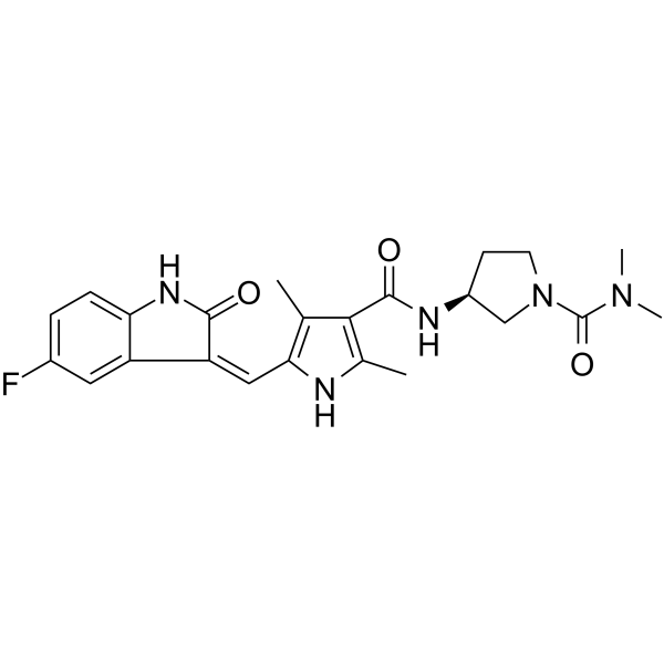
Vorolanib (CM082) is an orally active, potent multikinase VEGFR/PDGFR inhibitor. Vorolanib is a potent ATP-binding cassette (ABC) transporter inhibitor. Vorolanib is an angiogenesis inhibitor and has antitumor activity combined with ZD1839 (HY-50895).
For research use only. We do not sell to patients.
- Purity : 99.78%
- CAS No.: 1013920-15-4
- Formula: C23H26FN5O3
- Molecular Weight:439.48
-
Storage:Powder -20°C, 3 years , 4°C, 2 years ; In solvent -80°C, 2 years , -20°C, 1 year

To place orders, for customer services and technical support, please contact: MedChemExpress USA
Tel: 609-228-6898 E-mail: [email protected] [email protected]


-
 Biological Activity
-
Chemical Information
-
Solvent & Solubility
- Protocols
- Purity & Documentation
- References
-
 Help & FAQs


-
HY-10065
AxitinibTyrosine Kinase Inhibitor -
HY-10255
Sunitinib MalateTyrosine Kinase Inhibitor -
HY-10255A
SunitinibMulti-targeted RTK Inhibitor -
HY-10981
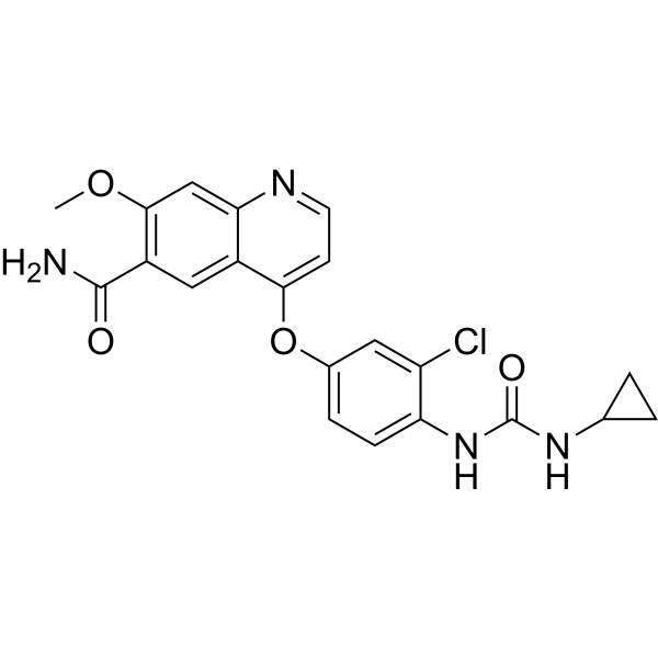
LenvatinibVEGFR/FGFR Inhibitor -
HY-10330
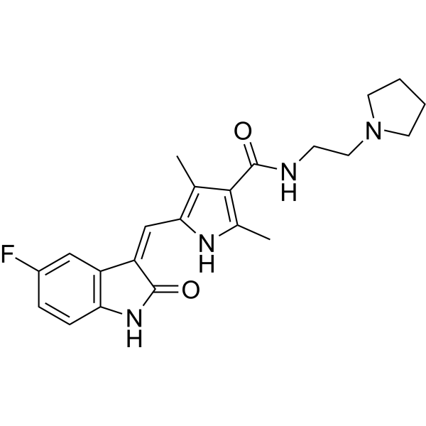
ToceranibRTK Inhibitor -
HY-50751
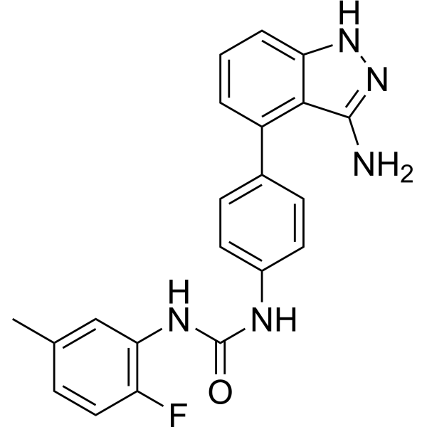
LinifanibMulti-Targeted RTK Inhibitor -
HY-12050
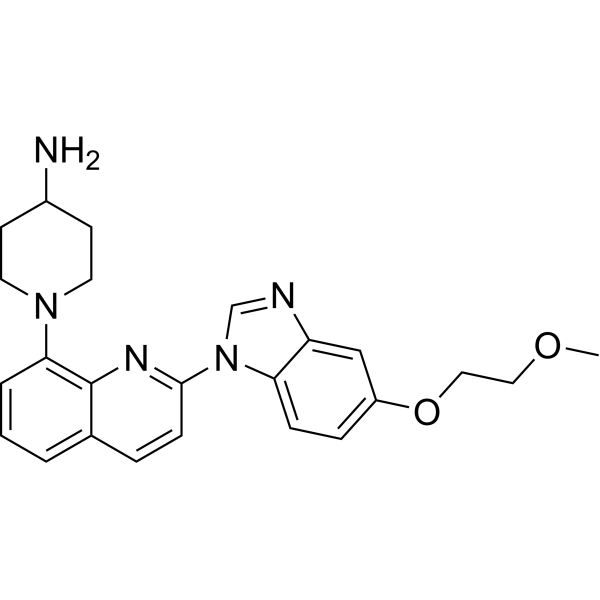
CP-673451PDGFR Inhibitor -

HY-108801
AfliberceptVEGFR Inhibitor -
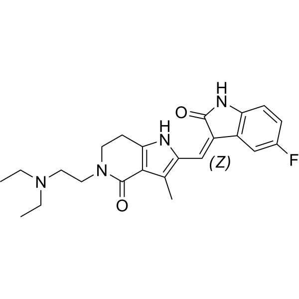
HY-108713
Famitinibc-kit/VEGFR/PDGFR Inhibitor -
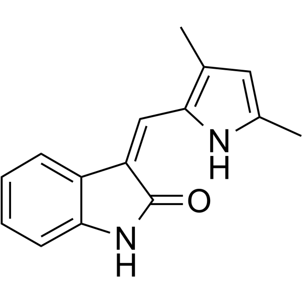
HY-10374
SemaxinibFlk-1/KDR Inhibitor -
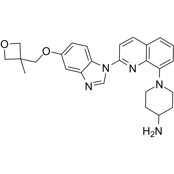
HY-13223
CrenolanibFLT3 Inhibitor -

HY-109019R
Vorolanib (Standard)Reference Standard
Publications Citing Use of MedChemExpress (MCE) Vorolanib
MoreAll VEGFR Isoforms
More

Biological Activity
Description
Cellular Effect
|
Cell Line

|
Type | Value | Description | References |
|---|---|---|---|---|
| HEK293 | IC50 |
0.0155 μM
Compound: CM082
|
Cytotoxicity against HEK293 cells expressing ABCG2-R-482 wildtype assessed as decrease in mitoxantrone half inhibitory concentration
Cytotoxicity against HEK293 cells expressing ABCG2-R-482 wildtype assessed as decrease in mitoxantrone half inhibitory concentration
|
[PMID: 35944339] |
| HEK293 | IC50 |
0.058 μM
Compound: CM082
|
Cytotoxicity against HEK293 cells expressing ABCG2-R-482 mutant assessed as decrease in mitoxantrone half inhibitory concentration
Cytotoxicity against HEK293 cells expressing ABCG2-R-482 mutant assessed as decrease in mitoxantrone half inhibitory concentration
|
[PMID: 35944339] |
| HUVEC | IC50 |
31.5 nM
Compound: CM082; X-82
|
Inhibition of VEGF-induced HUVECs proliferation
Inhibition of VEGF-induced HUVECs proliferation
|
[PMID: 37285684] |
In Vitro
Vorolanib (CM082; 1-100 μM) can specifically enhance the sensitivity of a substrate chemotherapeutical agent in overexpressing ABCG2 cells, but not in overexpressing ABCB1 cells. Vorolanib (1.25, 2.5, 5.0, 20 μM) does not influence the expression of ABCG2 in mRNA or protein Levels[1].
?
Vorolanib (0.001-10 μM) inhibits the growth of VEGF‐stimulated HUVECs (IC50=0.031 μM) and FBS‐stimulated HUVEC growth (IC50=29.9?μM)[2].
?
Vorolanib (0.01, 0.1, 1 μM) exhibits a concentration‐dependent inhibition on VEGF‐induced (40 ng/mL) phosphorylation of VEGFR2 and its downstream signaling molecules ERK1/2, AKT, and STAT3 in HUVECs. Vorolanib (0.1, 1 μM) inhibits FBS‐stimulated tube formation and cell migration of HUVECs in a concentration‐dependent manner[2].
MedChemExpress (MCE) has not independently confirmed the accuracy of these methods. They are for reference only.
In Vivo
? Vorolanib (80 mg/kg; twice daily; for 21 days) has antitumor activity combined with ZD1839 (10 mg/kg; q.d; for 21 days) on H3255 tumor xenograft (female BALB/c nude mice aged five weeks)[2].
MedChemExpress (MCE) has not independently confirmed the accuracy of these methods. They are for reference only.
Clinical Trial
| NCT Number | Sponsor | Condition | Start Date |
Phase
|
|---|---|---|---|---|
| NCT01329991 | 2011-05 | PHASE1 |


Chemical Information
-
CAS No. 1013920-15-4
-
Appearance Solid
-
Molecular Weight 439.48
-
Formula C23H26FN5O3
-
Color Light yellow to yellow
-
SMILES
O=C1/C(C2=CC(F)=CC=C2N1)=C/C(NC(C)=C3C(N[C@@H]4CN(C(N(C)C)=O)CC4)=O)=C3C
-
Synonyms
CM082; X-82
-
Shipping
Room temperature in continental US; may vary elsewhere.
-
Storage
Powder -20°C 3 years 4°C 2 years In solvent -80°C 2 years -20°C 1 year


Publications (1)
-
Journal Impact Factor
-
Most Recent


Solvent & Solubility
In Vitro:
DMSO : 27 mg/mL (61.44 mM; Need ultrasonic; Hygroscopic DMSO has a significant impact on the solubility of product, please use newly opened DMSO)
Please refer to the solubility information to select the appropriate solvent. Once prepared, please aliquot and store the solution to prevent product inactivation from repeated freeze-thaw cycles.
Storage method and period of stock solution: -80°C, 2 years; -20°C, 1 year. When stored at -80°C, please use it within 2 years. When stored at -20°C, please use it within 1 year.
Please refer to the solubility information to select the appropriate solvent. Once prepared, please aliquot and store the solution to prevent product inactivation from repeated freeze-thaw cycles.
Storage method and period of stock solution: -80°C, 2 years; -20°C, 1 year. When stored at -80°C, please use it within 2 years. When stored at -20°C, please use it within 1 year.
Concentration (start) × Volume (start) = Concentration (final) × Volume (final)
In Vivo:
Select the appropriate dissolution method based on your experimental animal and administration route.
- For the following dissolution methods, please ensure to first prepare a clear stock solution using an In Vitro approach and then sequentially add co-solvents:
- To ensure reliable experimental results, the clarified stock solution can be appropriately stored based on storage conditions. As for the working solution for In Vivo experiments, it is recommended to prepare freshly and use it on the same day.
- The percentages shown for the solvents indicate their volumetric ratio in the final prepared solution. If precipitation or phase separation occurs during preparation, heat and/or sonication can be used to aid dissolution.
Add each solvent one by one: 10% DMSO 40% PEG300 5% Tween-80 45% Saline
Solubility: ≥ 2.25 mg/mL (5.12 mM); Clear solution
This protocol yields a clear solution of ≥ 2.25 mg/mL (saturation unknown).
Taking 1 mL working solution as an example, add 100 μL DMSO stock solution (22.5 mg/mL) to 400 μL PEG300, and mix evenly; then add 50 μL Tween-80 and mix evenly; then add 450 μL Saline to adjust the volume to 1 mL.
Preparation of Saline: Dissolve 0.9 g sodium chloride in ddH₂O and dilute to 100 mL to obtain a clear Saline solution.
In Vivo Dissolution Calculator
Please enter the basic information of animal experiments:
-
-
-
-
Recommended: Prepare an additional quantity of animals to account for potential losses during experiments.
Please enter your animal formula composition:
-
%DMSO +
Recommended: Keep the proportion of DMSO in working solution below 2% if your animal is weak.
-
%+
-
+%Tween-80 + +
-
%Saline +
The co-solvents required include: DMSO, . All of co-solvents are available by MedChemExpress (MCE). , Tween 80. All of co-solvents are available by MedChemExpress (MCE).
Working solution concentration: 0.22 mg/mL
Method for preparing stock solution: mg drug dissolved in μL DMSO. Stock solution concentration: mg/mL.
1. Take μL DMSO stock solution;
2. Add μL .
μL , mix evenly;
3. Then add μL Tween 80, mix evenly;
4. Then add μL
Please ensure that the stock solution in the first step is dissolved to a clear state, and add co-solvents in sequence. You can use ultrasonic heating (ultrasonic cleaner, recommended frequency 20-40 kHz), vortexing, etc. to assist dissolution.


Purity & Documentation
-
Data Sheet (272 KB)
-
SDS (252 KB)
- English - EN (252 KB)
- Français - FR (252 KB)
- Deutsch - DE (252 KB)
- Norwegian - NO (252 KB)
- Español - ES (252 KB)
- Swedish - SV (252 KB)
- Italian - IT (252 KB)
- Korean - KR (252 KB)
- Portuguese - PT (252 KB)
-
Handling Instructions (2659 KB)
References
[1]. Lejia Xu, et al. CM082 Enhances the Efficacy of Chemotherapeutic Drugs by Inhibiting the Drug Efflux Function of ABCG2. Mol Ther Oncolytics. 2019 Dec 27;16:100-110. [Content Brief]
[2]. Kun Zhang, et al. CM082, a novel angiogenesis inhibitor, enhances the antitumor activity of ZD1839 on epidermal growth factor receptor mutant non-small cell lung cancer in vitro and in vivo. Thorac Cancer. 2020 Jun;11(6):1566-1577. [Content Brief]


Complete Stock Solution Preparation Table
Please refer to the solubility information to select the appropriate solvent. Once prepared, please aliquot and store the solution to prevent product inactivation from repeated freeze-thaw cycles.
Storage method and period of stock solution: -80°C, 2 years; -20°C, 1 year. When stored at -80°C, please use it within 2 years. When stored at -20°C, please use it within 1 year.
| Optional Solvent | Concentration Solvent Mass | 1 mg | 5 mg | 10 mg | 25 mg |
|---|---|---|---|---|---|
| DMSO | 1 mM | 2.2754 mL | 11.3771 mL | 22.7542 mL | 56.8854 mL |
| 5 mM | 0.4551 mL | 2.2754 mL | 4.5508 mL | 11.3771 mL | |
| 10 mM | 0.2275 mL | 1.1377 mL | 2.2754 mL | 5.6885 mL | |
| 15 mM | 0.1517 mL | 0.7585 mL | 1.5169 mL | 3.7924 mL | |
| 20 mM | 0.1138 mL | 0.5689 mL | 1.1377 mL | 2.8443 mL | |
| 25 mM | 0.0910 mL | 0.4551 mL | 0.9102 mL | 2.2754 mL | |
| 30 mM | 0.0758 mL | 0.3792 mL | 0.7585 mL | 1.8962 mL | |
| 40 mM | 0.0569 mL | 0.2844 mL | 0.5689 mL | 1.4221 mL | |
| 50 mM | 0.0455 mL | 0.2275 mL | 0.4551 mL | 1.1377 mL | |
| 60 mM | 0.0379 mL | 0.1896 mL | 0.3792 mL | 0.9481 mL |


Vorolanib Related Classifications
HY-109019 Related Classifications
 Powered by Bioz