



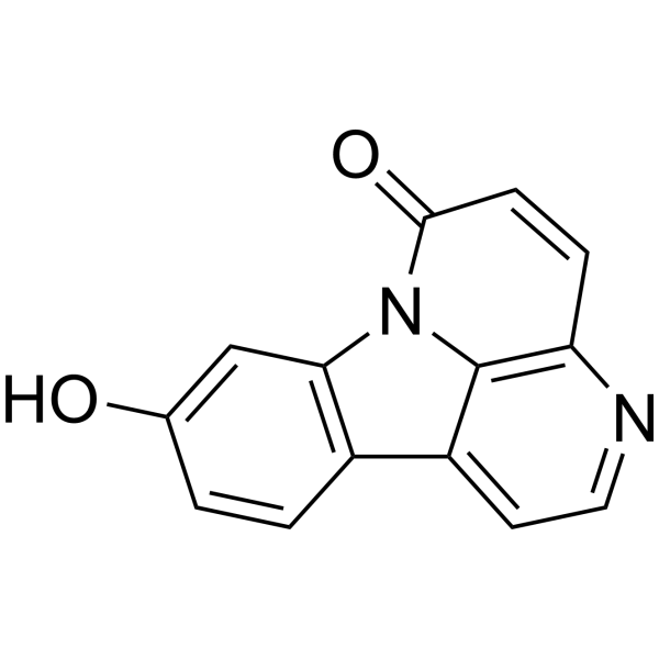
9-Hydroxycanthin-6-one
9-Hydroxycanthin-6-one is a β-carboline alkaloid. 9-Hydroxycanthin-6-one can be isolated from the roots of E. longifolia. 9-Hydroxycanthin-6-one inhibits TNF-α-induced activation of the NF-κB pathway. 9-Hydroxycanthin-6-one activates GSK3β independently of CK1α, drives phosphorylation and degradation of β-catenin, and inhibits the Wnt signaling pathway. 9-Hydroxycanthin-6-one exerts selective cytotoxicity against Wnt-dependent colon cancer cells. 9-Hydroxycanthin-6-one can be used in studies related to colon cancer.
Nos produits utilisent uniquement pour la recherche. Nous ne vendons pas aux patients.
- CAS No.: 138544-91-9
- Formule: C14H8N2O2
- Masse moléculaire:236.23
-
Stockage:
4°C, protect from light
* In solvent : -80°C, 6 months; -20°C, 1 month (protect from light)

To place orders, for customer services and technical support, please contact: MedChemExpress USA
Tel: 609-228-6898 E-mail: [email protected] [email protected]


-
 Activité biologique
-
Chemical Information
- Protocole
- Pureté et documentation
- Références
-
 Help & FAQs




Activité biologique
Description
IC50 & Target
[1]|
GSK3β |
Cellular Effect
|
Cell Line

|
Type | Value | Description | References |
|---|---|---|---|---|
| A549 | ED50 |
10 μg/mL
Compound: 9-hydroxycanthin-6-one
|
Cytotoxicity against human A549 cells by SRB assay
Cytotoxicity against human A549 cells by SRB assay
|
[PMID: 14575431] |
| Breast carcinoma cell | IC50 |
1.5 μg/mL
Compound: 31
|
Cytotoxicity against human Breast cancer cell
Cytotoxicity against human Breast cancer cell
|
[PMID: 34332400] |
| Calu-3 | CC50 |
>100 μM
Compound: Table 1, R10C2
|
Cytotoxicity against human Calu-3 cells assessed as reduction in cell viability incubated for 48 hrs by PrestoBlue reagent based assay
Cytotoxicity against human Calu-3 cells assessed as reduction in cell viability incubated for 48 hrs by PrestoBlue reagent based assay
|
[PMID: 36399766] |
| Cancer cell lines | ED50 |
2.6 μg/mL
Compound: 3
|
Cytotoxicity against human breast cancer cells
Cytotoxicity against human breast cancer cells
|
[PMID: 1800638] |
| Cancer cell lines | ED50 |
3.8 μg/mL
Compound: 3
|
Cytotoxicity against human lung cancer cells
Cytotoxicity against human lung cancer cells
|
[PMID: 1800638] |
| Cancer cell lines | ED50 |
4.8 μg/mL
Compound: 3
|
Cytotoxicity against human colon cancer cells
Cytotoxicity against human colon cancer cells
|
[PMID: 1800638] |
| Colon cancer cell line | IC50 |
1.5 μg/mL
Compound: 31
|
Cytotoxicity against human Colon cancer cell line
Cytotoxicity against human Colon cancer cell line
|
[PMID: 34332400] |
| DLD-1 | IC50 |
>40 μM
Compound: 1
|
Cytotoxicity against Wnt-dependent human DLD1 cells assessed as cell viability after 24 hrs by fluorescence assay
Cytotoxicity against Wnt-dependent human DLD1 cells assessed as cell viability after 24 hrs by fluorescence assay
|
[PMID: 25905468] |
| Fibrosarcoma cell line | IC50 |
1.5 μg/mL
Compound: 31
|
Cytotoxicity against human Fibrosarcoma cell line
Cytotoxicity against human Fibrosarcoma cell line
|
[PMID: 34332400] |
| HCT-116 | IC50 |
30 μM
Compound: 1
|
Cytotoxicity against Wnt-dependent human HCT116 cells assessed as cell viability after 24 hrs by fluorescence assay
Cytotoxicity against Wnt-dependent human HCT116 cells assessed as cell viability after 24 hrs by fluorescence assay
|
[PMID: 25905468] |
| HCT-8 | CC50 |
60.88 μM
Compound: Table 1, R10C2
|
Cytotoxicity against human HCT-8 cells assessed as redcution in cell viability incubated for 96 hrs by PrestoBlue reagent based assay
Cytotoxicity against human HCT-8 cells assessed as redcution in cell viability incubated for 96 hrs by PrestoBlue reagent based assay
|
[PMID: 36399766] |
| HEK293 | IC50 |
36.7 μM
Compound: 1
|
Cytotoxicity against HEK293 cells assessed as cell viability after 24 hrs by fluorescence assay
Cytotoxicity against HEK293 cells assessed as cell viability after 24 hrs by fluorescence assay
|
[PMID: 25905468] |
| HEK293 | IC50 |
6.8 μM
Compound: 1
|
Activation of GSK-3beta in HEK293 cells assessed as suppression of TCF/beta-catenin transcriptional activity after 24 hrs by SuperTOPFlash reporter gene assay
Activation of GSK-3beta in HEK293 cells assessed as suppression of TCF/beta-catenin transcriptional activity after 24 hrs by SuperTOPFlash reporter gene assay
|
[PMID: 25905468] |
| HEK-293T | IC50 |
>40 μM
Compound: 1
|
Cytotoxicity against HEK293T cells assessed as cell viability after 24 hrs by fluorescence assay
Cytotoxicity against HEK293T cells assessed as cell viability after 24 hrs by fluorescence assay
|
[PMID: 25905468] |
| HepG2 | CC50 |
>100 μM
Compound: Table 1, R10C2
|
Cytotoxicity against human HepG2 cells assessed as reduction in cell viability incubated for 48 hrs by PrestoBlue reagent based assay
Cytotoxicity against human HepG2 cells assessed as reduction in cell viability incubated for 48 hrs by PrestoBlue reagent based assay
|
[PMID: 36399766] |
| KB | ED50 |
2 μg/mL
Compound: 3
|
Cytotoxicity against human KB cells
Cytotoxicity against human KB cells
|
[PMID: 1800638] |
| KB | IC50 |
1.5 μg/mL
Compound: 31
|
Cytotoxicity against human Vincristine-resistant KB cells
Cytotoxicity against human Vincristine-resistant KB cells
|
[PMID: 34332400] |
| KB-V1 | ED50 |
>20 μg/mL
Compound: 3
|
Cytotoxicity against human KBV1 cells
Cytotoxicity against human KBV1 cells
|
[PMID: 1800638] |
| Lung cancer cell line | IC50 |
1.5 μg/mL
Compound: 31
|
Cytotoxicity against human Lung cancer cell line
Cytotoxicity against human Lung cancer cell line
|
[PMID: 34332400] |
| MCF7 | ED50 |
19.6 μg/mL
Compound: 9-hydroxycanthin-6-one
|
Cytotoxicity against human MCF7 cells by SRB assay
Cytotoxicity against human MCF7 cells by SRB assay
|
[PMID: 14575431] |
| Melanoma cell | ED50 |
5.4 μg/mL
Compound: 3
|
Cytotoxicity against human melanoma cells
Cytotoxicity against human melanoma cells
|
[PMID: 1800638] |
| Melanoma cell line | IC50 |
1.5 μg/mL
Compound: 31
|
Cytotoxicity against human Melanoma cell line
Cytotoxicity against human Melanoma cell line
|
[PMID: 34332400] |
| MRC5 | CC50 |
>100 μM
Compound: Table 1, R10C2
|
Cytotoxicity against human MRC5 cells assessed as reduction in cell viability incubated for 48 hrs by PrestoBlue reagent based assay
Cytotoxicity against human MRC5 cells assessed as reduction in cell viability incubated for 48 hrs by PrestoBlue reagent based assay
|
[PMID: 36399766] |
| P388 | ED50 |
1.5 μg/mL
Compound: 3
|
Cytotoxicity against mouse P388 cells
Cytotoxicity against mouse P388 cells
|
[PMID: 1800638] |
| RKO | IC50 |
>40 μM
Compound: 1
|
Cytotoxicity against Wnt-independent human RKO cells assessed as cell viability after 24 hrs by fluorescence assay
Cytotoxicity against Wnt-independent human RKO cells assessed as cell viability after 24 hrs by fluorescence assay
|
[PMID: 25905468] |
| SW480 | IC50 |
17.4 μM
Compound: 1
|
Cytotoxicity against Wnt-independent human SW480 cells assessed as cell viability after 24 hrs by fluorescence assay
Cytotoxicity against Wnt-independent human SW480 cells assessed as cell viability after 24 hrs by fluorescence assay
|
[PMID: 25905468] |
| Vero | CC50 |
>100 μM
Compound: Table 1, R10C2
|
Cytotoxicity against African green monkey Vero cells assessed as reduction in cell viability incubated for 48 hrs by PrestoBlue reagent based assay
Cytotoxicity against African green monkey Vero cells assessed as reduction in cell viability incubated for 48 hrs by PrestoBlue reagent based assay
|
[PMID: 36399766] |
In Vitro
9-Hydroxycanthin-6-one (3.8-30 μM; 30 min pretreatment, 4 h TNF-α stimulation) potently inhibits TNF-α-induced NF-κB activation in HEK-293/NF-κB-luc cells with an IC50 of 3.8 μM, and exhibits no cytotoxicity at 30 μM[1].
9-Hydroxycanthin-6-one (5-20 μM; 24 h) potently and selectively inhibits TCF/β-catenin transcriptional activity in STF/293 cells with an IC50 of 6.8 μM, while preserving high cell viability[2].
9-Hydroxycanthin-6-one (24 h) selectively inhibits the viability of Wnt-dependent human colon cancer cells (SW480, HCT116) with IC50 values of 17.4 μM and 30.0 μM, respectively, while sparing Wnt-independent and noncancer cell lines[2].
9-Hydroxycanthin-6-one (10-30 μM; 24 h) dose-dependently reduces total β-catenin levels (in whole cell, cytosolic, and nuclear fractions) and increases β-catenin phosphorylation at GSK3β target sites (Ser33/Ser37/Thr41) in SW480 cells, without altering CK1α-mediated β-catenin phosphorylation or total GSK3β/CK1α levels[2].
9-Hydroxycanthin-6-one (30 μM; 24 h post-siRNA transfection) inhibits Wnt signaling in SW480 cells through GSK3β activation independent of CK1α, as it reduces β-catenin and increases GSK3β-mediated β-catenin phosphorylation even when CK1α is knocked down[2].
MedChemExpress (MCE) has not independently confirmed the accuracy of these methods. They are for reference only.
-
Cell Line:SW480 cells
-
Concentration:10, 20, 30 μM
-
Incubation Time:24 h
-
Result:Dose-dependently decreased total β-catenin protein levels in whole cellular, cytosolic, and nuclear fractions.
Dose-dependently increased phosphorylated β-catenin at Ser33/Ser37/Thr41 (GSK3β phosphorylation sites).
Had no effect on phosphorylated β-catenin at Ser45 (CK1α phosphorylation site) or total GSK3β/CK1α protein levels.
-
Cell Line:SW480 cells
-
Concentration:30 μM
-
Incubation Time:24 h (post-siRNA transfection)
-
Result:Decreased β-catenin levels and increased phosphorylated β-catenin at Ser33/Ser37/Thr41 in cells transfected with control siRNA.
Attenuated reduction in β-catenin levels in cells transfected with GSK3β siRNA.
Decreased phosphorylated GSK3β at Ser9 (inactive form) protein levels.\nDecreased β-catenin levels and increased phosphorylated β-catenin at Ser33/Ser37/Thr41 in both control siRNA-transfected and CK1α siRNA-transfected cells.
Decreased phosphorylated GSK3β at Ser9 levels in CK1α siRNA-transfected cells.
In Vivo
MedChemExpress (MCE) has not independently confirmed the accuracy of these methods. They are for reference only.
-
Animal Model:TOPdGFP transgenic strain; wild-type AB strain (embryos)[2]
-
Dosage:300 μM (TOPdGFP activity, Wnt target gene expression, pigment cell assessment); 100 μM (eyeless phenotype rescue)
-
Administration:immersion in E3 medium; 10 hpf to 24 hpf (TOPdGFP activity); immersion in E3 medium; 4 hpf to 24 hpf (Wnt target gene expression); immersion in E3 medium; 4 hpf to 30 hpf (pigment cell assessment); immersion in E3 medium; 4 hpf to 30 hpf, co-administered with 0.5 μM BIO (eyeless phenotype rescue)
-
Result:Reduced TOPdGFP activity in the midbrain tectum of 24 hpf embryos without inducing excess cell death (control MO group: 100% activity, n=10; p53 MO group: 100% activity, n=10).
Reduced zic2a expression in the midbrain tectum of 24 hpf embryos (control MO group: 55% expression, n=6; p53 MO group: 54% expression, n=7).
Reduced mitf expression in neural-crest cells of 24 hpf embryos (control MO group: 50% expression, n=7; p53 MO group: 60% expression, n=9).
Reduced the number of pigment cells in the tails of 30 hpf embryos (100% reduction, n=10).
Rescued the eyeless phenotype induced by 0.5 μM BIO in 30 hpf embryos, with 46% showing fully rescued eyes (n=6) and 54% showing slightly rescued eyes (n=7).


Chemical Information
-
CAS No. 138544-91-9
-
Appearance Solid
-
Masse moléculaire 236.23
-
Formule C14H8N2O2
-
Color Light yellow to yellow
-
SMILES
O=C1N2C3=C(N=CC=C3C4=C2C=C(O)C=C4)C=C1
-
Structure Classification
-
Initial Source
-
Livraison
Room temperature in continental US; may vary elsewhere.
-
Stockage
4°C, protect from light
* In solvent : -80°C, 6 months; -20°C, 1 month (protect from light)


Pureté et documentation
-
Fiche technique (283 KB)
-
SDS (252 KB)
- English - EN (252 KB)
- Français - FR (252 KB)
- Deutsch - DE (252 KB)
- Norwegian - NO (252 KB)
- Español - ES (252 KB)
- Swedish - SV (252 KB)
- Italian - IT (252 KB)
- Korean - KR (252 KB)
- Portuguese - PT (252 KB)
-
Instruction de manipulation (2659 KB)
Références
[1].
Thi Van Anh Tran, et al. NF-κB inhibitors from Eurycoma longifolia. J Nat Prod. 2014 Mar 28;77(3):483-8.
[Content Brief]
[2]. Ohishi K, et al. 9-Hydroxycanthin-6-one, a β-Carboline Alkaloid from Eurycoma longifolia, Is the First Wnt Signal Inhibitor through Activation of Glycogen Synthase Kinase 3β without Depending on Casein Kinase 1α. J Nat Prod. 2015 May 22;78(5):1139-46. [Content Brief]

Calculators
Concentration (start) × Volume (start) = Concentration (final) × Volume (final)


9-Hydroxycanthin-6-one Related Classifications
HY-N7183 Related Classifications
 Powered by Bioz