



CXCR2 Antibody (YA3904)
(Synonyms: CXCR2; IL8R2; IL8RA; IL8RB; CMKAR2; CDw128b)Based on 1 Customer Validation
CXCR2 Antibody (YA3904) is a Mouse-derived and non-conjugated IgG1 monoclonal antibody, targeting to CXCR2.
-
Host:
Mouse
-
Isotype:
IgG
-
Application:
IHC-P, ICC/IF, FC, ELISA
-
Reactivity :
Human
-
Formulation:
Supplied in PBS with 0.05% sodium azide
-
Conjugation:
Non-conjugated


To place orders, for customer services and technical support, please contact: MedChemExpress USA
Tel: 609-228-6898 E-mail: [email protected] [email protected]





Applications
| Application |
IHC-P

IHC-P: Immunohistochemistry-Paraffin
|
ICC/IF
ICC/IF: Immunocytochemistry/
Immunofluorescence |
FC
FC: Flow Cytometry
|
ELISA
ELISA: Enzyme Linked Immunosorbent Assay
|
|---|---|---|---|---|
| Dilution Ratio | 1:200-1:1000 | 1:100 | 1:200-1:400 | 1:10000 |


Product Details
CXCR2 Antibody (YA3904) is a Mouse-derived and non-conjugated IgG1 monoclonal antibody, targeting to CXCR2.
-
Host Mouse

-
Clonality Monoclonal

-
Species ReactivityHuman
-
Observed Molecular WeightObserved band size: 41 kDa
Note: Due to possible protein modifications or aggregation, the molecular weight should be confirmed by actual measurement, and the predicted value is for reference only.

-
Calculated Molecular Weight Predicted band size: 41 kDa

Purified recombinant fragment of human CD182 expressed in E. Coli.
affinity purified.
Non-conjugated
Unmodified
IgG


Product Properties
-
Appearance
Solution
-
Formulation
Supplied in PBS with 0.05% sodium azide
-
Concentration
Batch-dependent, Please check the COA for the concentration of each lot. Check Lot Concentration
-
Storage & Stability
Stored at -20°C for 1 year. Avoid repeated freeze / thaw cycles.
-
Shipping
Shipping with blue ice.


Verification Images
-
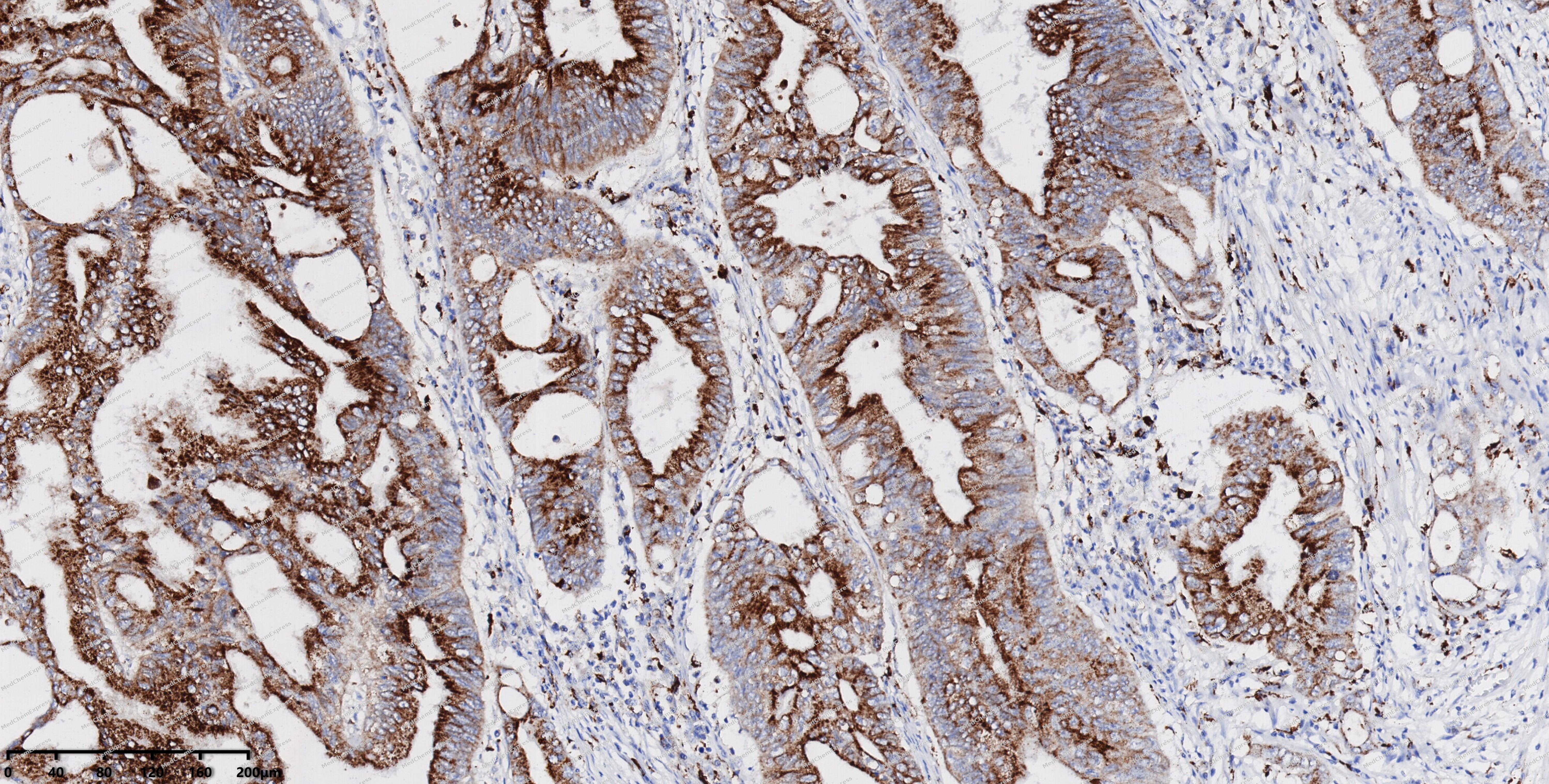 Immunohistochemical analysis of paraffin-embedded human colon cancer using CXCR2 antibody. The section was pre-treated using heat mediated antigen retrieval with EDTA (pH 9.0) for 14 minutes. The tissues were blocked in 5% BSA for 30 minutes at room temperature, washed with TBST, and then probed with the primary antibody (HY-P84207, 1/1000) for 30 minutes at room temperature. The detection was performed using Polymer HRP-conjugated Goat Anti-Mouse/Rabbit lgG(H&L) secondary antibody (HY-P83652). DAB was used as the chromogen. Tissues were counterstained with hematoxylin and mounted with DPX.
 -
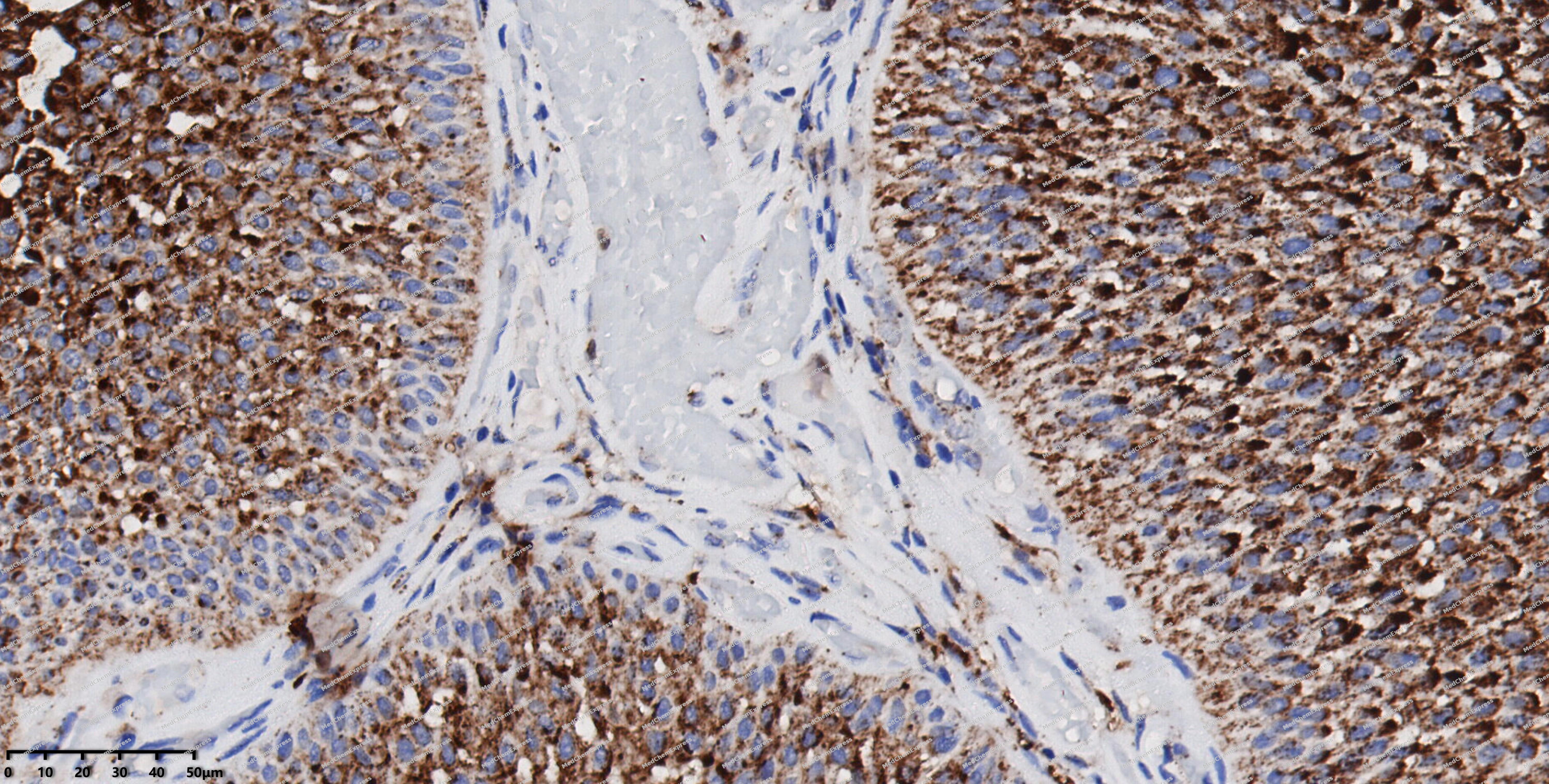 Immunohistochemical analysis of paraffin-embedded human bladder cancer using CXCR2 antibody. The section was pre-treated using heat mediated antigen retrieval with EDTA (pH 9.0) for 14 minutes. The tissues were blocked in 5% BSA for 30 minutes at room temperature, washed with TBST, and then probed with the primary antibody (HY-P84207, 1/1000) for 30 minutes at room temperature. The detection was performed using Polymer HRP-conjugated Goat Anti-Mouse/Rabbit lgG(H&L) secondary antibody (HY-P83652). DAB was used as the chromogen. Tissues were counterstained with hematoxylin and mounted with DPX.
 -
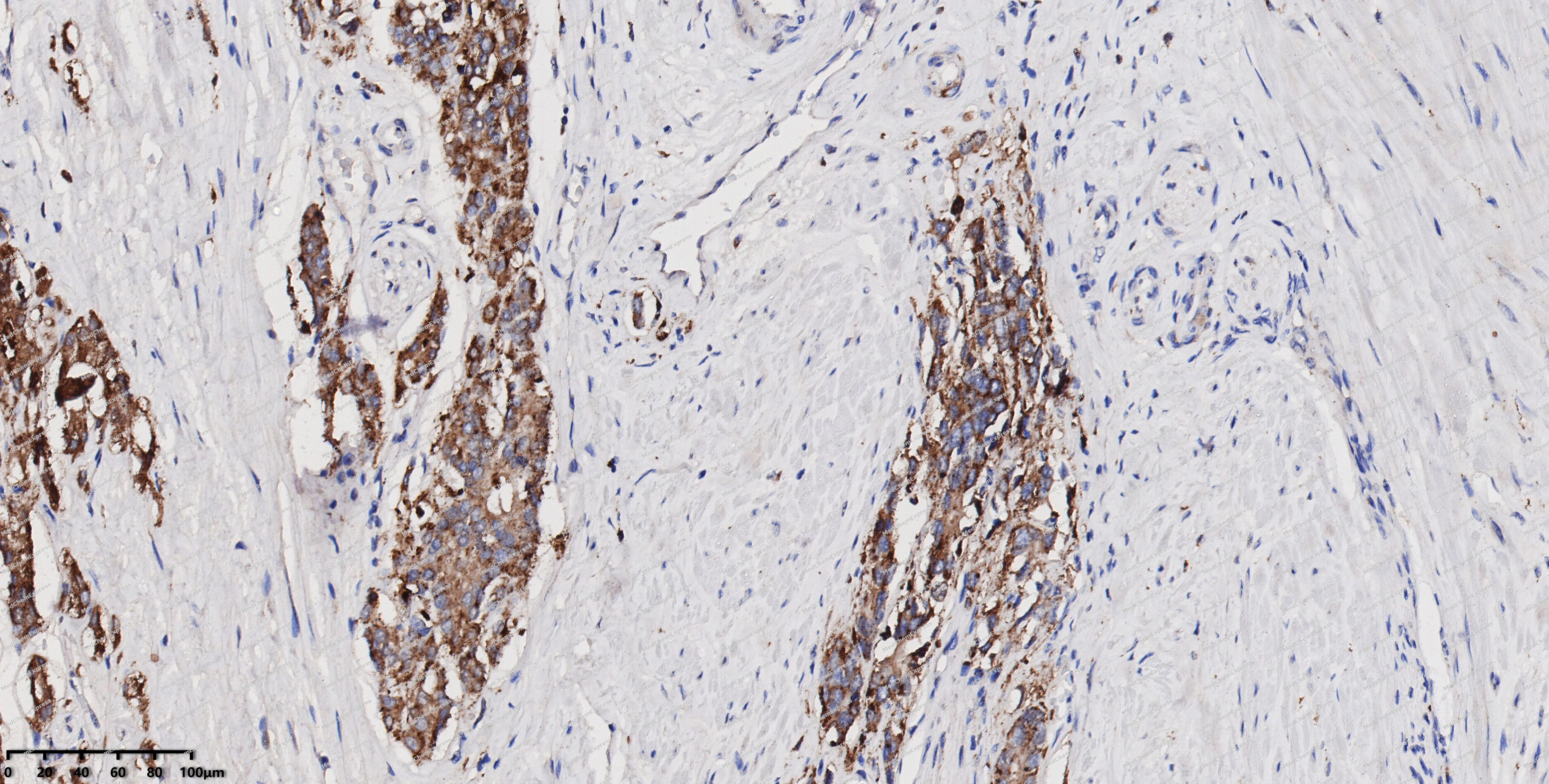 Immunohistochemical analysis of paraffin-embedded human prostate cancer using CXCR2 antibody. The section was pre-treated using heat mediated antigen retrieval with EDTA (pH 9.0) for 14 minutes. The tissues were blocked in 5% BSA for 30 minutes at room temperature, washed with TBST, and then probed with the primary antibody (HY-P84207, 1/1000) for 30 minutes at room temperature. The detection was performed using Polymer HRP-conjugated Goat Anti-Mouse/Rabbit lgG(H&L) secondary antibody (HY-P83652). DAB was used as the chromogen. Tissues were counterstained with hematoxylin and mounted with DPX.
 -
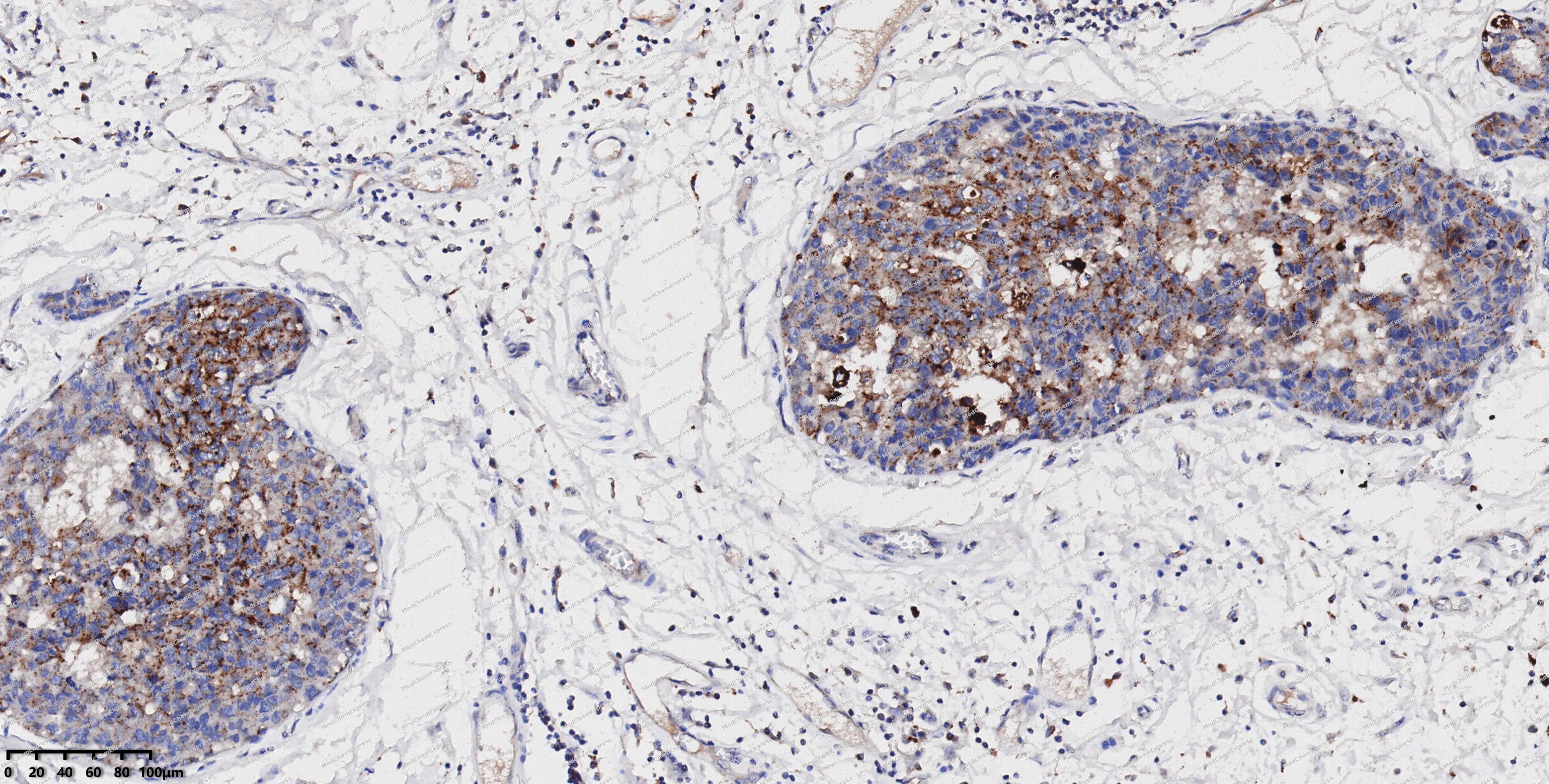 Immunohistochemical analysis of paraffin-embedded human breast cancer using CXCR2 antibody. The section was pre-treated using heat mediated antigen retrieval with EDTA (pH 9.0) for 14 minutes. The tissues were blocked in 5% BSA for 30 minutes at room temperature, washed with TBST, and then probed with the primary antibody (HY-P84207, 1/1000) for 30 minutes at room temperature. The detection was performed using Polymer HRP-conjugated Goat Anti-Mouse/Rabbit lgG(H&L) secondary antibody (HY-P83652). DAB was used as the chromogen. Tissues were counterstained with hematoxylin and mounted with DPX.
 -
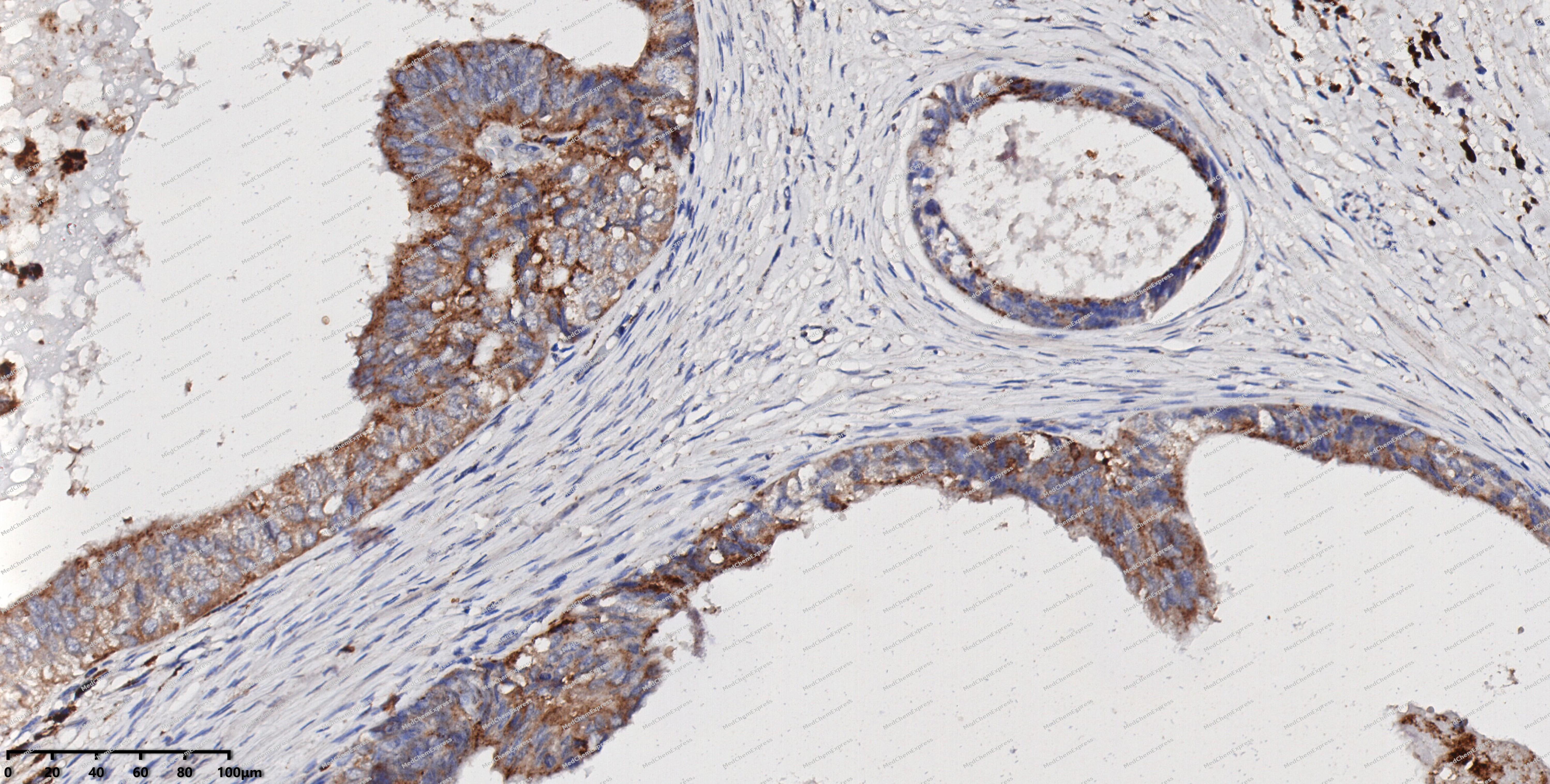 Immunohistochemical analysis of paraffin-embedded human cervical cancer using CXCR2 antibody. The section was pre-treated using heat mediated antigen retrieval with EDTA (pH 9.0) for 14 minutes. The tissues were blocked in 5% BSA for 30 minutes at room temperature, washed with TBST, and then probed with the primary antibody (HY-P84207, 1/1000) for 30 minutes at room temperature. The detection was performed using Polymer HRP-conjugated Goat Anti-Mouse/Rabbit lgG(H&L) secondary antibody (HY-P83652). DAB was used as the chromogen. Tissues were counterstained with hematoxylin and mounted with DPX.
 -
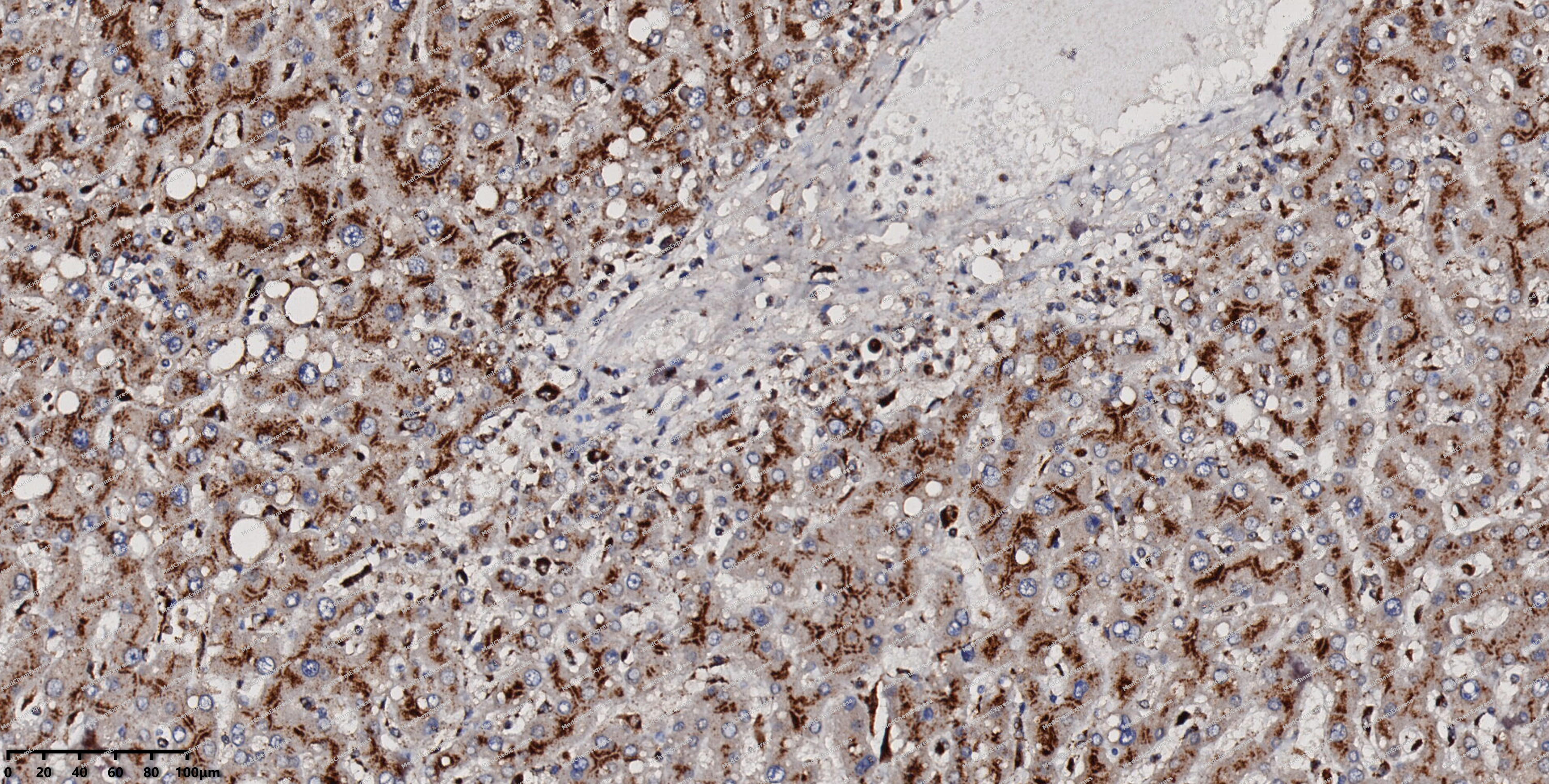 Immunohistochemical analysis of paraffin-embedded human liver using CXCR2 antibody. The section was pre-treated using heat mediated antigen retrieval with EDTA (pH 9.0) for 14 minutes. The tissues were blocked in 5% BSA for 30 minutes at room temperature, washed with TBST, and then probed with the primary antibody (HY-P84207, 1/1000) for 30 minutes at room temperature. The detection was performed using Polymer HRP-conjugated Goat Anti-Mouse/Rabbit lgG(H&L) secondary antibody (HY-P83652). DAB was used as the chromogen. Tissues were counterstained with hematoxylin and mounted with DPX.
 -
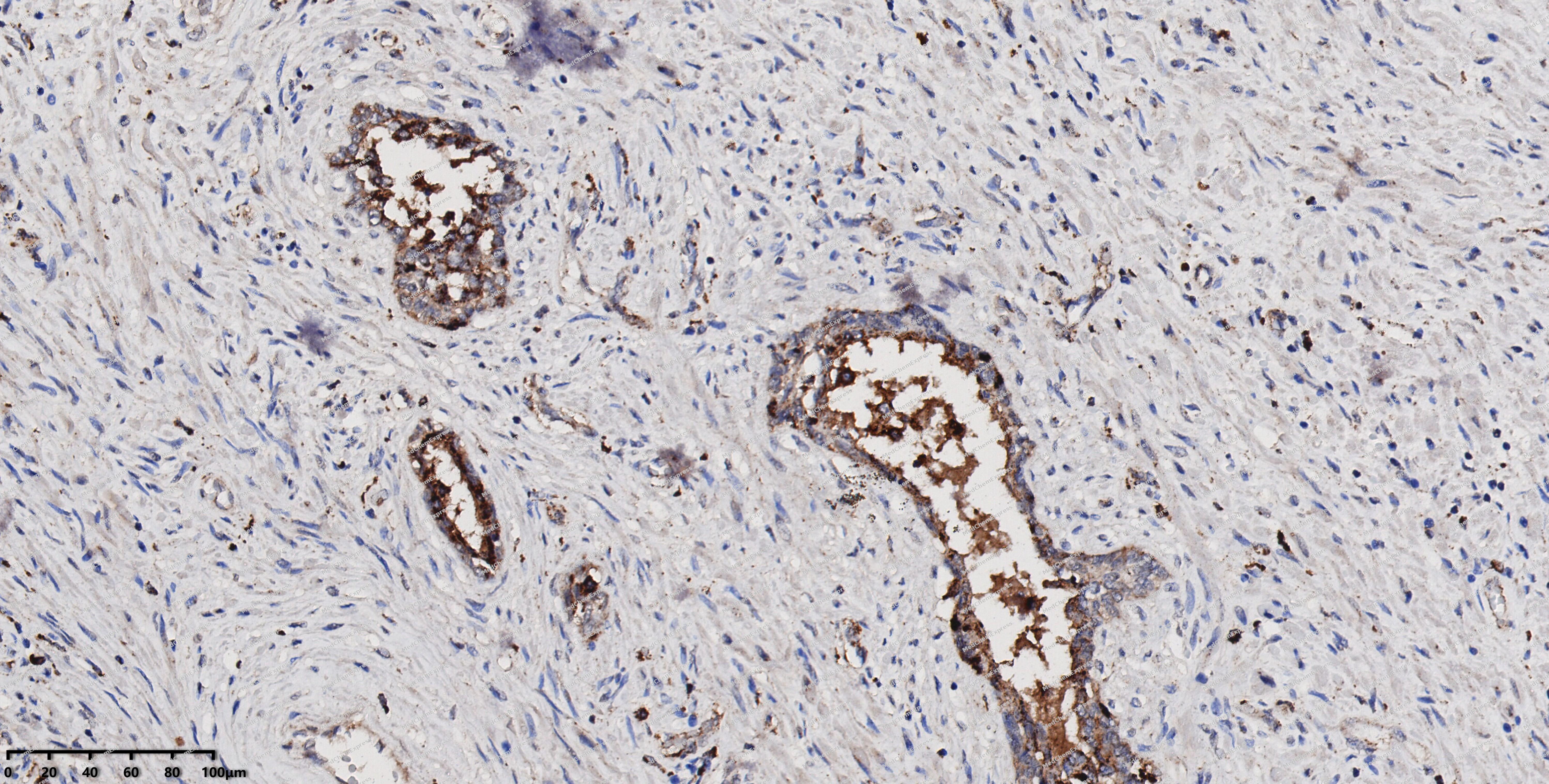 Immunohistochemical analysis of paraffin-embedded human prostate using CXCR2 antibody. The section was pre-treated using heat mediated antigen retrieval with EDTA (pH 9.0) for 14 minutes. The tissues were blocked in 5% BSA for 30 minutes at room temperature, washed with TBST, and then probed with the primary antibody (HY-P84207, 1/1000) for 30 minutes at room temperature. The detection was performed using Polymer HRP-conjugated Goat Anti-Mouse/Rabbit lgG(H&L) secondary antibody (HY-P83652). DAB was used as the chromogen. Tissues were counterstained with hematoxylin and mounted with DPX.
 -
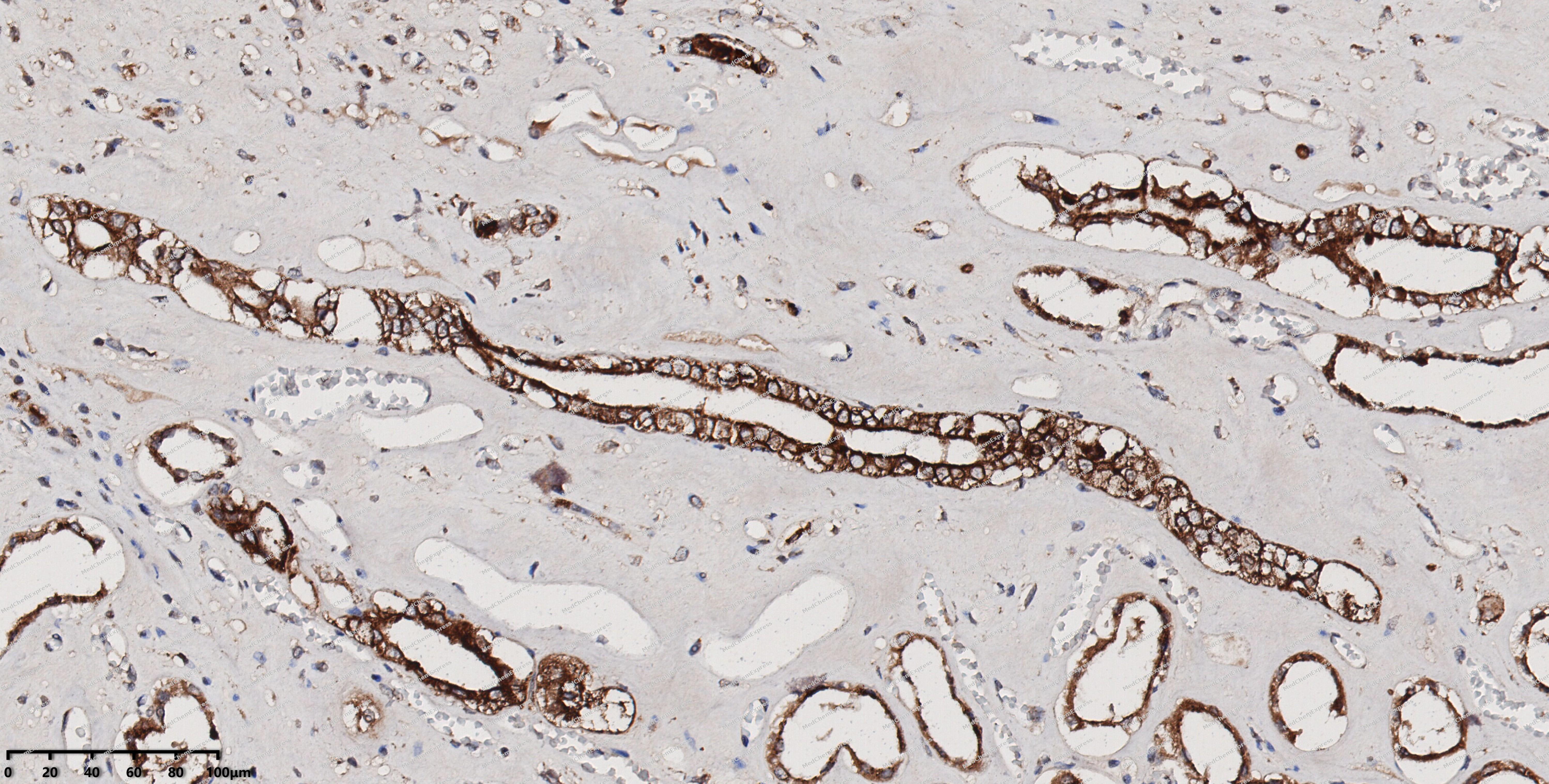 Immunohistochemical analysis of paraffin-embedded human kidney using CXCR2 antibody. The section was pre-treated using heat mediated antigen retrieval with EDTA (pH 9.0) for 14 minutes. The tissues were blocked in 5% BSA for 30 minutes at room temperature, washed with TBST, and then probed with the primary antibody (HY-P84207, 1/1000) for 30 minutes at room temperature. The detection was performed using Polymer HRP-conjugated Goat Anti-Mouse/Rabbit lgG(H&L) secondary antibody (HY-P83652). DAB was used as the chromogen. Tissues were counterstained with hematoxylin and mounted with DPX.


Background
-
Function
CXCR2 is a G protein-coupled chemokine receptor that predominantly regulates neutrophil recruitment, leukocyte trafficking, and inflammatory responses through binding multiple ELR+ CXC chemokines, including CXCL1 and CXCL8/IL-8[1][2]. Mechanistically, CXCR2 activation triggers chemokine-dependent signaling pathways that promote integrin activation, cell migration, adhesion, and tissue infiltration, thereby coordinating innate immune responses during infection and tissue injury[1]. Beyond physiological inflammation, the CXCL-CXCR2 signaling axis contributes to angiogenesis, tumor cell migration, invasion, and stromal remodeling in multiple cancer types, including pancreatic, colorectal, breast, lung, and ovarian cancers[3][4][5]. In disease models, elevated CXCR2 signaling promotes recruitment of neutrophils and myeloid-derived suppressor cells, supporting tumor progression and immunosuppressive microenvironments[3][4]. Compared with the related receptor CXCR1, which shares CXCL8 responsiveness, CXCR2 exhibits broader ligand recognition and mediates diverse inflammatory and tumor-associated chemokine networks across immune, endothelial, epithelial, and stromal cells[2][5]. Therefore, CXCR2 has emerged as a major experimental and therapeutic target, and multiple CXCR2 antagonists have demonstrated activity in preclinical cancer, cardiovascular, lung injury, and inflammatory disease models by reducing leukocyte recruitment, angiogenesis, and pathological inflammation[1][2][3].
-
Subcellular Localization
Cell membrane; Multi-pass membrane protein
-
Subunit
Interacts with IL8 (PubMed:1891716). Interacts with GNAI2 (PubMed:8662698)
-
SwissProt ID
-
Synonyms
CXCR2; IL8R2; IL8RA; IL8RB; CMKAR2; CDw128b


Documentation
-
Data Sheet (261 KB)
-
SDS (251 KB)
- English - EN (251 KB)
- Français - FR (251 KB)
- Deutsch - DE (251 KB)
- Norwegian - NO (251 KB)
- Español - ES (251 KB)
- Swedish - SV (251 KB)
- Italian - IT (251 KB)
- Korean - KR (251 KB)
- Portuguese - PT (251 KB)
-
User Guide for Antibodies (1077 KB)
[1]. Stadtmann A, et al. CXCR2: From Bench to Bedside. Front Immunol. 2012 Aug 24;3:263. [Content Brief]
[2]. Yaman I, et al. Advances in understanding cancer-associated neurogenesis and its implications on the neuroimmune axis in cancer. Pharmacol Ther. 2022 Nov;239:108199. [Content Brief]
[3]. Lazennec G, et al. CXCR2 chemokine receptor - a master regulator in cancer and physiology. Trends Mol Med. 2024 Jan;30(1):37-55. [Content Brief]
[5]. Baño V, et al. Characterization and Structural Performance in Bending of CLT Panels Made from Small-Diameter Logs of Loblolly/Slash Pine. Materials (Basel). 2018 Nov 30;11(12):2436. [Content Brief]

 Powered by Bioz
