



FXR1 Antibody (YA3513)
(Synonyms: FXR1; FXR1P; hFXR1p)Based on 1 publication(s) in Google Scholar
FXR1 Antibody (YA3513) is a Rabbit-derived and non-conjugated IgG monoclonal antibody, targeting to FXR1.
-
Host:
Rabbit
-
Isotype:
IgG
-
Application:
WB, IHC-P, ICC/IF, FC
-
Reactivity :
Human, Mouse, Rat
-
Formulation:
Supplied in Rabbit IgG in 10mM phosphate buffered saline , pH 7.4, 150mM sodium chloride, 0.05% BSA, 0.02% sodium azide and 50% glycerol.
-
Conjugation:
Non-conjugated


To place orders, for customer services and technical support, please contact: MedChemExpress USA
Tel: 609-228-6898 E-mail: [email protected] [email protected]



Publications Citing Use of MedChemExpress (MCE) FXR1 Antibody (YA3513)
More

Applications
| Application |
WB

WB: Western Blot
|
IHC-P
IHC-P: Immunohistochemistry-Paraffin
|
ICC/IF
ICC/IF: Immunocytochemistry/
Immunofluorescence |
FC
FC: Flow Cytometry
|
|---|---|---|---|---|
| Dilution Ratio | 1:500-1:1000 | 1:50-1:100 | 1:50-1:200 | 1:50-1:100 |


Product Details
FXR1 Antibody (YA3513) is a Rabbit-derived and non-conjugated IgG monoclonal antibody, targeting to FXR1.
-
Host Rabbit

-
Clonality Recombinant,Monoclonal

-
Species ReactivityHuman, Mouse, Rat
-
Observed Molecular WeightObserved band size: 70-85 kDa
Note: Due to possible protein modifications or aggregation, the molecular weight should be confirmed by actual measurement, and the predicted value is for reference only.

-
Calculated Molecular Weight Predicted band size: 70 kDa

Entrez Gene: 8087 Human ; 14359 Mouse ; 361927 Rat
SwissProt: P51114 Human ; Q61584 Mouse ; Q5XI81 Rat
OMIM: 618822 Human
A synthesized peptide derived from human FXR1 aa4-63 aa4-63/621.
Endogenous
Affinity Purified
Non-conjugated
Unmodified
IgG


Product Properties
-
Appearance
Solution
-
Formulation
Supplied in Rabbit IgG in 10mM phosphate buffered saline , pH 7.4, 150mM sodium chloride, 0.05% BSA, 0.02% sodium azide and 50% glycerol.
-
Concentration
Batch-dependent, Please check the COA for the concentration of each lot. Check Lot Concentration
-
Storage & Stability
Stored at -20°C for 1 year. Avoid repeated freeze / thaw cycles.
-
Shipping
Shipping with blue ice.


Publications (1)
-
Journal Impact Factor
-
Most Recent
-
Biochem Genet
2025 Nov 22. PMID: 41273664


Verification Images
-
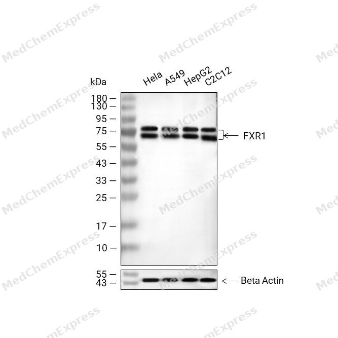 Western blot analysis of extracts from Hela (lane 2, 20μg), A549 (lane 3, 20μg), HepG2 (lane 4, 20μg) and C2C12 (lane 5, 20μg) using FXR1 Antibody (HY-P81004). Proteins were transferred to a PVDF membrane and blocked with 5% non-fat milk in TBST for 2 hours at room temperature. The primary antibody (1/1000) and Loading control antibody (Beta Actin, HY-P80993, 1/10,000) was used in 5% non-fat milk in TBST at 4°C overnight. Goat Anti-Rabbit IgG-HRP Secondary Antibody (HY-P8001, 1/10,000) was used for 1 hour at room temperature.
 -
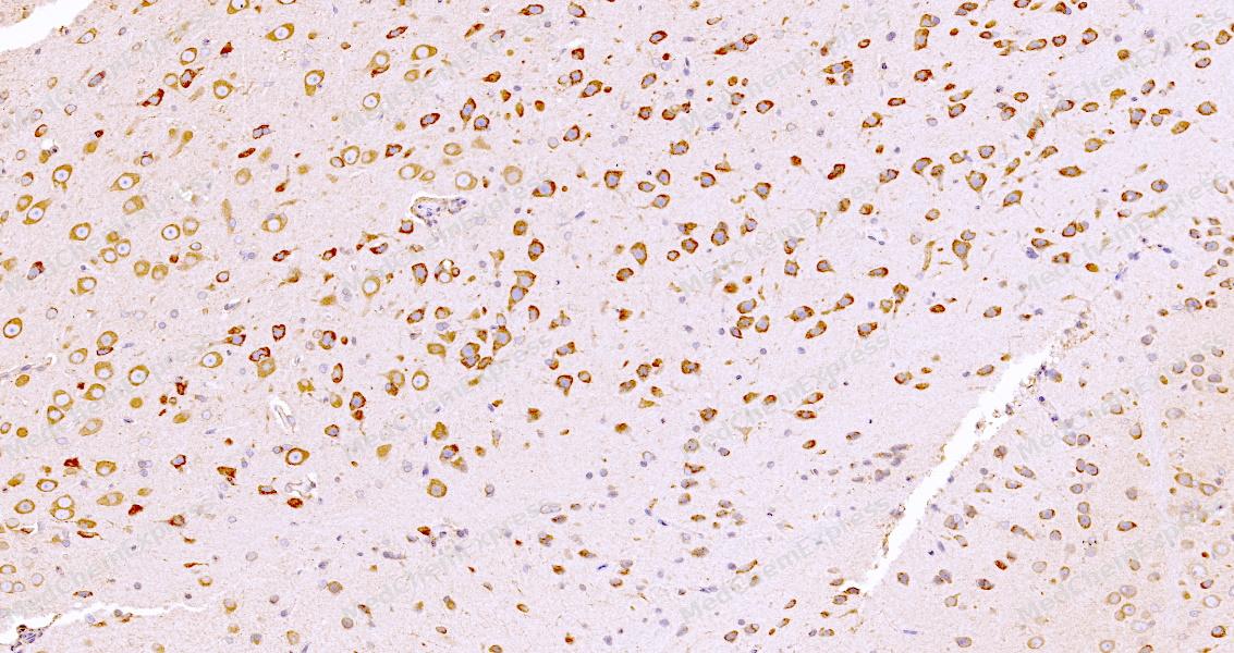 Immunohistochemical analysis of paraffin-embedded rat brain tissue using FXR1 Antibody. The section was pre-treated using heat mediated antigen retrieval with sodium citrate buffer (pH 6.0) for 8 minutes. The tissues were blocked in QuickBlock for 20 minutes at room temperature, washed with ddH2O and PBS, and then probed with the primary antibody at 1/100 dilution in 4℃ overnight. The detection was performed using an HRP conjugated compact polymer system. DAB was used as the chromogen. Tissues were counterstained with hematoxylin and mounted with DPX.
 -
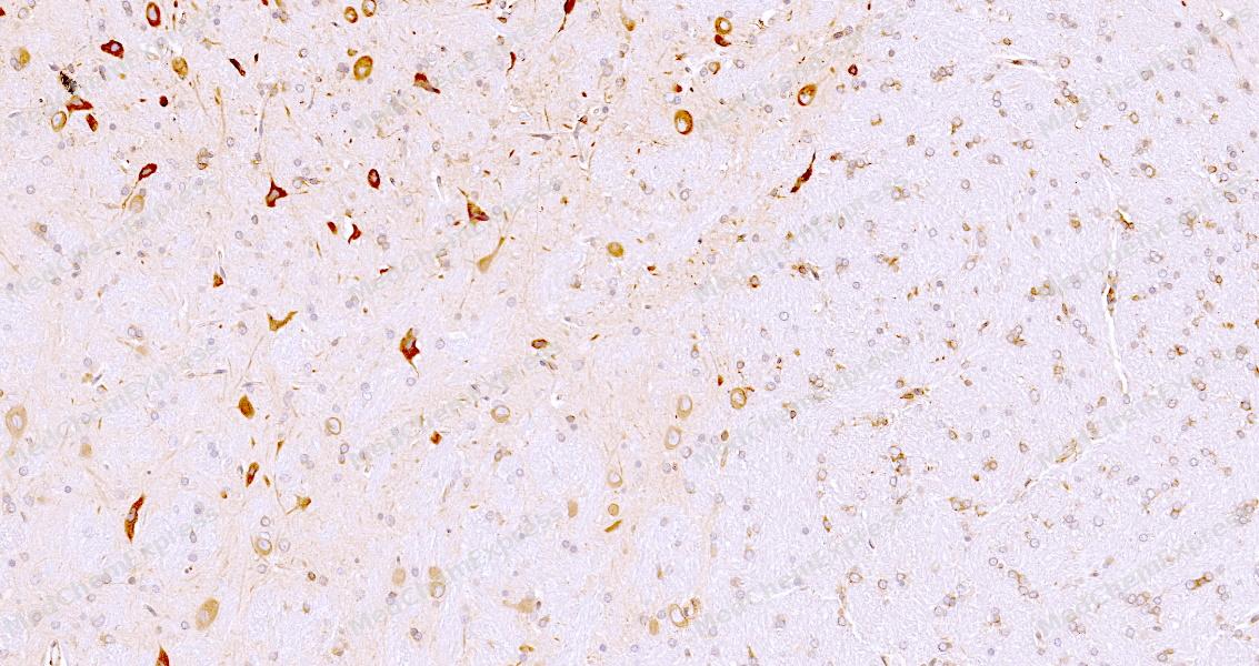 Immunohistochemical analysis of paraffin-embedded rat brain tissue using FXR1 Antibody. The section was pre-treated using heat mediated antigen retrieval with sodium citrate buffer (pH 6.0) for 8 minutes. The tissues were blocked in QuickBlock for 20 minutes at room temperature, washed with ddH2O and PBS, and then probed with the primary antibody at 1/100 dilution in 4℃ overnight. The detection was performed using an HRP conjugated compact polymer system. DAB was used as the chromogen. Tissues were counterstained with hematoxylin and mounted with DPX.
 -
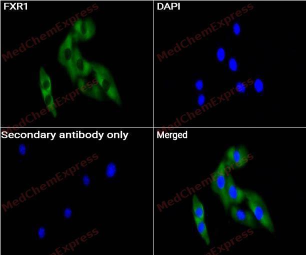 Immunocytochemistry analysis of C2C12 cells labeling FXR1 with FXR1 antibody (HY-P81004) at 1/50 dilution. Cells were fixed in 4% paraformaldehyde for 15 minutes at room temperature, permeabilized with 0.1% Triton X-100 for 10 minutes at room temperature, then blocked with QuickBlock™ Blocking Buffer for Immunol Staining for 10 min at room temperature. Cells were then incubated with FXR1 antibody (HY-P81004) at 1/50 dilution in QuickBlock™ Blocking Buffer for Immunol Staining at 4 ℃. Alexa Fluor® 488-conjugated AffiniPure Goat Anti-Rabbit IgG H&L(HY-P8002, Green) was used as the secondary antibody at 1/1,000 dilution. PBS instead of the primary antibody was used as the secondary antibody only control. The Nuclear counterstain was DAPI (Blue).
 -
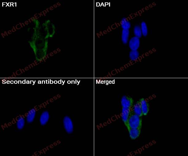 Immunocytochemistry analysis of HeLa cells labeling FXR1 with FXR1 antibody (HY-P81004) at 1/50 dilution. Cells were fixed in 4% paraformaldehyde for 15 minutes at room temperature, permeabilized with 0.1% Triton X-100 for 10 minutes at room temperature, then blocked with QuickBlock™ Blocking Buffer for Immunol Staining for 10 min at room temperature. Cells were then incubated with FXR1 antibody (HY-P81004) at 1/50 dilution in QuickBlock™ Blocking Buffer for Immunol Staining at 4 ℃. Alexa Fluor® 488-conjugated AffiniPure Goat Anti-Rabbit IgG H&L(HY-P8002, Green) was used as the secondary antibody at 1/1,000 dilution. PBS instead of the primary antibody was used as the secondary antibody only control. The Nuclear counterstain was DAPI (Blue).


Background
-
Function
FXR1 is a mRNA-binding protein that acts as a regulator of mRNAs translation and/or stability, and which is required for various processes, such as neurogenesis, muscle development and spermatogenesis. Specifically binds to AU-rich elements (AREs) in the 3'-UTR of target mRNAs. Promotes formation of some phase-separated membraneless compartment by undergoing liquid-liquid phase separation upon binding to AREs-containing mRNAs, leading to assemble mRNAs into cytoplasmic ribonucleoprotein granules that concentrate mRNAs with associated regulatory factors. Required to activate translation of stored mRNAs during late spermatogenesis: acts by undergoing liquid-liquid phase separation to assemble target mRNAs into cytoplasmic ribonucleoprotein granules that recruit translation initiation factor EIF4G3 to activate translation of stored mRNAs in late spermatids. Promotes translation of MYC transcripts by recruiting the eIF4F complex to the translation start site. Acts as a negative regulator of inflammation in response to IL19 by promoting destabilization of pro-inflammatory transcripts. Also acts as an inhibitor of inflammation by binding to TNF mRNA, decreasing TNF protein production. Acts as a negative regulator of AMPA receptor GRIA2/GluA2 synthesis during long-lasting synaptic potentiation of hippocampal neurons by binding to GRIA2/GluA2 mRNA, thereby inhibiting its translation. Regulates proliferation of adult neural stem cells by binding to CDKN1A mRNA and promoting its expression. Acts as a regulator of sleep and synaptic homeostasis by regulating translation of transcripts in neurons. Required for embryonic and postnatal development of muscle tissue by undergoing liquid-liquid phase separation to assemble target mRNAs into cytoplasmic ribonucleoprotein granules. Involved in the nuclear pore complex localization to the nuclear envelope by preventing cytoplasmic aggregation of nucleoporins: acts by preventing ectopic phase separation of nucleoporins in the cytoplasm via a microtubule-dependent mechanism. Plays a role in the stabilization of PKP2 mRNA and therefore protein abundance, via its interaction with PKP3. May also do the same for PKP2, PKP3 and DSP via its interaction with PKP1. Forms a cytoplasmic messenger ribonucleoprotein (mRNP) network by packaging long mRNAs, serving as a scaffold that recruits proteins and signaling molecules. This network facilitates signaling reactions by maintaining proximity between kinases and substrates, crucial for processes like actomyosin reorganization[1][2][3][4][5][6][7][8][9][10].
-
Subcellular Localization
Cytoplasm, Cytoplasmic ribonucleoprotein granule; Cytoplasm, Stress granule; Cytoplasm; Cell projection, dendrite; Cell projection, dendritic spine; Cell projection, axon; Nucleus envelope; Postsynapse
-
Expression
Tissue_specificity:It was expressed in all tissues examined, including the heart, brain, kidneys, and testes (PubMed:7781595, PubMed:9259278) . In the brain, it was found in higher concentrations in neurons, particularly in Purkinje cells at the granular-molecular junction (protein level) (PubMed:9259278) .
Induction:By Interleukin-19 (IL19) -
Isoforms & Post-Translational Modification
P51114 has 3 isomers: P51114-1: 69721 Da (predicted); P51114-2: 60792 Da (predicted); P51114-3: 59920 Da (predicted).
Phosphorylation at Ser-420 by PAK1 promotes its relocalization to stress granules and activity (PubMed:20417602). Phosphorylated by MAPK1/ERK2, promoting subsequent phosphorylation by GSK3B (By similarity). Phosphorylated by GSK3B, promoting ubiquitination and degradation by the proteasome (By similarity);Ubiquitinated by the SCF(FBXO4) complex, leading to its degradation by the proteasome: ubiquitination by the SCF(FBXO4) complex takes place following phosphorylation by GSK3B (PubMed:29142209). Ubiquitinated and degraded in a GSK3B-dependent manner in during both scaling and sleep deprivation (By similarity). Ubiquitinated via 'Lys-63'-linked ubiquitin, leading to its degradation: interaction with CEP63 inhibits 'Lys-63'-linked ubiquitination (PubMed:35989368) -
Subunit
Homodimer (via CC domains); homodiremization is required for FXR1-network nucleation (PubMed:39106863). Interacts with FMR1 (PubMed:11157796, PubMed:7489725, PubMed:8668200). Interacts with FRX2 (PubMed:7489725). Interacts with TDRD3 (PubMed:18664458). Interacts with HABP4 (PubMed:21771594). Interacts with CYFIP2 but not with CYFIP1 (By similarity). Interacts with EIF4G3; promoting translation of target mRNAs (By similarity). Interacts with ELAVL1 (PubMed:30067974). Interacts with CEP63; inhibiting 'Lys-63'-linked ubiquitination (PubMed:35989368). Interacts with PKP3; the interaction facilitates the binding of PKP3 to PKP2 mRNA (PubMed:25225333). Interacts with PKP1; the interaction may facilitate the binding of PKP1 to PKP2, PKP3 and DSP mRNA (PubMed:25225333)
-
SwissProt ID
-
Synonyms
FXR1; FXR1P; hFXR1p
-
Research Field
Immunology
Documentation
-
Data Sheet (234 KB)
-
SDS (251 KB)
- English - EN (251 KB)
- Français - FR (251 KB)
- Deutsch - DE (251 KB)
- Norwegian - NO (251 KB)
- Español - ES (251 KB)
- Swedish - SV (251 KB)
- Italian - IT (251 KB)
- Korean - KR (251 KB)
- Portuguese - PT (251 KB)
-
User Guide for Antibodies (1077 KB)

 Powered by Bioz
