



CD11a Antibody (YA2398)
(Synonyms: ITGAL; CD11A; Integrin alpha-L; CD11 antigen-like family member A; Leukocyte adhesion glycoprotein LFA-1 alpha chain; LFA-1A; Leukocyte function-associated molecule 1 alpha chain; CD11a)
Based on 1 Customer Validation
CD11a Antibody (YA2398) is a Rabbit-derived and non-conjugated IgG monoclonal antibody, targeting to CD11a.
-
Host:
Rabbit
-
Isotype:
IgG
-
Application:
WB, IHC-F, IHC-P, ICC/IF, FC, IP
-
Reactivity :
Human
-
Formulation:
Supplied in 50mM Tris-Glycine(pH 7.4), 0.15M NaCl, 40% Glycerol, 0.01% Sodium azide and 0.05% BSA
-
Conjugation:
Non-conjugated


To place orders, for customer services and technical support, please contact: MedChemExpress USA
Tel: 609-228-6898 E-mail: [email protected] [email protected]





Applications
| Application |
WB

WB: Western Blot
|
IHC-P
IHC-P: Immunohistochemistry-Paraffin
|
ICC/IF
ICC/IF: Immunocytochemistry/
Immunofluorescence |
FC
FC: Flow Cytometry
|
IP
IP: Immunoprecipitation
|
|---|---|---|---|---|---|
| Dilution Ratio | 1:1000-1:5000 | 1:200-1:1000 | 1:50-1:100 | 1:20-1:50 | 1:20-1:50 |


Product Details
CD11a Antibody (YA2398) is a Rabbit-derived and non-conjugated IgG monoclonal antibody, targeting to CD11a.
-
Host Rabbit

-
Clonality Recombinant,Monoclonal

-
Species ReactivityHuman
-
Observed Molecular WeightObserved band size: 150 kDa
Note: Due to possible protein modifications or aggregation, the molecular weight should be confirmed by actual measurement, and the predicted value is for reference only.

-
Calculated Molecular Weight Predicted band size: 129 kDa

A synthetic peptide of human CD11a
Endogenous
Affinity Purified
Non-conjugated
Unmodified
IgG


Product Properties
-
Appearance
Solution
-
Formulation
Supplied in 50mM Tris-Glycine(pH 7.4), 0.15M NaCl, 40% Glycerol, 0.01% Sodium azide and 0.05% BSA
-
Concentration
Batch-dependent, Please check the COA for the concentration of each lot. Check Lot Concentration
-
Storage & Stability
Stored at -20°C for 1 year. Avoid repeated freeze / thaw cycles.
-
Shipping
Shipping with blue ice.


Verification Images
-
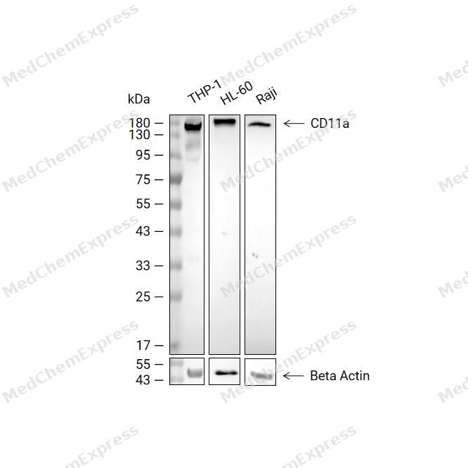 Western blot analysis of extracts from THP-1 (lane 2, 20ug), HL-60 (lane 3, 20ug) and Raji (lane 4, 20ug) using CD11a Antibody (HY-P82653). Proteins were transferred to a PVDF membrane and blocked with 5% non-fat milk in TBST at 4°C overnight. The primary antibody (1/2000) and Loading control antibody (Beta Actin, HY-P80993, 1/10,000) was used in 5% non-fat milk in TBST for 2 hour at room temperature. Goat Anti-Rabbit IgG-HRP Secondary Antibody (HY-P8001, 1/10,000) was used for 1 hour at room temperature.
 -
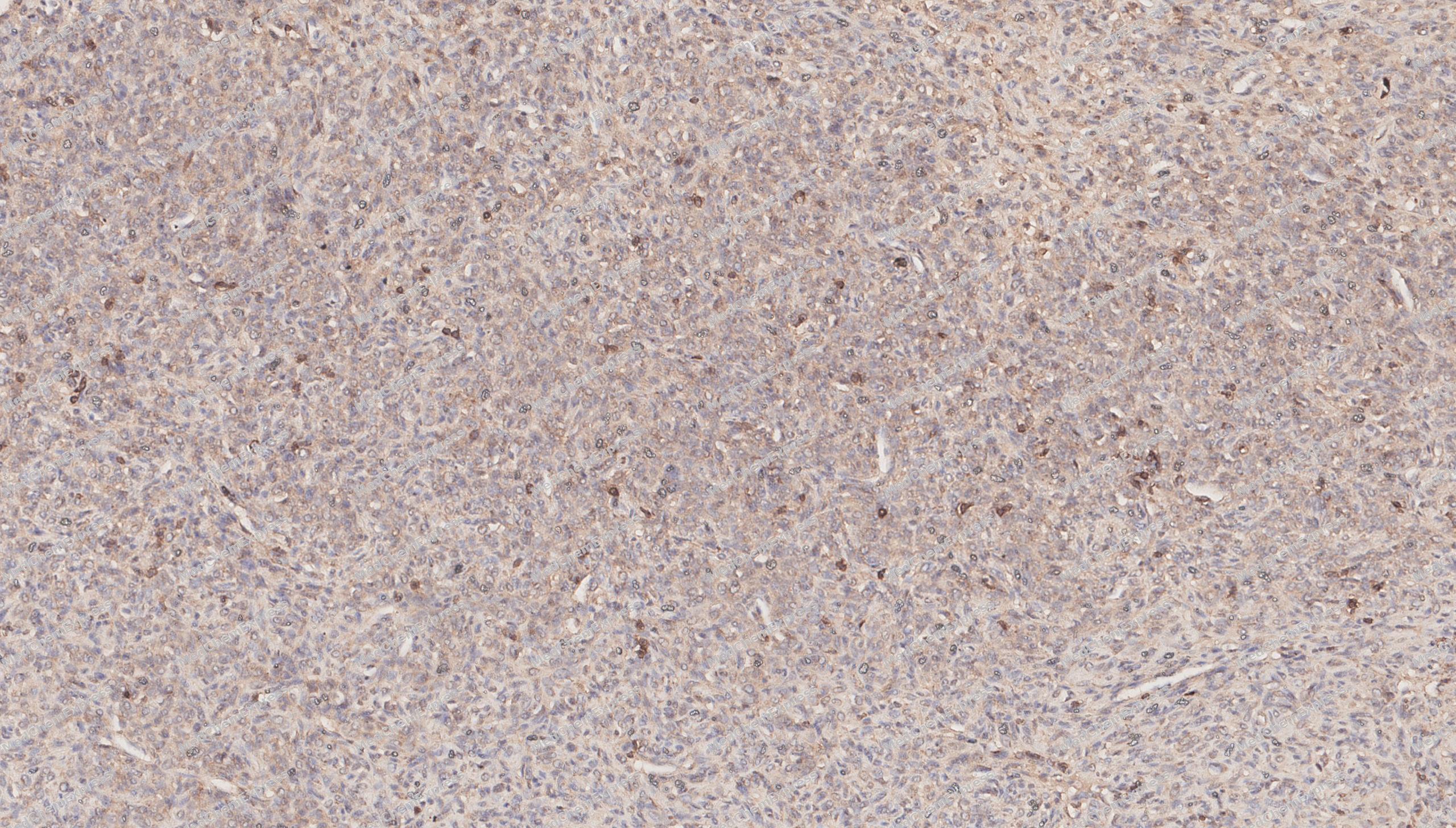 Immunohistochemical analysis of paraffin-embedded human solitary fibrous tumor tissue using CD11a antibody was performed. The section was pretreated using high-temperature mediated EDTA antigen retrieval buffer (pH 9.0), for 20 minutes. The tissues were incubated with primary antibody (HY-P82653, 1:100 dilution) at room temperature for 20 minutes. Detection was performed using an HRP conjugated compact polymer system. DAB was used as the chromogen. The tissues were counterstained with hematoxylin and mounted with neutral balsam mounting medium.
 -
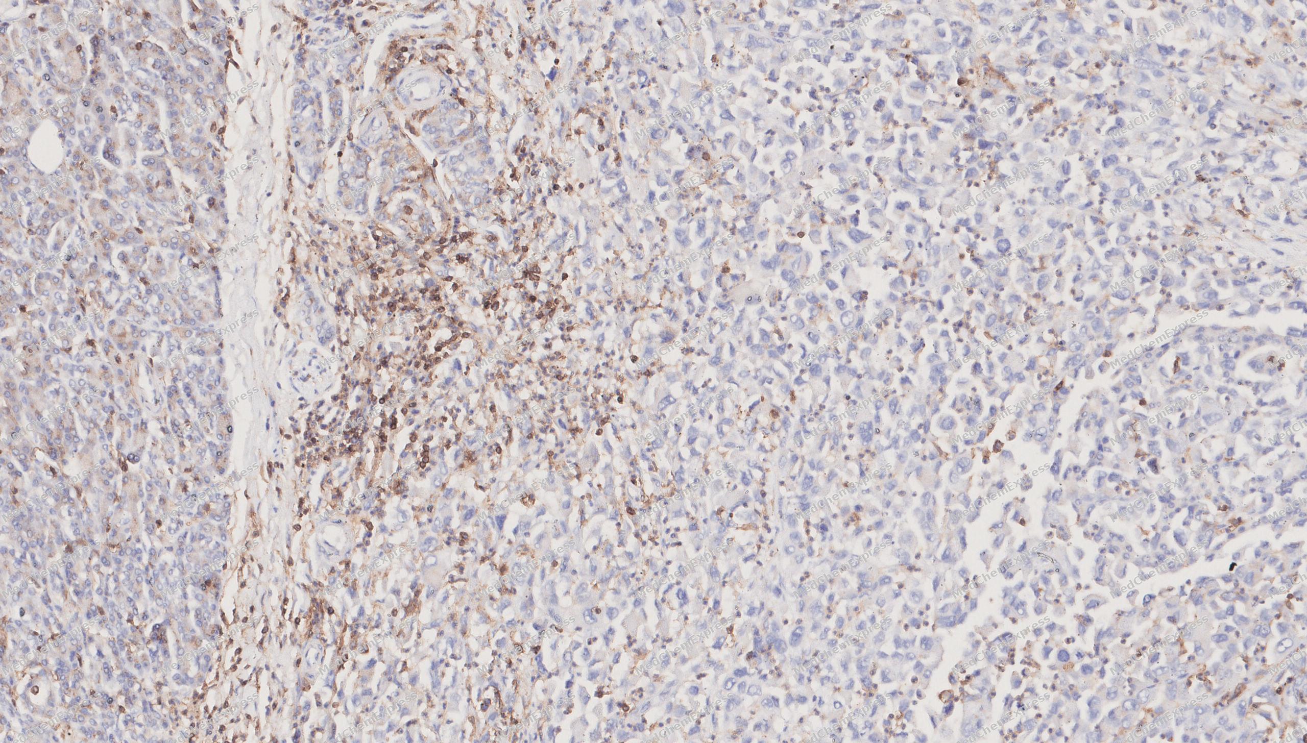 Immunohistochemical analysis of paraffin-embedded human Pancreatic cancer tissue using CD11a antibody was performed. The section was pretreated using high-temperature mediated EDTA antigen retrieval buffer (pH 9.0), for 20 minutes. The tissues were incubated with primary antibody (HY-P82653, 1:100 dilution) at room temperature for 20 minutes. Detection was performed using an HRP conjugated compact polymer system. DAB was used as the chromogen. The tissues were counterstained with hematoxylin and mounted with neutral balsam mounting medium.
 -
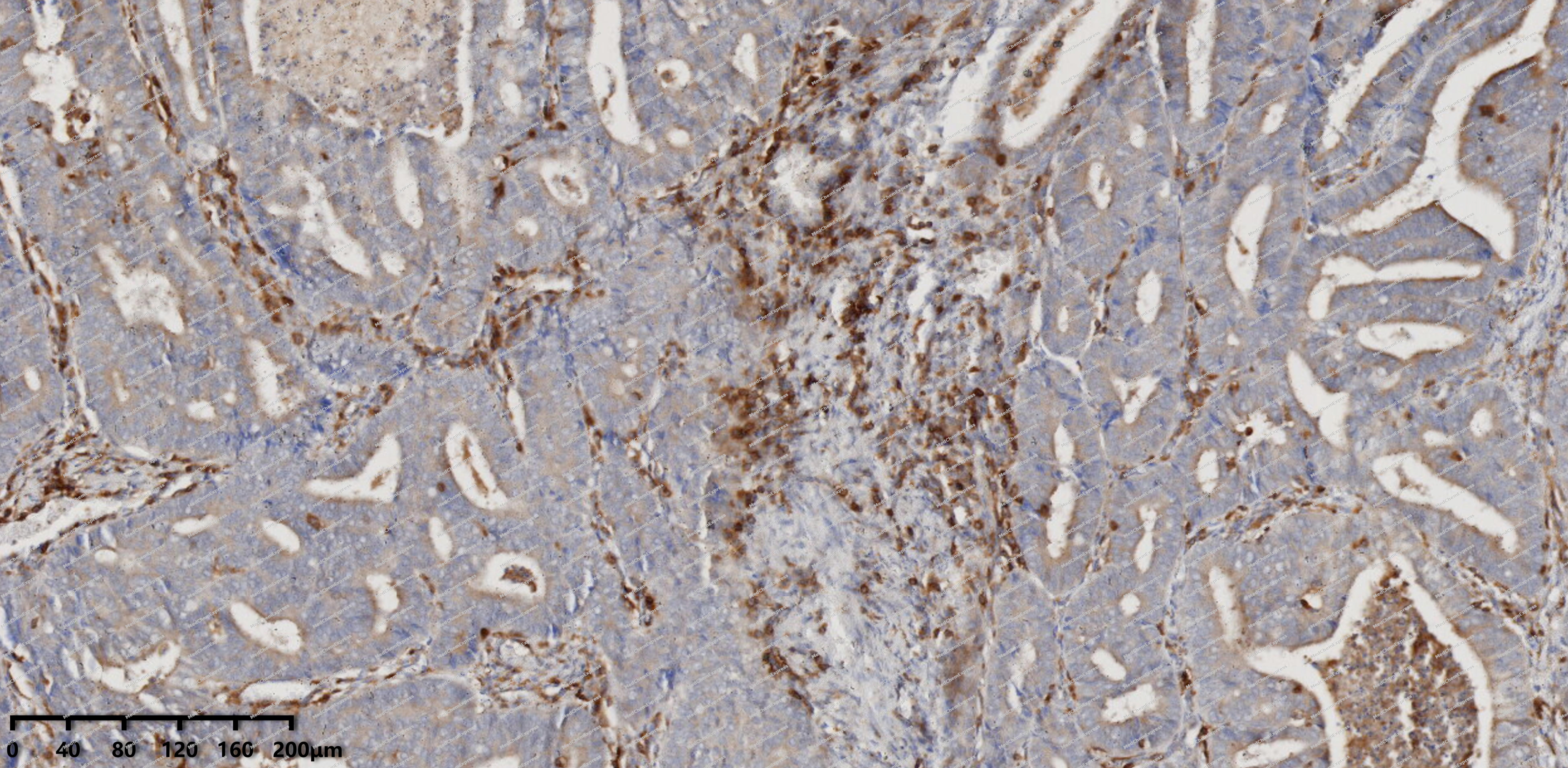 Immunohistochemical analysis of paraffin-embedded human Endometrial carcinoma tissue using CD11a antibody was performed. The section was pretreated using high-temperature mediated EDTA antigen retrieval buffer (pH 9.0), for 20 minutes. The tissues were incubated with primary antibody (HY-P82653, 1:100 dilution) at room temperature for 20 minutes. Detection was performed using an HRP conjugated compact polymer system. DAB was used as the chromogen. The tissues were counterstained with hematoxylin and mounted with neutral balsam mounting medium.
 -
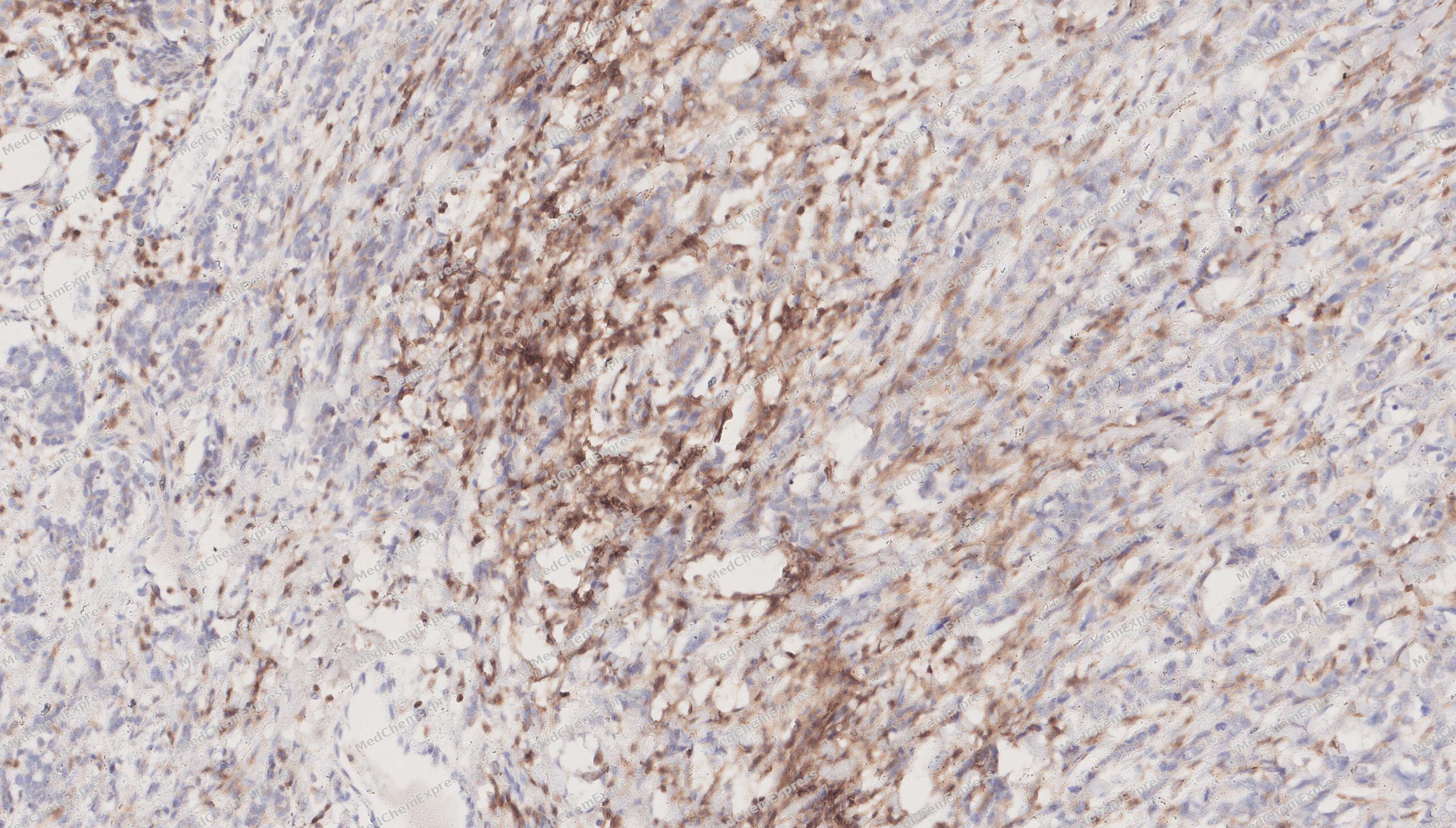 Immunohistochemical analysis of paraffin-embedded human breast cancer tissue using CD11a antibody was performed. The section was pretreated using high-temperature mediated EDTA antigen retrieval buffer (pH 9.0), for 20 minutes. The tissues were incubated with primary antibody (HY-P82653, 1:100 dilution) at room temperature for 20 minutes. Detection was performed using an HRP conjugated compact polymer system. DAB was used as the chromogen. The tissues were counterstained with hematoxylin and mounted with neutral balsam mounting medium.
 -
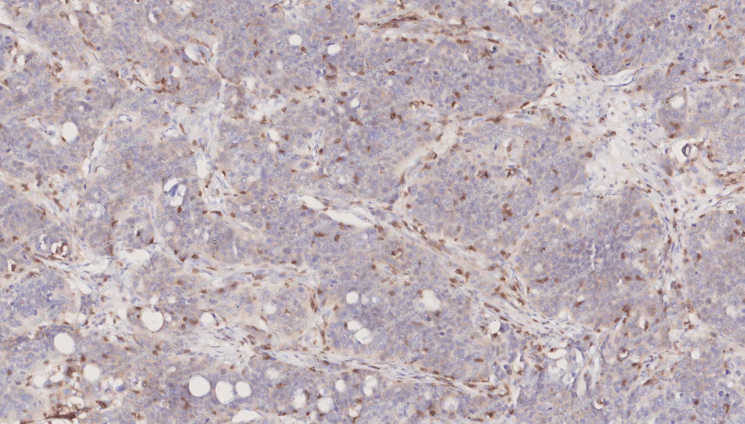 Immunohistochemical analysis of paraffin-embedded human gastric cancer tissue using CD11a antibody was performed. The section was pretreated using high-temperature mediated EDTA antigen retrieval buffer (pH 9.0), for 20 minutes. The tissues were incubated with primary antibody (HY-P82653, 1:100 dilution) at room temperature for 20 minutes. Detection was performed using an HRP conjugated compact polymer system. DAB was used as the chromogen. The tissues were counterstained with hematoxylin and mounted with neutral balsam mounting medium.
 -
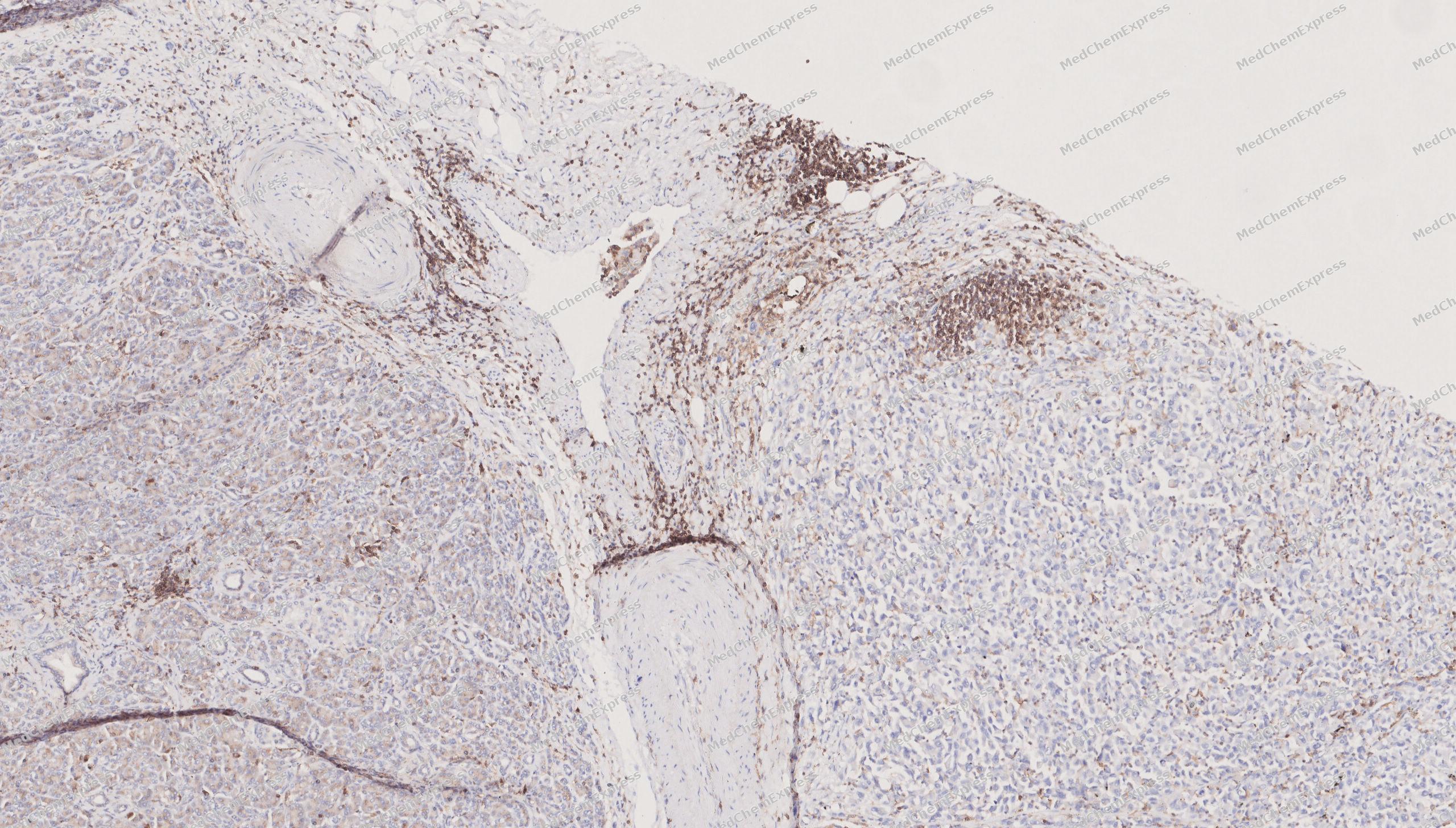 Immunohistochemical analysis of paraffin-embedded human Pancreatic cancer tissue using CD11a antibody was performed. The section was pretreated using high-temperature mediated EDTA antigen retrieval buffer (pH 9.0), for 20 minutes. The tissues were incubated with primary antibody (HY-P82653, 1:100 dilution) at room temperature for 20 minutes. Detection was performed using an HRP conjugated compact polymer system. DAB was used as the chromogen. The tissues were counterstained with hematoxylin and mounted with neutral balsam mounting medium.


Background
-
Function
CD11a is an Integrin ITGAL/ITGB2 is a receptor for ICAM1, ICAM2, ICAM3 and ICAM4. Integrin ITGAL/ITGB2 is a receptor for F11R. Integrin ITGAL/ITGB2 is a receptor for the secreted form of ubiquitin-like protein ISG15; the interaction is mediated by ITGAL. Involved in a variety of immune phenomena including leukocyte-endothelial cell interaction, cytotoxic T-cell mediated killing, and antibody dependent killing by granulocytes and monocytes. Contributes to natural killer cell cytotoxicity. Involved in leukocyte adhesion and transmigration of leukocytes including T-cells and neutrophils. Acts as a platform at the immunological synapse to translate TCR engagement and density of the ITGAL ligand ICAM1 into graded adhesion. Required for generation of common lymphoid progenitor cells in bone marrow, indicating a role in lymphopoiesis. Integrin ITGAL/ITGB2 in association with ICAM3, contributes to apoptotic neutrophil phagocytosis by macrophages[1][2][3][4][5][6].
-
Subcellular Localization
Cell membrane; Single-pass type I membrane protein
-
Expression
Tissue_specificity:leukocyte -
Isoforms & Post-Translational Modification
P20701 has 3 isomers: P20701-1: 128770 Da (predicted); P20701-2: 134427 Da (predicted); P20701-3: 119224 Da (predicted).
In resting T-cells, up to 40% of surface ITGAL is constitutively phosphorylated. Phosphorylation causes conformational changes needed for ligand binding and is necessary for activation by some physiological agents -
Subunit
Heterodimer of an alpha and a beta subunit (PubMed:12526797). The ITGAL alpha subunit associates with the ITGB2 beta subunit (PubMed:12526797). Interacts with THBD (PubMed:27055590). Interacts with CD226 (PubMed:15684041, PubMed:38195629)
-
SwissProt ID
-
Synonyms
ITGAL; CD11A; Integrin alpha-L; CD11 antigen-like family member A; Leukocyte adhesion glycoprotein LFA-1 alpha chain; LFA-1A; Leukocyte function-associated molecule 1 alpha chain; CD11a
-
Research Field
Tags & Cell Markers


Documentation
-
Data Sheet (261 KB)
-
SDS (252 KB)
- English - EN (252 KB)
- Français - FR (252 KB)
- Deutsch - DE (252 KB)
- Norwegian - NO (252 KB)
- Español - ES (252 KB)
- Swedish - SV (252 KB)
- Italian - IT (252 KB)
- Korean - KR (252 KB)
- Portuguese - PT (252 KB)
-
User Guide for Antibodies (1077 KB)
[1]. Ostermann G, et al. JAM-1 is a ligand of the beta(2) integrin LFA-1 involved in transendothelial migration of leukocytes. Nat Immunol. 2002 Feb;3(2):151-8. [Content Brief]
[2]. Fraemohs L, et al. The functional interaction of the beta 2 integrin lymphocyte function-associated antigen-1 with junctional adhesion molecule-A is mediated by the I domain. J Immunol. 2004 Nov 15;173(10):6259-64. [Content Brief]
[3]. Swaim CD, et al. Extracellular ISG15 Signals Cytokine Secretion through the LFA-1 Integrin Receptor. Mol Cell. 2017 Nov 2;68(3):581-590.e5. [Content Brief]
[4]. Barber DF, et al. LFA-1 contributes an early signal for NK cell cytotoxicity. J Immunol. 2004 Sep 15;173(6):3653-9. [Content Brief]
[5]. Lacouture C, et al. LFA-1 nanoclusters integrate TCR stimulation strength to tune T-cell cytotoxic activity. Nat Commun. 2024 Jan 9;15(1):407. [Content Brief]
[6]. Kristóf E, et al. Novel role of ICAM3 and LFA-1 in the clearance of apoptotic neutrophils by human macrophages. Apoptosis. 2013 Oct;18(10):1235-51. [Content Brief]

 Powered by Bioz
