



Damnacanthal

Based on 1 Customer Validation
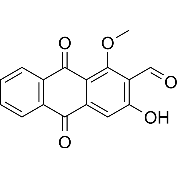
Damnacanthal is an anthraquinone isolated from the root of Morinda citrifolia. Damnacanthal is a highly potent, selective inhibitor of p56lck tyrosine kinase activity. Natural Damnacanthal inhibits p56 lck autophosphorylation and phosphorylation of exogenous substrates with IC50s of 46 nM and 220 nM, respectively. Damnacanthal is a potent inducer of apoptosis with anticancer activity. Damnacanthal also has antinociceptive, anti-inflammatory effects in mice and anti-fungal activity against Candida albicans.
For research use only. We do not sell to patients.
- Purity : 99.40%
- CAS No.: 477-84-9
- Formula: C16H10O5
- Molecular Weight:282.25
-
Storage:
4°C, protect from light
* In solvent : -80°C, 6 months; -20°C, 1 month (protect from light)

To place orders, for customer services and technical support, please contact: MedChemExpress USA
Tel: 609-228-6898 E-mail: [email protected] [email protected]


-
 Biological Activity
-
Chemical Information
-
Solvent & Solubility
- Protocols
- Purity & Documentation
- References
-
 Help & FAQs


-
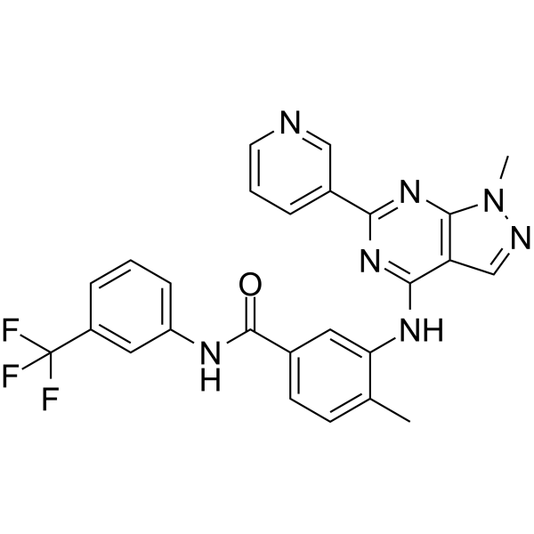
HY-13258A
NVP-BHG712EphB4 Inhibitor -
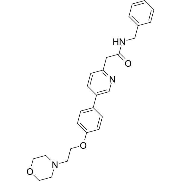
HY-10340
TirbanibulinSrc Inhibitor -
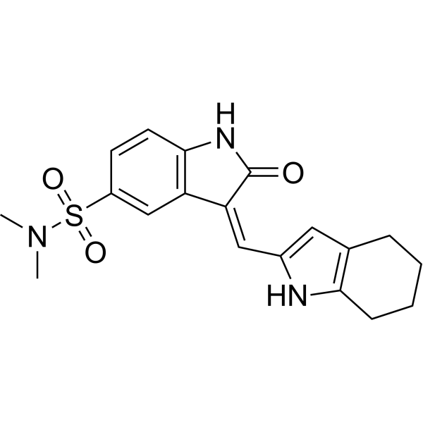
HY-B0789
SU6656Src Inhibitor -
HY-13804
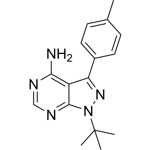
PP1Src Inhibitor -
HY-13785
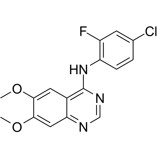
ZM 306416VEGFR Inhibitor -
HY-10181A
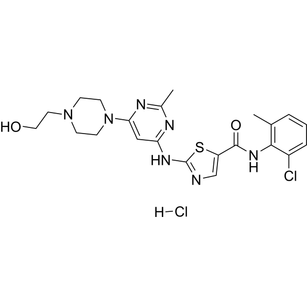
Dasatinib hydrochlorideSrc/Bcr-Abl Inhibitor -
HY-10340A
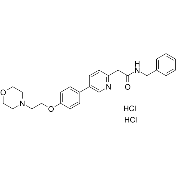
Tirbanibulin dihydrochloride -
HY-12005
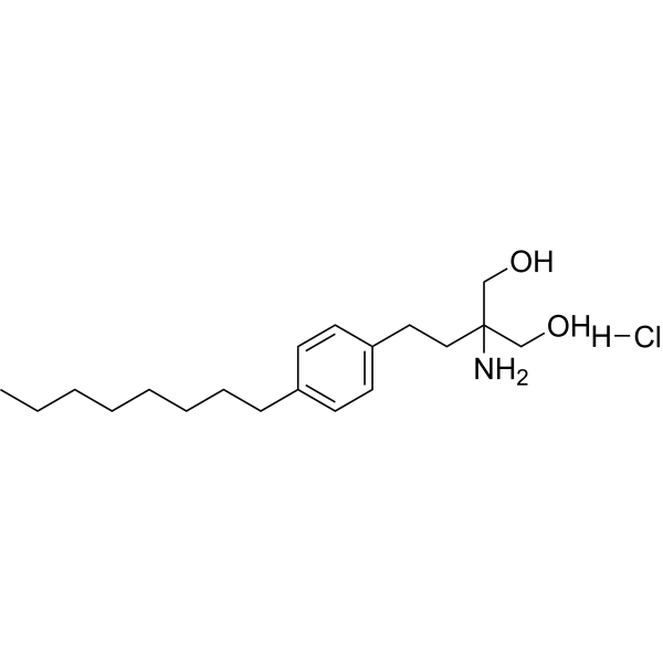
Fingolimod hydrochlorideS1P Receptors Modulator -
HY-13805
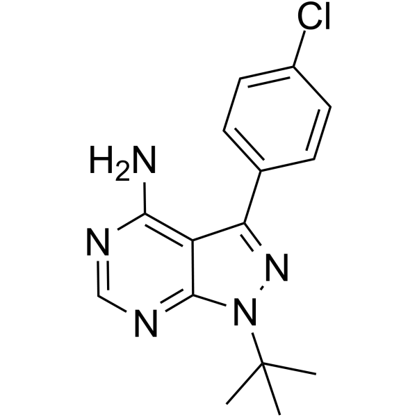
PP2Src Inhibitor -
HY-101053
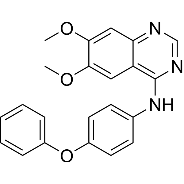
Src Inhibitor 1Src Inhibitor -
HY-12299
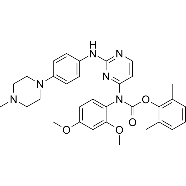
WH-4-023Src Inhibitor -
HY-59047
TolimidoneSrc Activator


Biological Activity
Description
IC50 & Target
IC50: 46 nM (p56 lck autophosphorylation) and 220 nM (phosphorylation of exogenous substrates by p56 lck)[1];
Apoptosis[2];
Candida albicans[2]
Cellular Effect
|
Cell Line

|
Type | Value | Description | References |
|---|---|---|---|---|
| DU-145 | IC50 |
26 μM
Compound: 26, damnacanthal
|
Cytotoxicity against Homo sapiens (human) DU145 cells assessed as inhibition of cell survival after 96 hr by MTT assay
Cytotoxicity against Homo sapiens (human) DU145 cells assessed as inhibition of cell survival after 96 hr by MTT assay
|
10.1007/s00044-012-0197-5 |
| Jurkat | IC50 |
17 nM
Compound: Damnacanthal
|
Inhibition of p56 Lck tyrosine kinase in Jurkat cells where p56lck autophosphorylation is inhibited.
Inhibition of p56 Lck tyrosine kinase in Jurkat cells where p56lck autophosphorylation is inhibited.
|
10.1016/S0960-894X(97)00034-6 |
| MCF7 | IC50 |
11 μM
Compound: 26, damnacanthal
|
Cytotoxicity against Homo sapiens (human) MCF7 cells assessed as inhibition of cell survival after 96 hr by MTT assay
Cytotoxicity against Homo sapiens (human) MCF7 cells assessed as inhibition of cell survival after 96 hr by MTT assay
|
10.1007/s00044-012-0197-5 |
| NCI-H460 | IC50 |
25 μM
Compound: 26, damnacanthal
|
Cytotoxicity against Homo sapiens (human) H460 cells assessed as inhibition of cell survival after 96 hr by MTT assay
Cytotoxicity against Homo sapiens (human) H460 cells assessed as inhibition of cell survival after 96 hr by MTT assay
|
10.1007/s00044-012-0197-5 |
| THP-1 | CC50 |
31.47 μM
Compound: 5
|
Cytotoxicity against human THP1 cells assessed as cell viability after 72 hrs by MTT assay
Cytotoxicity against human THP1 cells assessed as cell viability after 72 hrs by MTT assay
|
[PMID: 26906638] |
| THP-1 | IC50 |
39.51 μM
Compound: 5
|
Antiprotozoan activity against intracellular amastigote stage of Trypanosoma cruzi Tulahuen infected in human THP1 cells assessed as reduction in parasite viability after 48 hrs by microplate based assay
Antiprotozoan activity against intracellular amastigote stage of Trypanosoma cruzi Tulahuen infected in human THP1 cells assessed as reduction in parasite viability after 48 hrs by microplate based assay
|
[PMID: 26906638] |
In Vitro
Damnacanthal has > 100-fold selectivity for p56lck over the serine/threonine kinases, protein kinase A and protein kinase C, and > 40-fold selectivity for p56lck over four receptor tyrosine kinases. Damnacanthal also demonstrates modest (7-20-fold), but highly statistically significant, selectivity for p56lck over the homologous enzymes p60src and p59fyn[1].
Damnacanthal (0.1-100 μM; 1-4 days; HCT-116 and SW480 cells) treatment results in a significant reduction of cell proliferation in a concentration- and time-dependent manner[2].
Damnacanthal (1-50 μM; 72 hours; HCT-116 cells) treatment results in a significant enrichment in the number of cells in the S/G1 and G2/G1 phases at concentration of 50 μM[2].
Damnacanthal (10 μM; 24 hours; HCT-116 cells) treatment significantly increases caspase 3/7 activity. Damnacanthal-induced apoptosis[2].
Damnacanthal (0.1-10 μM; 24 hours; HCT-116 cells) treatment induces NAG-1 expression in HCT-116 cells. Cyclin D1 expression is reduced at 10 μM of Damnacanthal, whereas p21 and p53 does not alter their expression. PARP cleavage is seen at 10 μM Damnacanthal treatment only in HCT-116 cells, where NAG-1 is induced[2].
Damnacanthal treatment for 2 weeks shows significant decreasing colony number in HCT-116 cells in a concentration-dependent manner. Damnacanthal-treated cells show a dramatic inhibition of clonogenic capacity. Damnacanthal-treated (1-50 μM; 48 hours) cells significantly inhibits the migration of HCT-116 cells in a concentration-dependent manner[2].
MedChemExpress (MCE) has not independently confirmed the accuracy of these methods. They are for reference only.
-
Cell Line:HCT-116 and SW480 cells
-
Concentration:0.1 μM, 1 μM, 10 μM, 100 μM
-
Incubation Time:1, 2, and 4 days
-
Result:Resulted in a significant reduction of cell proliferation in a concentration- and time-dependent manner.
-
Cell Line:HCT-116 cells
-
Concentration:1 μM, 10 μM and 50 μM
-
Incubation Time:72 hours
-
Result:Resulted in a significant enrichment in the number of cells in the S/G1 and G2/G1 phases at concentration of 50 μM.
-
Cell Line:HCT-116 cells
-
Concentration:10 μM
-
Incubation Time:24 hours
-
Result:Significantly increased caspase 3/7 activity.
-
Cell Line:HCT-116 cells
-
Concentration:0.1 μM, 1 μM and 10 μM
-
Incubation Time:24 hours
-
Result:NAG-1 was induced in HCT-116 cells in a dose- and time-dependent manner. Cyclin D1 expression was reduced at 10 μM.
In Vivo
MedChemExpress (MCE) has not independently confirmed the accuracy of these methods. They are for reference only.
-
Animal Model:Male ddY mice (5-6 weeks) injected with formalin or Histamine[4]
-
Dosage:10 mg/kg, 30 mg/kg and 100 mg/kg
-
Administration:Oral administration; for 10 minutes, 30 minutes, 60 minutes or 300 minutes
-
Result:Significantly reduced the growth of human lung tumor without acute toxicity.
- Kinase activity and phosphorylation assays
- Western Blot
- Detection of Apoptosis
- TUNEL staining for apoptotic DNA fragmentation
- Annexin V plus membrane-impermeant dye apoptosis staining
- Apoptosis Solutions
- Protocol for Kinase activity and phosphorylation assays
- Research Protocol for Inflammation-related Diseases


Chemical Information
-
CAS No. 477-84-9
-
Appearance Solid
-
Molecular Weight 282.25
-
Formula C16H10O5
-
Color Light yellow to yellow
-
SMILES
O=CC(C(OC)=C1C2=O)=C(O)C=C1C(C3=C2C=CC=C3)=O
-
Structure Classification
-
Initial Source
-
Shipping
Room temperature in continental US; may vary elsewhere.
-
Storage
4°C, protect from light
* In solvent : -80°C, 6 months; -20°C, 1 month (protect from light)


Solvent & Solubility
In Vitro:
DMSO : 5 mg/mL (17.71 mM; ultrasonic and warming and heat to 60°C; Hygroscopic DMSO has a significant impact on the solubility of product, please use newly opened DMSO)
Please refer to the solubility information to select the appropriate solvent. Once prepared, please aliquot and store the solution to prevent product inactivation from repeated freeze-thaw cycles.
Storage method and period of stock solution: -80°C, 6 months; -20°C, 1 month (protect from light). When stored at -80°C, please use it within 6 months. When stored at -20°C, please use it within 1 month.
Please refer to the solubility information to select the appropriate solvent. Once prepared, please aliquot and store the solution to prevent product inactivation from repeated freeze-thaw cycles.
Storage method and period of stock solution: -80°C, 6 months; -20°C, 1 month (protect from light). When stored at -80°C, please use it within 6 months. When stored at -20°C, please use it within 1 month.
Concentration (start) × Volume (start) = Concentration (final) × Volume (final)
In Vivo:
For the following dissolution methods, please prepare the working solution directly:
It is recommended to prepare fresh solutions and use them promptly within a short period of time.
The percentages shown for the solvents indicate their volumetric ratio in the final prepared solution. If precipitation or phase separation occurs during preparation, heat and/or sonication can be used to aid dissolution.


Purity & Documentation
-
Data Sheet (293 KB)
-
SDS (392 KB)
- English - EN (392 KB)
- Français - FR (392 KB)
- Deutsch - DE (392 KB)
- Norwegian - NO (392 KB)
- Español - ES (392 KB)
- Swedish - SV (392 KB)
- Italian - IT (392 KB)
- Korean - KR (392 KB)
- Portuguese - PT (392 KB)
-
Handling Instructions (2659 KB)
References
[1]. Faltynek CR, et al. Damnacanthal is a highly potent, selective inhibitor of p56lck tyrosine kinase activity. Biochemistry. 1995 Sep 26;34(38):12404-10. [Content Brief]
[2]. Nualsanit T, et al. Damnacanthal, a noni component, exhibits antitumorigenic activity in human colorectal cancer cells. J Nutr Biochem. 2012 Aug;23(8):915-23. [Content Brief]
[3]. Aziz MY, et al. Damnacanthal is a potent inducer of apoptosis with anticancer activity by stimulating p53 and p21 genes in MCF-7 breast cancer cells. Oncol Lett. 2014 May;7(5):1479-1484. [Content Brief]
[4]. Okusada K, et al. The antinociceptive and anti-inflammatory action of the CHCl3-soluble phase and its main active component, damnacanthal, isolated from the root of Morinda citrifolia. Biol Pharm Bull. 2011;34(1):103-7. [Content Brief]


Complete Stock Solution Preparation Table
Please refer to the solubility information to select the appropriate solvent. Once prepared, please aliquot and store the solution to prevent product inactivation from repeated freeze-thaw cycles.
Storage method and period of stock solution: -80°C, 6 months; -20°C, 1 month (protect from light). When stored at -80°C, please use it within 6 months. When stored at -20°C, please use it within 1 month.
| Optional Solvent | Concentration Solvent Mass | 1 mg | 5 mg | 10 mg | 25 mg |
|---|---|---|---|---|---|
| DMSO | 1 mM | 3.5430 mL | 17.7148 mL | 35.4296 mL | 88.5740 mL |
| 5 mM | 0.7086 mL | 3.5430 mL | 7.0859 mL | 17.7148 mL | |
| 10 mM | 0.3543 mL | 1.7715 mL | 3.5430 mL | 8.8574 mL | |
| 15 mM | 0.2362 mL | 1.1810 mL | 2.3620 mL | 5.9049 mL |


Damnacanthal Related Classifications
HY-108485 Related Classifications
 Powered by Bioz