



Emavusertib
Based on 8 publication(s) in Google Scholar
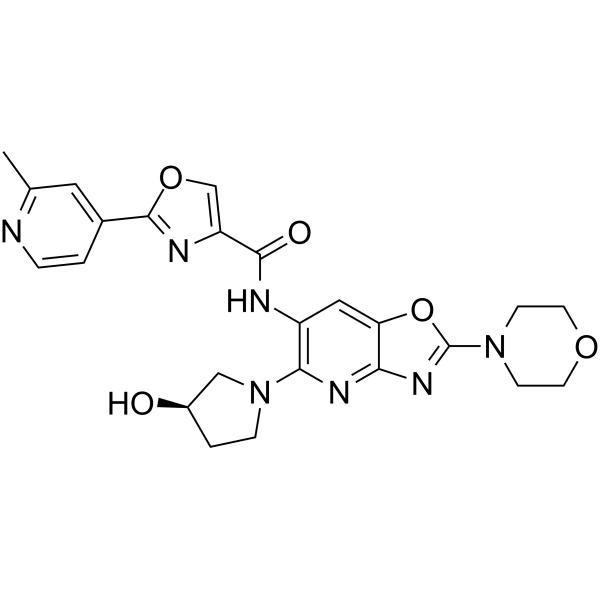
Emavusertib is an orally active inhibitor for IRAK4 (IC50=57 nM) and FLT3. Emavusertib inhibits NF-κB and MyD88 signaling pathways, reduces the generation of pro-inflammatory cytokines like IL-6 and IL-10, thereby exhibiting anti-inflammatory and anti-proliferative activities against cancer cells, leading to cell apoptosis. Emavusertib exhibits antitumor activity in mouse model
Nos produits utilisent uniquement pour la recherche. Nous ne vendons pas aux patients.
- Pureté : 99.97%
- CAS No.: 1801344-14-8
- Formule: C24H25N7O5
- Masse moléculaire:491.50
-
Stockage:Powder -20°C, 3 years , 4°C, 2 years ; In solvent -80°C, 1 year , -20°C, 6 months

To place orders, for customer services and technical support, please contact: MedChemExpress USA
Tel: 609-228-6898 E-mail: [email protected] [email protected]


-
 Activité biologique
-
Chemical Information
-
Solvant et solubilité
- Protocole
- Pureté et documentation
- Références
-
 Help & FAQs


-
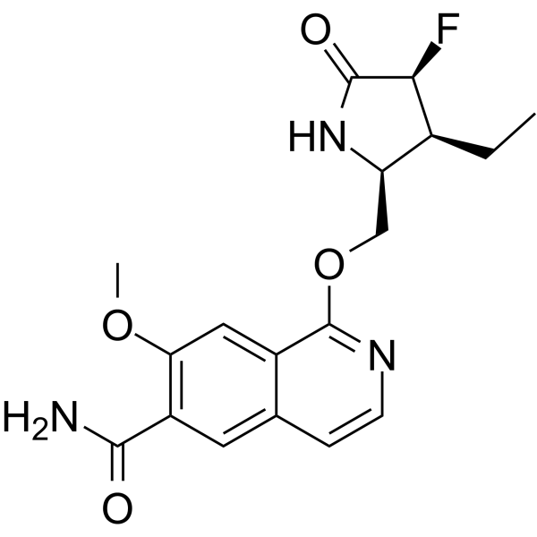
HY-19836
ZimlovisertibIRAK4 Inhibitor -
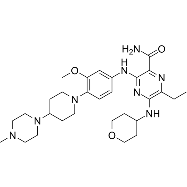
HY-12432
GilteritinibFLT3/AXL Inhibitor -
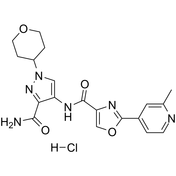
HY-18992
AS2444697IRAK-4 Inhibitor -
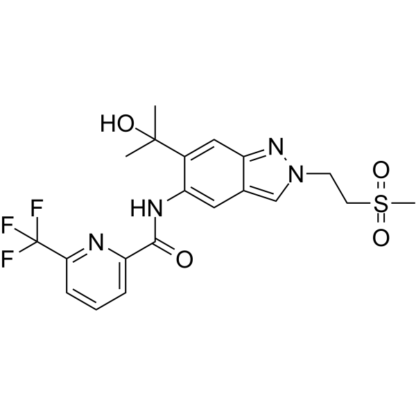
HY-139374
ZabedosertibIRAK4 Inhibitor -
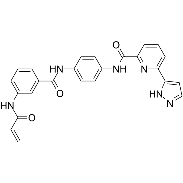
HY-103017A
JH-X-119-01IRAK1 Inhibitor -
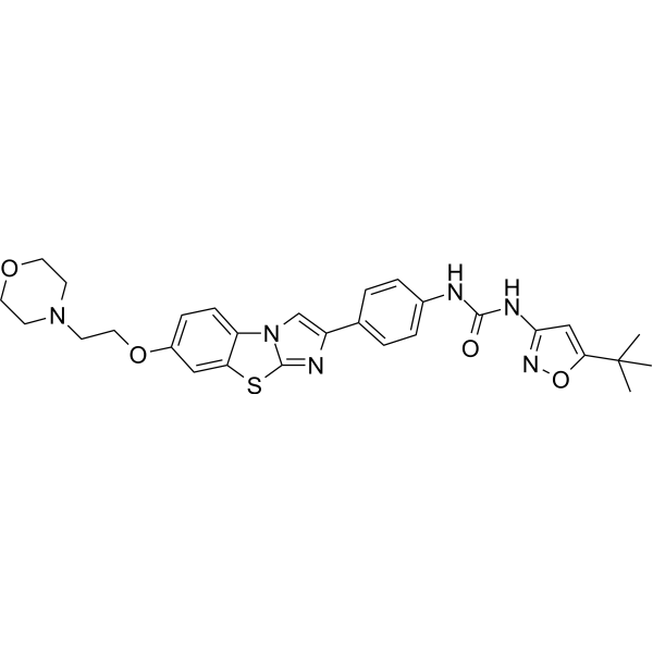
HY-13001
QuizartinibFLT3 Inhibitor -
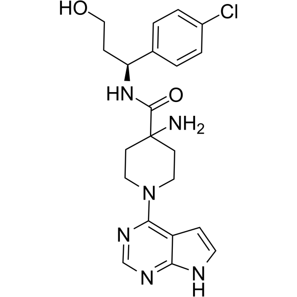
HY-15431
CapivasertibAkt Inhibitor -
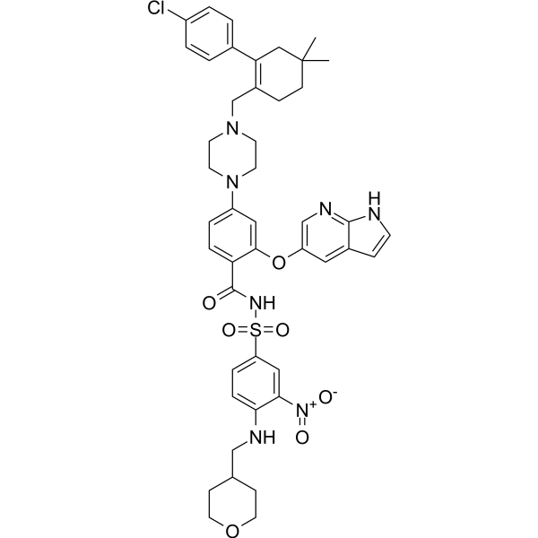
HY-15531
VenetoclaxBCL-2 Inhibitor -
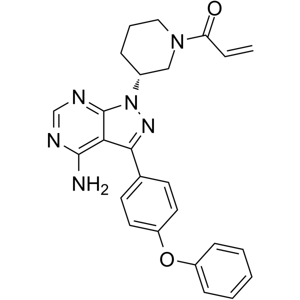
HY-10997
IbrutinibBtk Inhibitor -
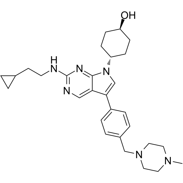
HY-101549
MRX-2843MERTK/FLT3 Inhibitor -
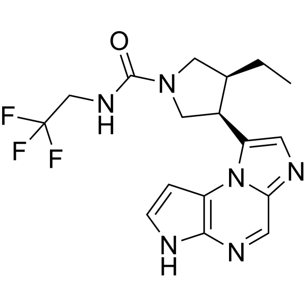
HY-19569
UpadacitinibJAK1 Inhibitor -
HY-10224
PanobinostatHDAC Inhibitor
Publications Citing Use of MedChemExpress (MCE) Emavusertib
More- Blood. 2023 Sep 14;142(11):989-1007. [Abstract]
- Nat Commun. 2026 Aug 22;17(1):10258.
- Leukemia. 2025 Sep;39(9):2163-2173. [Abstract]
- Cell Rep. 2025 Sep 24;44(10):116347. [Abstract]
- Front Immunol. 2021 Mar 9:12:637659. [Abstract]
- Curr Issues Mol Biol. 2024 Mar 29;46(4):2946-2960. [Abstract]
- bioRxiv. 2026 Feb 3.
- bioRxiv. 2026 Jan 9.
 Customer Validation & Images

-
Cell Proliferation/Viability Assay
-
WB
-
In Vivo Efficacy Study
-
IHC
-
Bio/Physico-chemical Assay


Activité biologique
Description
IC50 & Target
[1]|
IRAK4 57 nM (IC50) |
Cellular Effect
|
Cell Line

|
Type | Value | Description | References |
|---|---|---|---|---|
| OCI-Ly10 | IC50 |
1.5 μM
Compound: 24; CA-4948
|
Growth inhibition of human OCI-LY10 harboring mutant MYD88
Growth inhibition of human OCI-LY10 harboring mutant MYD88
|
[PMID: 33335659] |
In Vitro
Emavusertib (CA-4948) is over 500-fold more selective for IRAK-4 compared to IRAK-1. Emavusertib reduces TNF-α, IL-1β, IL-6 and IL-8 release from TLR-Stimulated THP-1 Cells with an IC50 <250 nM. Emavusertib also has antiproliferative activity due to inhibition of receptor-type tyrosine-protein kinase FLT3[1].
Emavusertib exhibits >350-fold higher binding affinity for IRAK-4 than that observed for IRAKs 1, 2 and 3[3].
Emavusertib (10 μM, 72 h) decreases the percentage of proliferating cells and induces a moderate increase in the sub-G0 fraction in marginal zone lymphomas (MZL) cell lines[3].
Emavusertib (10 μM, 72 h) induces a significant increase in the apoptotic cell population of MZL cells, particularly when combined with Ibrutinib (HY-10997) compared to ibrutinib and emavusertib alone[3].
MedChemExpress (MCE) has not independently confirmed the accuracy of these methods. They are for reference only.
In Vivo
Emavusertib (25-150 mg/kg, Orally, once daily, for 14 consecutive days) induces tumor growth inhibition in mice[3].
MedChemExpress (MCE) has not independently confirmed the accuracy of these methods. They are for reference only.
-
Animal Model:Mice bearing OCI-LY10 tumors[3]
-
Dosage:25, 50, or 150 mg/kg (once daily), 12.5, 25, or 50 mg/kg (twice daily)
-
Administration:Orally, once daily or twice daily, for 14 consecutive days
-
Result:Induced tumor growth inhibition. Emavusertib administered as a twice-daily divided dose was equivalent to the corresponding once-daily dose with regards to antitumor activity, i.e., 12.5 mg/kg BID versus 25 mg/kg QD.
- Detection of Apoptosis
- TUNEL staining for apoptotic DNA fragmentation
- LPS-Induced Endotoxemia/Systemic Inflammation
- Annexin V plus membrane-impermeant dye apoptosis staining
- Apoptosis Solutions
- Research Protocol for Inflammation-related Diseases
- Subcutaneous Cell-Line-Derived Xenograft
- Cell Viability Determination by MTT Colorimetric Assay
- How to Choose the Right Model Animal
Essai clinique
| NCT Number | Sponsor | Condition | Start Date |
Phase
|
|---|---|---|---|---|
| NCT01329991 | Plexxikon| | 2011-05 | PHASE1 |


Chemical Information
-
CAS No. 1801344-14-8
-
Appearance Solid
-
Masse moléculaire 491.50
-
Formule C24H25N7O5
-
Color Light yellow to green yellow
-
SMILES
O=C(C1=COC(C2=CC(C)=NC=C2)=N1)NC3=C(N4C[C@H](O)CC4)N=C5C(OC(N6CCOCC6)=N5)=C3
-
Synonyms
CA-4948
-
Livraison
Room temperature in continental US; may vary elsewhere.
-
Stockage
Powder -20°C 3 years 4°C 2 years In solvent -80°C 1 year -20°C 6 months


Publications (8)
-
Journal Impact Factor
-
Most Recent
-
Blood
2023 Sep 14;142(11):989-1007. PMID: 37172199
Emavusertib purchased from MedChemExpress. Usage Cited in: Blood. 2023 Sep 14;142(11):989-1007. [Abstract]
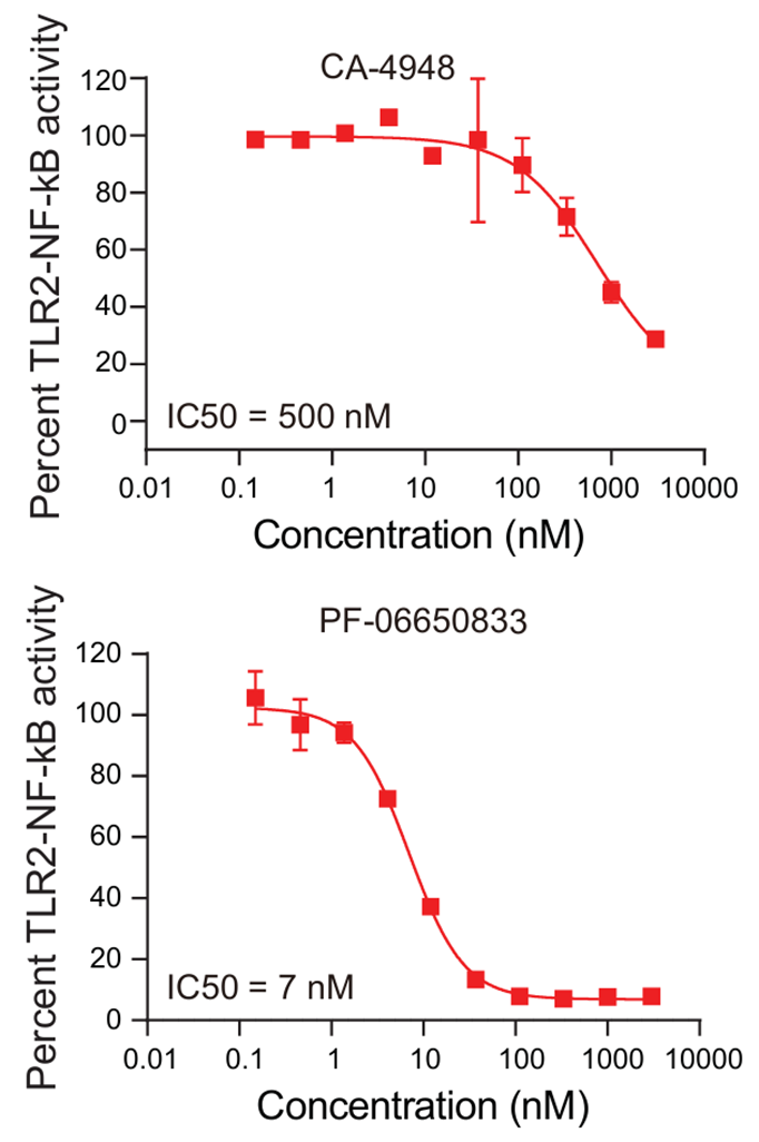
IC50 curves for CA-4948 (Emavusertib) (0.01-10000 nM; 24 h) and PF-06650833 in an assay measuring NF-kB activity upon TLR2 stimulation with PAM3CSK4 in THP1 NF-kB reporter cells.
Emavusertib purchased from MedChemExpress. Usage Cited in: Blood. 2023 Sep 14;142(11):989-1007. [Abstract]
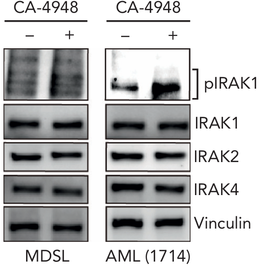
Immunoblots for phospho-IRAK1, total IRAK1, IRAK2, and IRAK4 in MDSL and AML (1714) treated for 24 hours with CA-4948 (Emavusertib) (10 μM). The results showed that treatment with CA-4948 resulted in increased IRAK1 phosphorylation.
-
-
Leukemia
Targeting of IRAK4 and GSPT1 enhances therapeutic efficacy in AML via c-Myc destabilization. [Abstract]2025 Sep;39(9):2163-2173. PMID: 40670672
Emavusertib purchased from MedChemExpress. Usage Cited in: Leukemia. 2025 Sep;39(9):2163-2173. [Abstract]
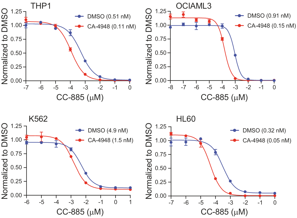
Cell viability was determined in THP1 and HL60 cells cultured with 10 µM CA-4948 (Emavusertib) or in OCI-AML3 and K562 cultured with 1 µM CA-4948 or vehicle (n = 4) for 14 days and then treated with CC-885 (n = 4).
Emavusertib purchased from MedChemExpress. Usage Cited in: Leukemia. 2025 Sep;39(9):2163-2173. [Abstract]
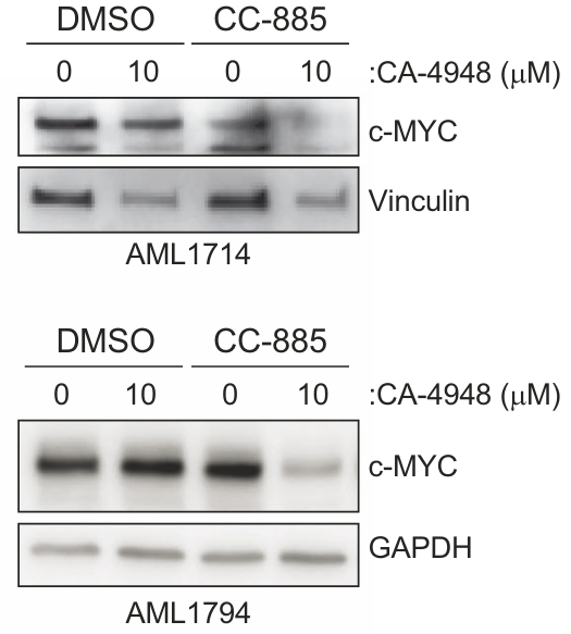
Immunoblots for c-MYC in the patient-derived AML samples (AML1794, AML1714) cultured with CA-4948 (Emavusertib) (10 µM) or vehicle for 14 days and then treated with CC-885 (50 nM) for 4 h.
-
Cell Rep
Disruption of fibroblast MYD88 signaling promotes antitumor immunity in pancreatic ductal adenocarcinoma. [Abstract]2025 Sep 24;44(10):116347. PMID: 41004339
Emavusertib purchased from MedChemExpress. Usage Cited in: Cell Rep. 2025 Sep 24;44(10):116347. [Abstract]
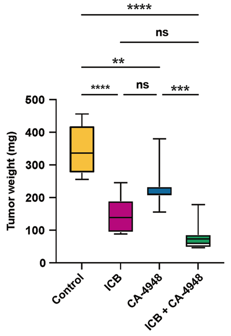
Harvested tumor weights by treatment condition (n = 8 per condition).Tumor growth studies demonstrated statistically significant inhibition with both CA-4948 (Emavusertib) (50 mg/kg; oral gavage; once daily) alone and ICB alone.
Emavusertib purchased from MedChemExpress. Usage Cited in: Cell Rep. 2025 Sep 24;44(10):116347. [Abstract]
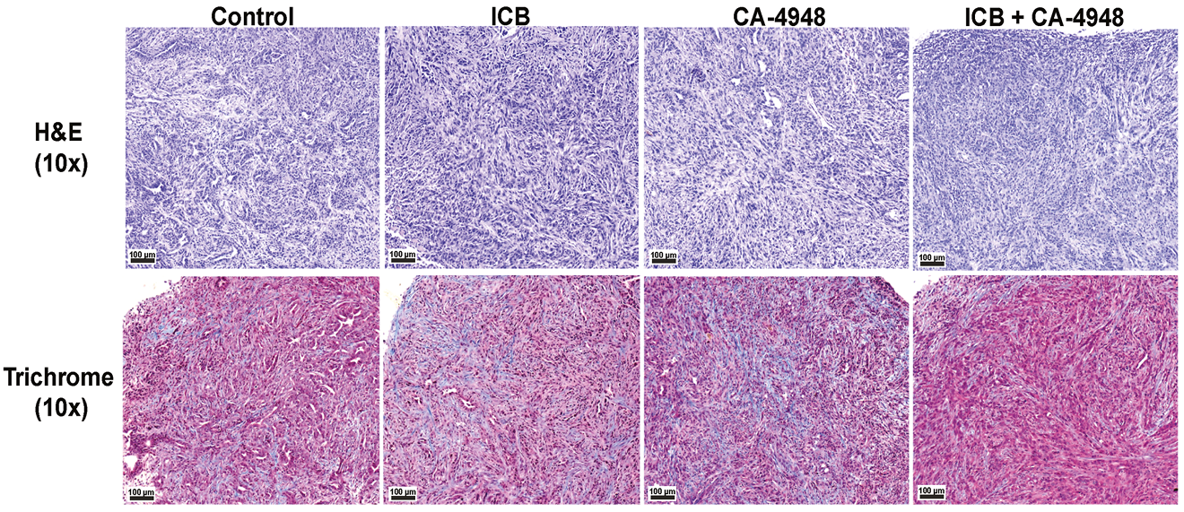
H&E and trichrome staining of tumors by treatment condition (Emavusertib (50 mg/kg; oral gavage; once daily), ect.) at 10× magnification. The scale bar represents 100 μm.
-
Front Immunol
Activation of Toll-Like Receptor 7 Signaling Pathway in Primary Sjögren's Syndrome-Associated Thrombocytopenia. [Abstract]2021 Mar 9:12:637659. PMID: 33767707 -
Curr Issues Mol Biol
FLT3 and IRAK4 Inhibitor Emavusertib in Combination with BH3-Mimetics in the Treatment of Acute Myeloid Leukemia. [Abstract]2024 Mar 29;46(4):2946-2960. PMID: 38666914 -
-


Solvant et solubilité
In Vitro:
DMSO : 50 mg/mL (101.73 mM; Need ultrasonic; Hygroscopic DMSO has a significant impact on the solubility of product, please use newly opened DMSO)
Please refer to the solubility information to select the appropriate solvent. Once prepared, please aliquot and store the solution to prevent product inactivation from repeated freeze-thaw cycles.
Storage method and period of stock solution: -80°C, 1 year; -20°C, 6 months. When stored at -80°C, please use it within 1 year. When stored at -20°C, please use it within 6 months.
Please refer to the solubility information to select the appropriate solvent. Once prepared, please aliquot and store the solution to prevent product inactivation from repeated freeze-thaw cycles.
Storage method and period of stock solution: -80°C, 1 year; -20°C, 6 months. When stored at -80°C, please use it within 1 year. When stored at -20°C, please use it within 6 months.
Concentration (start) × Volume (start) = Concentration (final) × Volume (final)
In Vivo:
Select the appropriate dissolution method based on your experimental animal and administration route.
- For the following dissolution methods, please ensure to first prepare a clear stock solution using an In Vitro approach and then sequentially add co-solvents:
- To ensure reliable experimental results, the clarified stock solution can be appropriately stored based on storage conditions. As for the working solution for In Vivo experiments, it is recommended to prepare freshly and use it on the same day.
- The percentages shown for the solvents indicate their volumetric ratio in the final prepared solution. If precipitation or phase separation occurs during preparation, heat and/or sonication can be used to aid dissolution.
Add each solvent one by one: 10% DMSO 40% PEG300 5% Tween-80 45% Saline
Solubility: ≥ 2.08 mg/mL (4.23 mM); Clear solution
This protocol yields a clear solution of ≥ 2.08 mg/mL (saturation unknown).
Taking 1 mL working solution as an example, add 100 μL DMSO stock solution (20.8 mg/mL) to 400 μL PEG300, and mix evenly; then add 50 μL Tween-80 and mix evenly; then add 450 μL Saline to adjust the volume to 1 mL.
Preparation of Saline: Dissolve 0.9 g sodium chloride in ddH₂O and dilute to 100 mL to obtain a clear Saline solution.
Add each solvent one by one: 10% DMSO 90% (20% SBE-β-CD in Saline)
Solubility: 2.08 mg/mL (4.23 mM); Suspended solution; Need ultrasonic
This protocol yields a suspended solution of 2.08 mg/mL. Suspended solution can be used for oral and intraperitoneal injection.
Taking 1 mL working solution as an example, add 100 μL DMSO stock solution (20.8 mg/mL) to 900 μL 20% SBE-β-CD in Saline, and mix evenly.
Preparation of 20% SBE-β-CD in Saline (4°C, storage for one week): 2 g SBE-β-CD powder is dissolved in 10 mL Saline, completely dissolve until clear.
In Vivo Dissolution Calculator
Please enter the basic information of animal experiments:
-
-
-
-
Recommended: Prepare an additional quantity of animals to account for potential losses during experiments.
Please enter your animal formula composition:
-
%DMSO +
Recommended: Keep the proportion of DMSO in working solution below 2% if your animal is weak.
-
%+
-
+%Tween-80 + +
-
%Saline +
The co-solvents required include: DMSO, . All of co-solvents are available by MedChemExpress (MCE). , Tween 80. All of co-solvents are available by MedChemExpress (MCE).
Working solution concentration: 0.22 mg/mL
Method for preparing stock solution: mg drug dissolved in μL DMSO. Stock solution concentration: mg/mL.
1. Take μL DMSO stock solution;
2. Add μL .
μL , mix evenly;
3. Then add μL Tween 80, mix evenly;
4. Then add μL
Please ensure that the stock solution in the first step is dissolved to a clear state, and add co-solvents in sequence. You can use ultrasonic heating (ultrasonic cleaner, recommended frequency 20-40 kHz), vortexing, etc. to assist dissolution.


Pureté et documentation
-
Fiche technique (277 KB)
-
SDS (393 KB)
- English - EN (393 KB)
- Français - FR (393 KB)
- Deutsch - DE (393 KB)
- Norwegian - NO (393 KB)
- Español - ES (393 KB)
- Swedish - SV (393 KB)
- Italian - IT (393 KB)
- Korean - KR (393 KB)
- Portuguese - PT (393 KB)
-
Instruction de manipulation (2659 KB)
Références
[1]. Wiese MD, et al. Investigational IRAK-4 inhibitors for the treatment of rheumatoid arthritis. Expert Opin Investig Drugs. 2020 Apr 17:1-8. [Content Brief]
[2]. Guidetti F, et al. Targeting IRAK4 with Emavusertib in Lymphoma Models with Secondary Resistance to PI3K and BTK Inhibitors. J Clin Med. 2023 Jan 4;12(2):399. [Content Brief]
[3]. Parrondo RD, et al. IRAK-4 inhibition: emavusertib for the treatment of lymphoid and myeloid malignancies. Front Immunol. 2023 Oct 26;14:1239082. [Content Brief]


Complete Stock Solution Preparation Table
Please refer to the solubility information to select the appropriate solvent. Once prepared, please aliquot and store the solution to prevent product inactivation from repeated freeze-thaw cycles.
Storage method and period of stock solution: -80°C, 1 year; -20°C, 6 months. When stored at -80°C, please use it within 1 year. When stored at -20°C, please use it within 6 months.
| Optional Solvent | Concentration Solvent Mass | 1 mg | 5 mg | 10 mg | 25 mg |
|---|---|---|---|---|---|
| DMSO | 1 mM | 2.0346 mL | 10.1729 mL | 20.3459 mL | 50.8647 mL |
| 5 mM | 0.4069 mL | 2.0346 mL | 4.0692 mL | 10.1729 mL | |
| 10 mM | 0.2035 mL | 1.0173 mL | 2.0346 mL | 5.0865 mL | |
| 15 mM | 0.1356 mL | 0.6782 mL | 1.3564 mL | 3.3910 mL | |
| 20 mM | 0.1017 mL | 0.5086 mL | 1.0173 mL | 2.5432 mL | |
| 25 mM | 0.0814 mL | 0.4069 mL | 0.8138 mL | 2.0346 mL | |
| 30 mM | 0.0678 mL | 0.3391 mL | 0.6782 mL | 1.6955 mL | |
| 40 mM | 0.0509 mL | 0.2543 mL | 0.5086 mL | 1.2716 mL | |
| 50 mM | 0.0407 mL | 0.2035 mL | 0.4069 mL | 1.0173 mL | |
| 60 mM | 0.0339 mL | 0.1695 mL | 0.3391 mL | 0.8477 mL | |
| 80 mM | 0.0254 mL | 0.1272 mL | 0.2543 mL | 0.6358 mL | |
| 100 mM | 0.0203 mL | 0.1017 mL | 0.2035 mL | 0.5086 mL |


Emavusertib Related Classifications
HY-135317 Related Classifications
 Powered by Bioz