



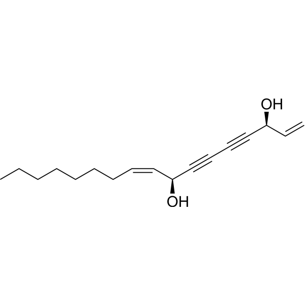
Falcarindiol
Based on 3 publication(s) in Google Scholar
Falcarindiol, an orally active polyacetylenic oxylipin, activates PPARγ and increases the expression of the cholesterol transporter ABCA1 in cells. Falcarindiol induces apoptosis and autophagy. Falcarindiol has anti-inflammatory, antifungal, anticancer and antidiabetic properties. Falcarindiol is a click chemistry reagent, it contains an Alkyne group and can undergo copper-catalyzed azide-alkyne cycloaddition (CuAAc) with molecules containing Azide groups.
For research use only. We do not sell to patients.
- Purity : 97.62%
- CAS No.: 55297-87-5
- Formula: C17H24O2
- Molecular Weight:260.37
-
Storage:
4°C, sealed storage, away from moisture and light
* In solvent : -80°C, 6 months; -20°C, 1 month (sealed storage, away from moisture and light)

To place orders, for customer services and technical support, please contact: MedChemExpress USA
Tel: 609-228-6898 E-mail: [email protected] [email protected]


-
 Biological Activity
-
Chemical Information
-
Solvent & Solubility
- Protocols
- Purity & Documentation
- References
-
 Help & FAQs


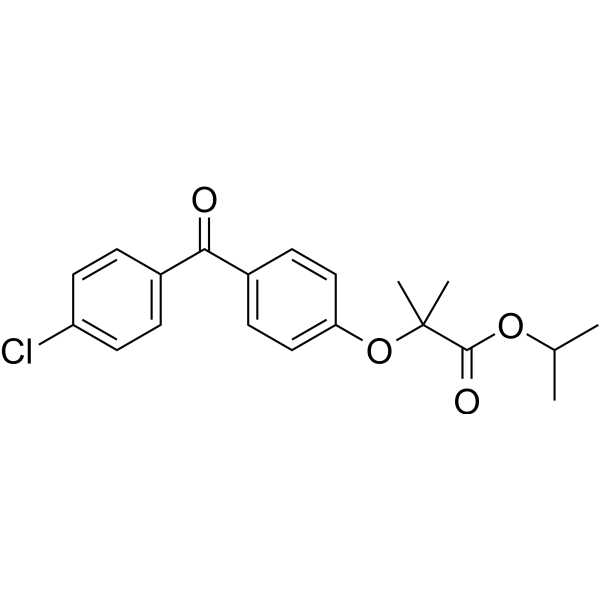
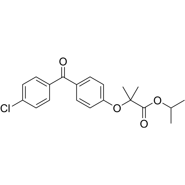
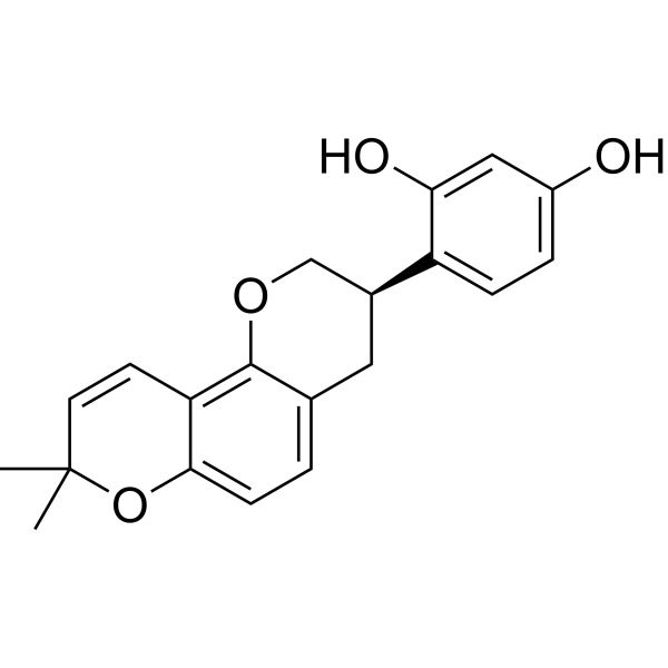
Publications Citing Use of MedChemExpress (MCE) Falcarindiol
More Customer Validation & Images

-
WB


Biological Activity
Description
IC50 & Target
[1]|
PPARγ |
Cellular Effect
|
Cell Line

|
Type | Value | Description | References |
|---|---|---|---|---|
| B16 | IC50 |
2.9 μg/mL
Compound: 8
|
Inhibition of alpha-MSH-stimulated melanogenesis in mouse B16 cells assessed as melanin release after 72 hrs
Inhibition of alpha-MSH-stimulated melanogenesis in mouse B16 cells assessed as melanin release after 72 hrs
|
[PMID: 22450129] |
| CHO | IC50 |
117.5 μM
Compound: 12
|
Displacement of [3H]LSD from human recombinant 5HT7 receptor expressed in CHO cells by liquid scintillation counting
Displacement of [3H]LSD from human recombinant 5HT7 receptor expressed in CHO cells by liquid scintillation counting
|
[PMID: 16643021] |
| HEK293 | EC50 |
3.2 μM
Compound: Ref 6, Cpd 2
|
Activation of human PPARgamma expressed in HEK293 cells incubated for 18 hrs by luciferase reporter assay relative to control
Activation of human PPARgamma expressed in HEK293 cells incubated for 18 hrs by luciferase reporter assay relative to control
|
[PMID: 32129622] |
| HL-60 | IC50 |
8.9 μM
Compound: 5
|
Cytotoxicity against human HL60 cells after 72 hrs by MTT assay
Cytotoxicity against human HL60 cells after 72 hrs by MTT assay
|
[PMID: 15387667] |
| HT-1080 | IC50 |
13 μM
Compound: 5
|
Cytotoxicity against human HT1080 cells after 72 hrs by MTT assay
Cytotoxicity against human HT1080 cells after 72 hrs by MTT assay
|
[PMID: 15387667] |
| LoVo | IC50 |
>40 μM
Compound: 5
|
Cytotoxicity against human doxorubicin-resistant LoVo cells after 72 hrs by MTT assay
Cytotoxicity against human doxorubicin-resistant LoVo cells after 72 hrs by MTT assay
|
[PMID: 15387667] |
| LoVo | IC50 |
4.3 μM
Compound: 5
|
Cytotoxicity against human LoVo cells after 72 hrs by MTT assay
Cytotoxicity against human LoVo cells after 72 hrs by MTT assay
|
[PMID: 15387667] |
| RAW264.7 | IC50 |
5.4 μM
Compound: 25
|
Anti-inflammatory activity in mouse RAW264.7 cells assessed as inhibition of LPS-induced NO production preincubated for 1 hr followed by LPS stimulation and measured after 24 hrs by Griess reagent based assay
Anti-inflammatory activity in mouse RAW264.7 cells assessed as inhibition of LPS-induced NO production preincubated for 1 hr followed by LPS stimulation and measured after 24 hrs by Griess reagent based assay
|
[PMID: 33476145] |
In Vitro
Falcarindiol (3, 6, 12, 24 μM; for 24 hours) significantly decreases cell viability of MDA-MB-231 and MDA-MB-468 cells. Cell viability of MCF-10A cells is unchanged until the dose of Falcarindiol reaches to 24 uM. Falcarindiol preferentially induces cell death in breast cancer cells[1].
Falcarindiol (6 uM; for 2 hours) induces autophagy and causes significant level of LC3-I converted to LC3-II in MDA-MB-231, MDA-MB-468 and SKBR3 cells[1].
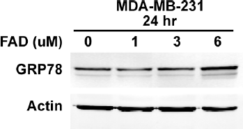
Falcarindiol (6 uM; for 2, 4, 8, 24 hours) increases the level of GRP78 in MDA-MB-231 cells in dose- and time-dependent manner[1].
Falcarindiol (1-20 μM) has no effect on hMSCs and HT-29 cell viability. Falcarindiol with only concentrations above 50 μM exhibits a toxic effect on the cells[2].
Falcarindiol (5 μM; 10 min, 1 h and 24 h) causes a significant upregulation on PPARγ2 expression at 24 h[2].
MedChemExpress (MCE) has not independently confirmed the accuracy of these methods. They are for reference only.
In Vivo
MedChemExpress (MCE) has not independently confirmed the accuracy of these methods. They are for reference only.
- RNA extraction experimental
- EdU Incorporation Assay (Click Chemistry-Based DNA Synthesis Measurement)
- Detection of Apoptosis
- TUNEL staining for apoptotic DNA fragmentation
- Annexin V plus membrane-impermeant dye apoptosis staining
- Autophagy
- Apoptosis Solutions
- Lysosome and acidic-vesicle live-cell staining
- Research Protocol for Inflammation-related Diseases
- Macroautophagy Solutions


Chemical Information
-
CAS No. 55297-87-5
-
Appearance Liquid (Density: 1.000±0.06 g/cm3)
-
Molecular Weight 260.37
-
Formula C17H24O2
-
Color Light yellow to brown
-
SMILES
C=C[C@H](O)C#CC#C[C@@H](O)/C=C\CCCCCCC
-
Structure Classification
-
Shipping
Room temperature in continental US; may vary elsewhere.
-
Storage
4°C, sealed storage, away from moisture and light
* In solvent : -80°C, 6 months; -20°C, 1 month (sealed storage, away from moisture and light)


Publications (3)
-
Journal Impact Factor
-
Most Recent
-
Sci Rep
Falcarindiol alleviates airway inflammation and oxidative stress in asthma through Nrf2 pathway activation. [Abstract]2026 Feb 21;16(1):10140. PMID: 41723162 -
Daru
Falcarindiol induces apoptosis, ROS accumulation, and cell cycle arrest via EGFR/mTOR pathway modulation: an integrated in silico and in vitro study in cervical cancer. [Abstract]2026 Jan 19;34(1):7. PMID: 41549146 -
PLoS One
Autophagy contributes to falcarindiol-induced cell death in breast cancer cells with enhanced endoplasmic reticulum stress. [Abstract]2017 Apr 25;12(4):e0176348. PMID: 28441457
Falcarindiol purchased from MedChemExpress. Usage Cited in: PLoS One. 2017 Apr 25;12(4):e0176348. [Abstract]
FAD-induced cell death in breast cancer cells is mediates by ER stress. The western blot analyzed the level of GRP78 in MDA-MB-231 cells treated with indicated concentration of FAD for 24 hours. FAD increases the level of GRP78 in MDA-MB-231 cells in dose- and time-dependent manner.


Solvent & Solubility
In Vitro:
DMSO : ≥ 33.33 mg/mL (128.01 mM; Hygroscopic DMSO has a significant impact on the solubility of product, please use newly opened DMSO)
* "≥" means soluble, but saturation unknown.
Please refer to the solubility information to select the appropriate solvent. Once prepared, please aliquot and store the solution to prevent product inactivation from repeated freeze-thaw cycles.
Storage method and period of stock solution: -80°C, 6 months; -20°C, 1 month (sealed storage, away from moisture and light). When stored at -80°C, please use it within 6 months. When stored at -20°C, please use it within 1 month.
Please refer to the solubility information to select the appropriate solvent. Once prepared, please aliquot and store the solution to prevent product inactivation from repeated freeze-thaw cycles.
Storage method and period of stock solution: -80°C, 6 months; -20°C, 1 month (sealed storage, away from moisture and light). When stored at -80°C, please use it within 6 months. When stored at -20°C, please use it within 1 month.
Concentration (start) × Volume (start) = Concentration (final) × Volume (final)
In Vivo:
Select the appropriate dissolution method based on your experimental animal and administration route.
- For the following dissolution methods, please ensure to first prepare a clear stock solution using an In Vitro approach and then sequentially add co-solvents:
- To ensure reliable experimental results, the clarified stock solution can be appropriately stored based on storage conditions. As for the working solution for In Vivo experiments, it is recommended to prepare freshly and use it on the same day.
- The percentages shown for the solvents indicate their volumetric ratio in the final prepared solution. If precipitation or phase separation occurs during preparation, heat and/or sonication can be used to aid dissolution.
Add each solvent one by one: 10% DMSO 40% PEG300 5% Tween-80 45% Saline
Solubility: 1.11 mg/mL (4.26 mM); Suspended solution; Need ultrasonic
This protocol yields a suspended solution of 1.11 mg/mL. Suspended solution can be used for oral and intraperitoneal injection.
Taking 1 mL working solution as an example, add 100 μL DMSO stock solution (11.1 mg/mL) to 400 μL PEG300, and mix evenly; then add 50 μL Tween-80 and mix evenly; then add 450 μL Saline to adjust the volume to 1 mL.
Preparation of Saline: Dissolve 0.9 g sodium chloride in ddH₂O and dilute to 100 mL to obtain a clear Saline solution.
In Vivo Dissolution Calculator
Please enter the basic information of animal experiments:
-
-
-
-
Recommended: Prepare an additional quantity of animals to account for potential losses during experiments.
Please enter your animal formula composition:
-
%DMSO +
Recommended: Keep the proportion of DMSO in working solution below 2% if your animal is weak.
-
%+
-
+%Tween-80 + +
-
%Saline +
The co-solvents required include: DMSO, . All of co-solvents are available by MedChemExpress (MCE). , Tween 80. All of co-solvents are available by MedChemExpress (MCE).
Working solution concentration: 0.22 mg/mL
Method for preparing stock solution: mg drug dissolved in μL DMSO. Stock solution concentration: mg/mL. * In solvent : -80°C, 6 months; -20°C, 1 month (sealed storage, away from moisture and light)
1. Take μL DMSO stock solution;
2. Add μL .
μL , mix evenly;
3. Then add μL Tween 80, mix evenly;
4. Then add μL
Please ensure that the stock solution in the first step is dissolved to a clear state, and add co-solvents in sequence. You can use ultrasonic heating (ultrasonic cleaner, recommended frequency 20-40 kHz), vortexing, etc. to assist dissolution.


Purity & Documentation
-
Data Sheet (270 KB)
-
SDS (252 KB)
- English - EN (252 KB)
- Français - FR (252 KB)
- Deutsch - DE (252 KB)
- Norwegian - NO (252 KB)
- Español - ES (252 KB)
- Swedish - SV (252 KB)
- Italian - IT (252 KB)
- Korean - KR (252 KB)
- Portuguese - PT (252 KB)
-
Handling Instructions (2659 KB)
References
[1]. Tingting Lu, et al. Autophagy contributes to falcarindiol-induced cell death in breast cancer cells with enhanced endoplasmic reticulum stress. PLoS One. 2017 Apr 25;12(4):e0176348. [Content Brief]
[2]. Camilla Bertel Andersen, et al. Falcarindiol Purified From Carrots Leads to Elevated Levels of Lipid Droplets and Upregulation of Peroxisome Proliferator-Activated Receptor-γ Gene Expression in Cellular Models. Front Pharmacol. 2020 Aug 28;11:565524. [Content Brief]


Complete Stock Solution Preparation Table
Please refer to the solubility information to select the appropriate solvent. Once prepared, please aliquot and store the solution to prevent product inactivation from repeated freeze-thaw cycles.
Storage method and period of stock solution: -80°C, 6 months; -20°C, 1 month (sealed storage, away from moisture and light). When stored at -80°C, please use it within 6 months. When stored at -20°C, please use it within 1 month.
| Optional Solvent | Concentration Solvent Mass | 1 mg | 5 mg | 10 mg | 25 mg |
|---|---|---|---|---|---|
| DMSO | 1 mM | 3.8407 mL | 19.2034 mL | 38.4069 mL | 96.0172 mL |
| 5 mM | 0.7681 mL | 3.8407 mL | 7.6814 mL | 19.2034 mL | |
| 10 mM | 0.3841 mL | 1.9203 mL | 3.8407 mL | 9.6017 mL | |
| 15 mM | 0.2560 mL | 1.2802 mL | 2.5605 mL | 6.4011 mL | |
| 20 mM | 0.1920 mL | 0.9602 mL | 1.9203 mL | 4.8009 mL | |
| 25 mM | 0.1536 mL | 0.7681 mL | 1.5363 mL | 3.8407 mL | |
| 30 mM | 0.1280 mL | 0.6401 mL | 1.2802 mL | 3.2006 mL | |
| 40 mM | 0.0960 mL | 0.4801 mL | 0.9602 mL | 2.4004 mL | |
| 50 mM | 0.0768 mL | 0.3841 mL | 0.7681 mL | 1.9203 mL | |
| 60 mM | 0.0640 mL | 0.3201 mL | 0.6401 mL | 1.6003 mL | |
| 80 mM | 0.0480 mL | 0.2400 mL | 0.4801 mL | 1.2002 mL | |
| 100 mM | 0.0384 mL | 0.1920 mL | 0.3841 mL | 0.9602 mL |


Falcarindiol Related Classifications
HY-N0364 Related Classifications
 Powered by Bioz