



PF-04217903
Based on 2 publication(s) in Google Scholar
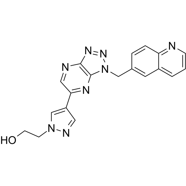
PF-04217903 is an orally active, highly selective ATP-competitive c-Met kinase inhibitor with a Ki value of 4.8 nM and a Kd value of 4.5 nM. PF-04217903 blocks c-Met and HGF signaling pathways, inhibits MET phosphorylation, and blocks downstream MAPK, PI3K/AKT and PLCγ1 pathways. PF-04217903 suppresses tumor proliferation, survival, migration, invasion, angiogenesis and metastasis, induces apoptosis, and enhances efferocytosis, Annexin A1 expression and resolution of inflammation. PF-04217903 retains activity against several c-Met mutants (M1131T, V1220I, H1094R). PF-04217903 increases the incidence of subarachnoid hemorrhage and reduces survival rate without altering aneurysm formation, and also prevents lymph node metastasis induced by VEGF inhibition. PF-04217903 is applicable to research related to tumors (pancreas, stomach, lung, brain, colon, breast, kidney, melanoma, etc.), intracranial aneurysms and inflammatory diseases (gouty arthritis, neutrophilic pleuritis).
연구목적의 판매만을 진행합니다. 환자를 대상으로 한 판매는 하지 않습니다.
- Purity: 99.85%
- CAS No.: 956905-27-4
- 화학식: C19H16N8O
- 분자량:372.38
-
보관:Powder -20°C, 3 years , 4°C, 2 years ; In solvent -80°C, 2 years , -20°C, 1 year

To place orders, for customer services and technical support, please contact: MedChemExpress USA
Tel: 609-228-6898 E-mail: [email protected] [email protected]


-
 Biological Activity
-
Chemical Information
-
용액&용해도
- 순도&문서
- References
-
 Help & FAQs
-

Kinase Inhibitor Library
HY-L009
-
Protein Tyrosine Kinase Compound Library
HY-L016
-
Anti-Cancer Compound Library
HY-L025
-
Clinical Compound Library
HY-L026
-
Anti-Lung Cancer Compound Library
HY-L075
-
Angiogenesis-Related Compound Library
HY-L088
-
Targeted Diversity Library
HY-L099
-
Anti-Liver Cancer Compound Library
HY-L101
-
Anti-Colorectal Cancer Compound Library
HY-L103
-
Cancer Stem Cells Compound Library
HY-L135
-
Membrane Protein-targeted Compound Library
HY-L149
-
Membrane Receptor-targeted Compound Library
HY-L150
-
Cytokine Inhibitors Library
HY-L161
-
Anti-Ovarian Cancer Compound Library
HY-L173
-
Bioactive Compound Library Max
HY-L181
-
MCE Bioactive Compound Library
HY-L001V
-
Clinical Compound Library Plus
HY-L026P
-
Bioactive Compound Library
HY-L001
-
Anti-Gastric Cancer Compound Library
HY-L184
-
Anti-Brain Cancer Compound Library
HY-L188
-
Protein Kinase Compound Library
HY-L196
-
High-Throughput Bioactive Compound Library
HY-L205
-
Posttranslational Modification Library
HY-L226


Publications Citing Use of MedChemExpress (MCE) PF-04217903
MoreAll Phospholipase Isoforms
More

Biological Activity
|
c-Met 4.8 nM (Ki) |
c-Met 4.5 nM (Kd) |
PLC |
AnxA1 |
|
Cell Line

|
Type | Value | Description | References |
|---|---|---|---|---|
| A549 | IC50 |
>10000 nM
Compound: Chemical Probe: PF-04217903
|
Antiproliferative activity against human A549 cells over expressing c-Met assessed as reduction in cell viability after 48 to 72 hrs by MTT assay
Antiproliferative activity against human A549 cells over expressing c-Met assessed as reduction in cell viability after 48 to 72 hrs by MTT assay
|
[PMID: 22389468] |
| A549 | IC50 |
0.004 μM
Compound: 2, PF-04217903
|
Antagonist activity at c-MET receptor in human A549 cells assessed as inhibition of autophosphorylation after 1 hr by ELISA
Antagonist activity at c-MET receptor in human A549 cells assessed as inhibition of autophosphorylation after 1 hr by ELISA
|
[PMID: 22924734] |
| COLO 205 | IC50 |
>10000 nM
Compound: Chemical Probe: PF-04217903
|
Antiproliferative activity against human COLO 205 cells over expressing c-Met assessed as reduction in cell viability after 48 to 72 hrs by MTT assay
Antiproliferative activity against human COLO 205 cells over expressing c-Met assessed as reduction in cell viability after 48 to 72 hrs by MTT assay
|
[PMID: 22389468] |
| DLD-1 | IC50 |
>10000 nM
Compound: Chemical Probe: PF-04217903
|
Antiproliferative activity against human DLD-1 cells over expressing c-Met assessed as reduction in cell viability after 48 to 72 hrs by MTT assay
Antiproliferative activity against human DLD-1 cells over expressing c-Met assessed as reduction in cell viability after 48 to 72 hrs by MTT assay
|
[PMID: 22389468] |
| HCC1143 | IC50 |
>10000 nM
Compound: Chemical Probe: PF-04217903
|
Antiproliferative activity against human HCC1143 cells over expressing c-Met assessed as reduction in cell viability after 48 to 72 hrs by MTT assay
Antiproliferative activity against human HCC1143 cells over expressing c-Met assessed as reduction in cell viability after 48 to 72 hrs by MTT assay
|
[PMID: 22389468] |
| HCT-116 | IC50 |
>10000 nM
Compound: Chemical Probe: PF-04217903
|
Antiproliferative activity against human HCT-116 cells over expressing c-Met assessed as reduction in cell viability after 48 to 72 hrs by MTT assay
Antiproliferative activity against human HCT-116 cells over expressing c-Met assessed as reduction in cell viability after 48 to 72 hrs by MTT assay
|
[PMID: 22389468] |
| HT-29 | IC50 |
>10000 nM
Compound: Chemical Probe: PF-04217903
|
Antiproliferative activity against human HT-29 cells over expressing c-Met assessed as reduction in cell viability after 48 to 72 hrs by MTT assay
Antiproliferative activity against human HT-29 cells over expressing c-Met assessed as reduction in cell viability after 48 to 72 hrs by MTT assay
|
[PMID: 22389468] |
| HT-29 | IC50 |
7 nM
Compound: Chemical Probe: PF-04217903
|
Inhibition of cell invasion in HGF-stimulated human HT-29 cells over expressing c-Met incubated for 48 hrs by Matrigel based analysis
Inhibition of cell invasion in HGF-stimulated human HT-29 cells over expressing c-Met incubated for 48 hrs by Matrigel based analysis
|
[PMID: 22389468] |
| HUVEC | IC50 |
12.3 nM
Compound: Chemical Probe: PF-04217903
|
Antiproliferative activity against HGF-stimulated HUVEC cells assessed as reduction in cell viability after 48 to 72 hrs by MTT assay
Antiproliferative activity against HGF-stimulated HUVEC cells assessed as reduction in cell viability after 48 to 72 hrs by MTT assay
|
[PMID: 22389468] |
| HUVEC | IC50 |
27 nM
Compound: Chemical Probe: PF-04217903
|
Inhibition of cell invasion in HGF-stimulated HUVEC cells incubated for 48 hrs by Matrigel based analysis
Inhibition of cell invasion in HGF-stimulated HUVEC cells incubated for 48 hrs by Matrigel based analysis
|
[PMID: 22389468] |
| HUVEC | IC50 |
7.3 nM
Compound: Chemical Probe: PF-04217903
|
Induction of apoptosis in HGF-stimulated HUVEC cells assessed as increase in apoptotic cells incubated for 48 hrs by ELISA
Induction of apoptosis in HGF-stimulated HUVEC cells assessed as increase in apoptotic cells incubated for 48 hrs by ELISA
|
[PMID: 22389468] |
| MDA-MB-231 | IC50 |
>10000 nM
Compound: Chemical Probe: PF-04217903
|
Antiproliferative activity against human MDA-MB-231 cells over expressing c-Met assessed as reduction in cell viability after 48 to 72 hrs by MTT assay
Antiproliferative activity against human MDA-MB-231 cells over expressing c-Met assessed as reduction in cell viability after 48 to 72 hrs by MTT assay
|
[PMID: 22389468] |
| MDA-MB-468 | IC50 |
>10000 nM
Compound: Chemical Probe: PF-04217903
|
Antiproliferative activity against human MDA-MB-468 cells over expressing c-Met assessed as reduction in cell viability after 48 to 72 hrs by MTT assay
Antiproliferative activity against human MDA-MB-468 cells over expressing c-Met assessed as reduction in cell viability after 48 to 72 hrs by MTT assay
|
[PMID: 22389468] |
| NCI-H1993 | IC50 |
30 nM
Compound: Chemical Probe: PF-04217903
|
Antiproliferative activity against human NCI-H1993 cells over expressing c-Met assessed as reduction in cell viability after 48 to 72 hrs by MTT assay
Antiproliferative activity against human NCI-H1993 cells over expressing c-Met assessed as reduction in cell viability after 48 to 72 hrs by MTT assay
|
[PMID: 22389468] |
| NCI-H1993 | IC50 |
33.6 nM
Compound: PF-4217903
|
Cytotoxicity against human NCI-H1993 cells assessed as growth inhibition after 72 hrs by CCK-8 assay
Cytotoxicity against human NCI-H1993 cells assessed as growth inhibition after 72 hrs by CCK-8 assay
|
[PMID: 27448775] |
| NCI-H441 | IC50 |
11 nM
Compound: Chemical Probe: PF-04217903
|
Inhibition of cell migration in HGF-stimulated human NCI-H441 cells over expressing c-Met incubated for 48 hrs by Matrigel based analysis
Inhibition of cell migration in HGF-stimulated human NCI-H441 cells over expressing c-Met incubated for 48 hrs by Matrigel based analysis
|
[PMID: 22389468] |
| NCI-H441 | IC50 |
12.5 nM
Compound: Chemical Probe: PF-04217903
|
Inhibition of cell invasion in HGF-stimulated human NCI-H441 cells over expressing c-Met incubated for 48 hrs by Matrigel based analysis
Inhibition of cell invasion in HGF-stimulated human NCI-H441 cells over expressing c-Met incubated for 48 hrs by Matrigel based analysis
|
[PMID: 22389468] |
| PC-3 | IC50 |
>10000 nM
Compound: Chemical Probe: PF-04217903
|
Antiproliferative activity against human PC-3 cells over expressing c-Met assessed as reduction in cell viability after 48 to 72 hrs by MTT assay
Antiproliferative activity against human PC-3 cells over expressing c-Met assessed as reduction in cell viability after 48 to 72 hrs by MTT assay
|
[PMID: 22389468] |
| SW-620 | IC50 |
>10000 nM
Compound: Chemical Probe: PF-04217903
|
Antiproliferative activity against human SW620 cells over expressing c-Met assessed as reduction in cell viability after 48 to 72 hrs by MTT assay
Antiproliferative activity against human SW620 cells over expressing c-Met assessed as reduction in cell viability after 48 to 72 hrs by MTT assay
|
[PMID: 22389468] |
| U-87MG ATCC | IC50 |
>10000 nM
Compound: Chemical Probe: PF-04217903
|
Antiproliferative activity against human U-87 MG cells over expressing c-Met assessed as reduction in cell viability after 48 to 72 hrs by MTT assay
Antiproliferative activity against human U-87 MG cells over expressing c-Met assessed as reduction in cell viability after 48 to 72 hrs by MTT assay
|
[PMID: 22389468] |
PF-04217903 acts as a highly selective c-Met inhibitor, with >1000-fold greater potency for c-Met than for 208 other tested kinases[1].
PF-04217903 (1 μM) shows biochemical inhibition of IGF1R in kinase selectivity screens, but does not inhibit IGF1R activity in cell-based assays[8].
PF-04217903 (30 min) potently inhibits wild-type c-Met (Ki = 6.5-7.4 pM) and most oncogenic c-Met mutants, but exhibits drastically reduced potency against c-MetY1230C, c-MetY1230H, and c-MetD1228H mutants, and no measurable inhibition of c-MetD1228H[9].
PF-04217903 (1 h; 20 min) potently inhibits c-Met phosphorylation in cells expressing wild-type or most mutant c-Met (IC50 = 3.1-24 nM), but shows no measurable inhibition (IC50 >10000 nM) in NIH-3T3 cells expressing the c-MetY1230C mutant and drastically reduced potency in T47D cells expressing the c-MetY1235D mutant[9].
PF-04217903 (7.8-1000 nM; 1 min association, 5 min dissociation) binds tightly to purified c-Met kinase domain with a Kd of 4.5 nM and a residence time of 180 s[9].
PF-04217903 (5 μM; 30 min) significantly stabilizes wild-type c-Met and most oncogenic c-Met mutants (Tm shift = 11.6-17.0 °C), but shows minimal stabilization of c-MetY1230C (Tm shift = 2.1 °C) and c-MetY1230H (Tm shift = 4.4 °C) mutants[9].
PF-04217903 (6 μg/mL; 1 hour pre-incubation, 3 hours LPS treatment) blocks the HGF-mediated reduction of VCAM-1 and E-selectin expression in human aortic endothelial cells, but has no effect on inflammatory molecule expression in human aortic smooth muscle cells or THP-1 monocytes[3].
PF-04217903 (10-10000 nM; 6 days) potently inhibits growth of Met-amplified recurrent murine ATC cell lines 34286 (IC50 ~150 nM) and 36934 (IC50 ~50 nM), but shows minimal activity against primary murine ATC and Met-diploid recurrent murine ATC cell lines[4].
PF-04217903 (10-1000 nM; 1 hour) inhibits Met phosphorylation and downstream Akt/Erk signaling in Met-amplified recurrent murine ATC cell lines, but does not affect these signaling pathways in primary murine ATC or Met-diploid recurrent murine ATC cell lines[4].
PF-04217903 (153 pM-10 μM; 3 days) inhibits parental GTL16 gastric carcinoma cell viability with an IC50 of 10 nM, but does not inhibit viability in SND1-BRAF fusion-positive resistant GTL16R1 and GTL16R3 clones at concentrations up to 10 μM[8].
PF-04217903 (2.5 μM; 1 hour) inhibits c-MetY1235 phosphorylation in parental GTL16, GTL16R1, and GTL16R3 gastric carcinoma cells, confirming the inhibitor binds and inhibits c-Met catalytic activity in resistant clones[8].
PF-04217903 (200 nM; 4 hours) inhibits c-Met Y1349 and AKT S473 phosphorylation in parental GTL16, GTL16R1, and GTL16R3 gastric carcinoma cells; it completely blocks ERK phosphorylation in parental GTL16 cells but only partially reduces ERK phosphorylation in resistant clones, with combined treatment with RAFi achieving greater ERK inhibition in resistant clones[8].
PF-04217903 (≥39 nM/2.5 nM; 3 days) synergizes with RAF inhibitor PF-04880594 or MEK inhibitor PD-0325901 to inhibit viability in SND1-BRAF fusion-positive resistant GTL16R1 and GTL16R3 gastric carcinoma clones, but does not enhance growth inhibition in parental GTL16 cells compared to single-agent treatment[8].
PF-04217903 (0.5-2.5 μM; 4 months) generates PF-04217903-resistant cell clones from GTL16 gastric carcinoma cells[8].
PF-04217903 (30 µM; 1-6 h) inhibits LPS/HGF-induced and MSU-induced MET phosphorylation in primary human peripheral blood neutrophils, and induces caspase-dependent apoptosis by increasing cleaved caspase-3 levels and reducing Bcl-xL levels[5].
MedChemExpress (MCE) has not independently confirmed the accuracy of these methods. They are for reference only.
-
Cell Line:human aortic endothelial cells, human aortic smooth muscle cells, THP-1 human monocyte cells
-
Concentration:6 μg/mL
-
Incubation Time:1 h (pre-incubation); 3 hs (LPS treatment)
-
Result:Abolished the HGF-mediated attenuation of VCAM-1 and E-selectin expression induced by LPS in human aortic endothelial cells, such that VCAM-1 and E-selectin levels were similar to those in cells treated with LPS alone.
Showed no significant effects on TNF-α, IL-1β, MCP-1, COX-1, COX-2, TGF-β, or ICAM-1 expression in human aortic endothelial cells.
Had no significant effect on any measured inflammatory molecule expression in human aortic smooth muscle cells and THP-1 monocytes when combined with HGF and LPS, compared to LPS alone or HGF plus LPS.
-
Cell Line:murine primary ATCs and Met-amplified/Met-diploid recurrent murine ATCs
-
Concentration:10, 50, 200 and 1000 nM
-
Incubation Time:1 h
-
Result:Inhibited phosphorylation of Met (Y1234/Y1235) only in Met-amplified recurrent ATC cell lines 34286 and 36934, with reduced pMet visible at doses as low as 10 nM.
Blocked downstream phosphorylation of Akt (S473) and Erk (T202/Y204) in these Met-amplified lines, but had no effect on pMet, pAkt, or pErk levels in primary ATC or Met-diploid recurrent ATC cell lines.
-
Cell Line:Primary human peripheral blood neutrophils
-
Concentration:30 µM
-
Incubation Time:1, 2 and 6 h
-
Result:Selective HGF/MET inhibition blocked MET phosphorylation in human primary neutrophils stimulated by LPS/HGF or MSU crystals.
MET blockade increased neutrophil apoptosis at 6 hours, evidenced by chromatin condensation and nuclear fragmentation.
This apoptosis was caspase-dependent, as zVAD-fmk abolished the effect at 2 hours.
Mechanistically, MET suppression elevated cleaved caspase-3 and reduced Bcl-xL expression.
The effect was neutrophil-specific, as MET inhibition did not induce macrophage apoptosis in vivo.
Additionally, MET blockade reversed the pro-survival effect of combined LPS/HGF stimulation.
-
Cell Line:parental GTL16 gastric carcinoma cells, METi-resistant GTL16R1, GTL16R3 clones
-
Concentration:200 nM (single agent); 2.5 μM (combination treatment assays)
-
Incubation Time:4 h (single agent)
-
Result:Inhibited phosphorylation of c-Met Y1349 in all three cell lines, despite moderately increased total c-Met protein levels in treated samples.
Completely inhibited ERK phosphorylation in parental GTL16 cells, but only caused a slight decrease in ERK phosphorylation in GTL16R1 and GTL16R3 clones.
Reduced AKT S473 phosphorylation to ~10% of baseline levels in all cell lines.
When combined with 200 nM RAFi, further inhibited ERK phosphorylation in GTL16R1 and GTL16R3 to 38% and 10.5% of untreated GTL16 levels, respectively.
PF-04217903 (30 mg/kg; p.o.; once daily; for 3 consecutive weeks) enhances the anti-tumor growth and anti-invasion effects of Sunitinib in a nude mouse orthotopic Panc-1 pancreatic adenocarcinoma model, and eliminates micrometastases observed with Sunitinib monotherapy[1].
PF-04217903 (30-45 mg/kg; p.o.; once daily administration) (active compound PF-04217903) exerts potent and selective in vivo growth inhibitory effects on Met-amplified recurrent anaplastic thyroid carcinoma xenografts in nude mice[4].
PF-04217903 (40 mg/kg; p.o.; once daily; for 25 consecutive days) achieves a 45% tumor growth inhibition (TGI) rate in c-Met-overexpressing SW620 xenograft models, while sustaining inhibition of c-Met phosphorylation and upregulating total c-Met protein expression after treatment[6].
PF-04217903 (40-50 mg/kg; p.o.; once daily; for 14-31 consecutive days) inhibits tumor growth in c-Met-overexpressing Colo205, MDA-MB-231, and H292 xenograft models[6].
PF-04217903 (10 mg/kg; p.o.; daily administration; for 21 consecutive days) does not alter intracranial aneurysm formation in a mouse model of intracranial aneurysm, but increases the incidence of subarachnoid hemorrhage to 94% and reduces the survival rate to 25%[3].
PF-04217903 (40 mg/kg; p.o.; single administration; 12 h after MSU challenge) induces caspase-dependent neutrophil apoptosis and efferocytosis, alleviates joint inflammation, tissue damage and hyperalgesia, and promotes the expression of the pro-resolving protein annexin A1 in a mouse model of MSU-induced gouty arthritis[5].
PF-04217903 (40 mg/kg; p.o.; single administration; 4 h after LPS challenge) promotes the resolution of inflammation in a mouse model of LPS-induced neutrophilic pleurisy by enhancing neutrophil apoptosis and efferocytosis[5].
PF-04217903 (40 mg/kg; p.o.; single administration; 90 h after zymosan challenge) enhances the efferocytosis capacity of macrophages in a zymosan-induced peritonitis mouse model[5].
PF-04217903 (1-30 mg/kg; p.o.; once daily; 16 days; 0.15-15 mg/kg/d; s.c.; continuous infusion; 14 days; 0.3-10 mg/kg/d; p.o.; once daily; 4 days) exhibits potent, dose-dependent antitumor efficacy in MET-amplified GTL-16 xenograft models, with a tumor growth inhibition (TGI) rate of 100% at the oral dose of 30 mg/kg/d; its antitumor activity is directly associated with sustained inhibition of c-Met phosphorylation and downstream signaling pathways, as well as induction of tumor cell apoptosis[6].
PF-04217903 (0.3-30 mg/kg; p.o.; once daily; for 10 consecutive days; 5-50 mg/kg; p.o.; once daily; for 3 consecutive days) exhibits dose-dependent antitumor efficacy in U87MG xenograft models with an HGF/c-Met autocrine loop, achieving a tumor growth inhibition (TGI) rate of 84% at an oral dose of 30 mg/kg/d. Its activity is mediated by inhibition of the c-Met signaling pathway, reduction of tumor cell proliferation, induction of apoptosis, anti-angiogenic effects, and decreased secretion of pro-angiogenic cytokines[6].
As a monotherapy, PF-04217903 (50 mg/kg; p.o.; once daily; for 21 consecutive days) achieves a 40% TGI in a c-Met-overexpressing HT29 xenograft model, while RON shRNA alone achieves a 52% TGI; due to the blockade of the AKT signaling pathway and the induction of tumor cell apoptosis, the efficacy of the combination therapy is enhanced to a 77% TGI[6].
PF-04217903 (1-30 mg/kg/d; p.o.; multiple administrations) dose-dependently reduces the plasma levels of pro-angiogenic factors IL-8 and VEGFA in mice bearing GTL-16 xenograft tumors[6].
MedChemExpress (MCE) has not independently confirmed the accuracy of these methods. They are for reference only.
-
Animal Model:C57BL/6-Tg (RIP-Tag) 2Han mice (aged 14 weeks at treatment initiation, spontaneously developed pancreatic neuroendocrine tumors driven by RIP-promoter SV40 T antigen expression in β-cells)[1]
-
Dosage:30 mg/kg (single-agent; co-administered with anti-mouse VEGF antibody; co-administered with Sunitinib) (HY-10255A); 150 μg (anti-mouse VEGF antibody); 40 mg/kg (sunitinib)
-
Administration:p.o.; daily; 3 weeks; p.o.; daily; 6 weeks (survival study); i.p.; 3x/week; 3 weeks (anti-mouse VEGF antibody)
-
Result:Reduced amylase-positive area density from ~4.8% to ~3.2%.
Reduced liver metastases 98% (from 0.7 to 0.014 per mm2), with mean diameter reduced 40% (from 90 to 54 μm).
Prolonged median survival to 16.1 weeks compared to 14.7 weeks for vehicle.
Reduced trapped acinar cell area density from ~12% to ~3% when co-administered with anti-VEGF antibody.
Reduced liver metastases 92% (from 3.5 to 0.28 per mm2), with mean diameter reduced 50% (from 172 to 86 μm) when co-administered with anti-VEGF antibody.
Achieved 70% survival rate over 3 weeks (vs. 50% for anti-VEGF antibody alone) when co-administered with anti-VEGF antibody.
Prolonged median survival to 17.3 weeks in the 6-week study when co-administered with anti-VEGF antibody.
Reduced trapped acinar cell area density by 38% (from ~9% to ~3.0%) when co-administered with Sunitinib (HY-10255A).
Reduced liver metastases 99% (from 1.3 to 0.013 per mm2), with mean diameter reduced 81% (from 369 to 70 μm) when co-administered with Sunitinib.
Achieved 80% survival rate over 3 weeks (matching sunitinib alone) when co-administered with Sunitinib.
Did not alter vascular pruning or intratumoral hypoxia caused by anti-VEGF antibody or Sunitinib alone.
Prevented the increase in mesenchymal markers (Snail1, N-cadherin, vimentin) induced by VEGF inhibition.
-
Animal Model:Nu/Nu nude mice (orthotopically injected with luciferase-expressing Panc-1 human pancreatic adenocarcinoma cells into the pancreas allowed to grow for 2-3 weeks until detectable before treatment)
[1] -
Dosage:30 mg/kg (single-agent; co-administered with Sunitinib); 40 mg/kg (Sunitinib)
-
Administration:p.o.; daily; 3 weeks
-
Result:Had little effect on tumor growth, vascularity, or intratumoral hypoxia when administered alone.
Reduced tumor growth more than Sunitinib alone (bioluminescence tumor burden was lower at all time points over 21 days) when co-administered with Sunitinib.
Reduced trapped acinar cell area density from ~4% to ~1% when co-administered with Sunitinib.
Eliminated micrometastases (detected in 4 of 6 mice treated with Sunitinib alone, but 0 of 6 mice treated with the combination) when co-administered with Sunitinib.
Maintained vascular pruning and intratumoral hypoxia similar toSunitinib alone when co-administered with Sunitinib.
-
Animal Model:C57BL/6 mice (aged 4.8 months; induced by stereotactic intracranial injection of elastase (35 mU in 2.5 μL) into the basal cistern followed by subcutaneous implantation of an osmotic mini-pump delivering angiotensin II (1000 ng/kg/min))[3]
-
Dosage:10 mg/kg
-
Administration:p.o.; daily; 21 days or until euthanasia
-
Result:Detected intracranial aneurysms in 94% of treated mice, with no significant difference from vehicle-treated mice.
Increased subarachnoid hemorrhage prevalence to 94%, which was significantly higher than the 64% in vehicle-treated mice.
Reduced survival rate to 25%, which was significantly lower than the 57% in vehicle-treated mice.
Showed no significant effect on systolic blood pressure or weight loss compared to vehicle-treated mice.
-
Animal Model:Cells were trypsinized, resuspended in Coon’s F12with 5% FBS, 0.5% BBE and PSG, and injected into the right flank ofnude mice (5 × 10⁶ cells per mouse); treatment initiated when tumors reached 200 to 400 mm3)
[4] -
Dosage:30 mg/kg (single dose); 45 mg/kg (daily)
-
Administration:p.o.; single dose (30 mg/kg); daily (45 mg/kg)
-
Result:Equally inhibited Met phosphorylation and downstream pAkt and pERK signaling in Met-amplified ATC allografts at single oral doses of 30 mg/kg or 45 mg/kg.
Caused significant tumor growth inhibition with daily oral 45 mg/kg dosing: vehicle-treated allografts showed ~25-fold tumor volume change, while treated allografts showed ~8-fold change.
Reduced tumor growth by ~68% relative to vehicle at day 15 with daily oral 45 mg/kg dosing.
Had no significant effect on Met-diploid ATC allografts.
-
Animal Model:C57BL/6 (male, 8-10 weeks old, 23 g, gouty arthritis induced by intra-articular injection of MSU crystals)[5]
-
Dosage:40 mg/kg
-
Administration:p.o.; single dose; 12 h post-MSU challenge
-
Result:Reduced neutrophil accumulation in the synovial cavity.
Increased the percentage of neutrophils with apoptotic morphology.
Increased efferocytosis rates of apoptotic neutrophils by macrophages.
Reduced periarticular tissue myeloperoxidase (MPO) activity, CXCL1 levels, and IL-1β levels.
Decreased histopathological joint inflammation scores.
Increased paw withdrawal mechanical thresholds.
Increased Annexin A1 expression in periarticular tissue, synovial macrophages (F4/80+) and neutrophils (Ly6G+).
Abrogated all induced effects (reduced neutrophils, increased apoptosis, reduced MPO and CXCL1 levels) when pretreated with caspase inhibitor zVAD-fmk.
-
Animal Model:C57BL/6 (male, 8-10 weeks old, 23 g, neutrophilic pleurisy induced by intrapleural injection of LPS)[5]
-
Dosage:40 mg/kg
-
Administration:p.o.; single dose; 4 h post-LPS challenge; 90 h post-zymosan challenge
-
Result:Reduced neutrophil accumulation in the pleural cavity.
Increased neutrophil apoptosis.
Increased efferocytosis of apoptotic neutrophils by macrophages.
-
Animal Model:C57BL/6 (male, 8-10 weeks old, 23 g, peritonitis induced by intraperitoneal injection of zymosan)[5]
-
Dosage:40 mg/kg
-
Administration:p.o.; single dose; 90 h post-zymosan challenge
-
Result:Increased efferocytosis rates, shown by a higher percentage of F4/80+CFSE+ macrophages.
Increased MFI of CFSE in macrophages.
Increased percentage of macrophages with ingested apoptotic thymocytes via morphological counting.
-
Animal Model:Female nu/nu mice were injected subcutaneously in the right flank with GTL-16 human gastric carcinoma cells. When tumors reached approximately 250 mm3, mice were administered this compound by oral gavage in 0.5% methylcellulose suspension.
[6] -
Dosage:1, 3, 10 and 30 mg/kg (p.o., 16 days); 0.15, 1.5, 5 and15 mg/kg/d (s.c., 14 days); 0.3, 1, 3 and 10 mg/kg/d (p.o., 4 days)
-
Administration:p.o.; once daily; 16 days; s.c.; continuous infusion; 14 days; p.o.; once daily; 4 days
-
Result:Achieved 64%, 74%, 90%, and 100% tumor growth inhibition (TGI) at oral doses of 1 mg/kg/d, 3 mg/kg/d, 10 mg/kg/d, and 30 mg/kg/d, respectively.
Achieved 46%, 63%, 75%, and 93% TGI at continuous subcutaneous infusion doses of 0.5 mg/kg/d, 1.5 mg/kg/d, 5 mg/kg/d, and 15 mg/kg/d, respectively.
Inhibited c-Met phosphorylation with an EC50 of 10 nmol/L and EC90 of 45 nmol/L.
Reached EC50 of 13 nmol/L and EC90 of 80 nmol/L for TGI.
Correlated 90% inhibition of c-Met phosphorylation with 80% TGI.
Showed dose-dependent inhibition of downstream c-Met signaling molecules (phospho-Gab-1, phospho-AKT, phospho-Erk, phospho-PLCr1, phospho-STAT5).
Induced dose-dependent decrease in Ki67-positive proliferating cells.
Induced dose-dependent induction of cleaved caspase-3 (apoptosis marker).
-
Animal Model:Athymic nude mice were implanted subcutaneously in the right flank with U87MG human glioblastoma cells; treatment was initiated when tumors reached approximately 170 mm3[6]
-
Dosage:0.3, 1, 3, 10 and 30 mg/kg (p.o., 10 days); 5, 15 and 50 mg/kg (p.o., 3 days)
-
Administration:p.o.; once daily; 10 days; p.o.; once daily; 3 days
-
Result:Achieved 0%, 30%, 48%, 68%, and 84% TGI at oral doses of 0.3 mg/kg/d, 1 mg/kg/d, 3 mg/kg/d, 10 mg/kg/d, and 30 mg/kg/d, respectively.
Showed dose-dependent inhibition of c-Met, phospho-Gab-1, phospho-Erk1/2, and phospho-AKT.
Induced dose-dependent induction of cleaved caspase-3.
Observed strong dose-dependent increase in phospho-PDGFRβ levels, while phospho-EGFR levels remained unchanged.
Induced dose-dependent reductions in Ki67-positive proliferating cells and CD31-positive microvessel density (MVD), with significant reductions at 3, 10, and 30 mg/kg/d.
Showed dose-dependent reductions in plasma human IL-8 levels, with significant decreases at 3, 10, and 30 mg/kg/d.
-
Animal Model:Athymic nude mice were implanted subcutaneously in the right flank with SW620 human colon carcinoma cells; treatment was initiated when tumors reached approximately 200 mm3[6]
-
Dosage:40 mg/kg; 50 mg/kg
-
Administration:p.o.; once daily; 25 days (40 mg/kg); 21 days (50 mg/kg)
-
Result:Achieved 45% TGI.
Marked inhibition of c-Met phosphorylation maintained throughout the treatment period.
Increased total c-Met protein levels in tumors significantly following treatment, though no increase in MET gene copy number was observed.
-
Animal Model:Athymic nude mice were implanted subcutaneously in the right flank with HT29 human colon carcinoma cells; treatment was initiated when tumors reached approximately 200 mm3.[6]
-
Dosage:50 mg/kg
-
Administration:p.o.; once daily; 21 days
-
Result:Achieved 40% TGI as monotherapy, with significant inhibition of c-Met phosphorylation.
Increased phospho-AKT levels and did not induce tumor apoptosis as monotherapy.
Achieved 77% TGI when combined with RON shRNA knockdown, blocked phospho-AKT upregulation, and significantly increased cleaved caspase-3 levels.
RON shRNA alone achieved 52% TGI.
-
Animal Model:Athymic nude mice were implanted subcutaneously in the right flank with Colo205 human colon carcinoma cells; treatment was initiated when tumors reached approximately 200 mm3.[6]
-
Dosage:40 mg/kg
-
Administration:p.o.; once daily; 18 days
-
Result:Achieved 44% TGI.
-
Animal Model:Athymic nude mice were implanted subcutaneously in the right flank with MDA-MB-231 human breast carcinoma cells; treatment was initiated when tumors reached approximately 200 mm3.[6]
-
Dosage:40 mg/kg
-
Administration:p.o.; once daily; 14 days
-
Result:Achieved 43% TGI.
-
Animal Model:Athymic nude mice were implanted subcutaneously in the right flank with H292 human non-small cell lung carcinoma cells; treatment was initiated when tumors reached approximately 200 mm3.[6]
-
Dosage:50 mg/kg
-
Administration:p.o.; once daily; 31 days
-
Result:Achieved 39% TGI.
-
Animal Model:Female nu/nu mice were injected subcutaneously in the right flank with GTL-16 human gastric carcinoma cells. When tumors reached approximately 250 mm3, mice were administered this compound by oral gavage in 0.5% methylcellulose suspension.[6]
-
Dosage:1, 3, 10 and 30 mg/kg/d
-
Administration:p.o.; multiple doses
-
Result:Showed dose-dependent reductions in plasma human IL-8 and VEGFA levels.
Decreased plasma IL-8 levels significantly at 3 mg/kg/d and above.
Decreased plasma VEGFA levels significantly at 10 mg/kg/d and above.
| NCT Number | Sponsor | Condition | Start Date |
Phase
|
|---|---|---|---|---|
| NCT01329991 | Plexxikon| | 2011-05 | PHASE1 |


Chemical Information
-
CAS No. 956905-27-4
-
Appearance Solid
-
분자량 372.38
-
화학식 C19H16N8O
-
Color White to off-white
-
SMILES
OCCN1N=CC(C2=NC3=C(N=C2)N=NN3CC4=CC5=CC=CN=C5C=C4)=C1
-
선적
Room temperature in continental US; may vary elsewhere.
-
보관
Powder -20°C 3 years 4°C 2 years In solvent -80°C 2 years -20°C 1 year


Publications (2)
-
Journal Impact Factor
-
Most Recent
-
Sci Transl Med
PP2A inhibition is a druggable MEK inhibitor resistance mechanism in KRAS-mutant lung cancer cells. [Abstract]2018 Jul 18;10(450):eaaq1093. PMID: 30021885 -
JCI Insight
Injury-induced FoxM1 expression in mouse kidney drives epithelial proliferation by a Cyclin F dependent mechanism. [Abstract]2024 Jun 25;9(15):e175416. PMID: 38916959


용액&용해도
DMSO : 20 mg/mL (53.71 mM; Need ultrasonic; Hygroscopic DMSO has a significant impact on the solubility of product, please use newly opened DMSO)
Ethanol : < 1 mg/mL (insoluble)
Please refer to the solubility information to select the appropriate solvent. Once prepared, please aliquot and store the solution to prevent product inactivation from repeated freeze-thaw cycles.
Storage method and period of stock solution: -80°C, 2 years; -20°C, 1 year. When stored at -80°C, please use it within 2 years. When stored at -20°C, please use it within 1 year.
Please refer to the solubility information to select the appropriate solvent. Once prepared, please aliquot and store the solution to prevent product inactivation from repeated freeze-thaw cycles.
Storage method and period of stock solution: -80°C, 2 years; -20°C, 1 year. When stored at -80°C, please use it within 2 years. When stored at -20°C, please use it within 1 year.
Concentration (start) × Volume (start) = Concentration (final) × Volume (final)
Select the appropriate dissolution method based on your experimental animal and administration route.
- For the following dissolution methods, please ensure to first prepare a clear stock solution using an In Vitro approach and then sequentially add co-solvents:
- To ensure reliable experimental results, the clarified stock solution can be appropriately stored based on storage conditions. As for the working solution for In Vivo experiments, it is recommended to prepare freshly and use it on the same day.
- The percentages shown for the solvents indicate their volumetric ratio in the final prepared solution. If precipitation or phase separation occurs during preparation, heat and/or sonication can be used to aid dissolution.
Add each solvent one by one: 10% DMSO 40% PEG300 5% Tween-80 45% Saline
Solubility: 2 mg/mL (5.37 mM); Suspended solution; Need ultrasonic
This protocol yields a suspended solution of 2 mg/mL. Suspended solution can be used for oral and intraperitoneal injection.
Taking 1 mL working solution as an example, add 100 μL DMSO stock solution (20.0 mg/mL) to 400 μL PEG300, and mix evenly; then add 50 μL Tween-80 and mix evenly; then add 450 μL Saline to adjust the volume to 1 mL.
Preparation of Saline: Dissolve 0.9 g sodium chloride in ddH₂O and dilute to 100 mL to obtain a clear Saline solution.
Add each solvent one by one: 10% DMSO 90% (20% SBE-β-CD in Saline)
Solubility: 2 mg/mL (5.37 mM); Suspended solution; Need ultrasonic
This protocol yields a suspended solution of 2 mg/mL. Suspended solution can be used for oral and intraperitoneal injection.
Taking 1 mL working solution as an example, add 100 μL DMSO stock solution (20.0 mg/mL) to 900 μL 20% SBE-β-CD in Saline, and mix evenly.
Preparation of 20% SBE-β-CD in Saline (4°C, storage for one week): 2 g SBE-β-CD powder is dissolved in 10 mL Saline, completely dissolve until clear.
Please enter the basic information of animal experiments:
-
-
-
-
Recommended: Prepare an additional quantity of animals to account for potential losses during experiments.
Please enter your animal formula composition:
-
%DMSO +
Recommended: Keep the proportion of DMSO in working solution below 2% if your animal is weak.
-
%+
-
+%Tween-80 + +
-
%Saline +
The co-solvents required include: DMSO, . All of co-solvents are available by MedChemExpress (MCE). , Tween 80. All of co-solvents are available by MedChemExpress (MCE).
Working solution concentration: 0.22 mg/mL
Method for preparing stock solution: mg drug dissolved in μL DMSO. Stock solution concentration: mg/mL.
1. Take μL DMSO stock solution;
2. Add μL .
μL , mix evenly;
3. Then add μL Tween 80, mix evenly;
4. Then add μL
Please ensure that the stock solution in the first step is dissolved to a clear state, and add co-solvents in sequence. You can use ultrasonic heating (ultrasonic cleaner, recommended frequency 20-40 kHz), vortexing, etc. to assist dissolution.


순도&문서
-
Data Sheet (316 KB)
-
SDS (393 KB)
- English - EN (393 KB)
- Français - FR (393 KB)
- Deutsch - DE (393 KB)
- Norwegian - NO (393 KB)
- Español - ES (393 KB)
- Swedish - SV (393 KB)
- Italian - IT (393 KB)
- Korean - KR (393 KB)
- Portuguese - PT (393 KB)
-
Handling Instructions (2659 KB)
References
[6]. Zou HY, et al. Sensitivity of selected human tumor models to PF-04217903, a novel selective c-Met kinase inhibitor. Molecular cancer therapeutics. 2012 Apr;11(4):1036-47. [Content Brief]
[9]. Timofeevski SL, et al. Enzymatic characterization of c-Met receptor tyrosine kinase oncogenic mutants and kinetic studies with aminopyridine and triazolopyrazine inhibitors. Biochemistry. 2009 Jun 16;48(23):5339-49. [Content Brief]


Complete Stock Solution Preparation Table
Please refer to the solubility information to select the appropriate solvent. Once prepared, please aliquot and store the solution to prevent product inactivation from repeated freeze-thaw cycles.
Storage method and period of stock solution: -80°C, 2 years; -20°C, 1 year. When stored at -80°C, please use it within 2 years. When stored at -20°C, please use it within 1 year.
| Optional Solvent | Concentration Solvent Mass | 1 mg | 5 mg | 10 mg | 25 mg |
|---|---|---|---|---|---|
| DMSO | 1 mM | 2.6854 mL | 13.4271 mL | 26.8543 mL | 67.1357 mL |
| 5 mM | 0.5371 mL | 2.6854 mL | 5.3709 mL | 13.4271 mL | |
| 10 mM | 0.2685 mL | 1.3427 mL | 2.6854 mL | 6.7136 mL | |
| 15 mM | 0.1790 mL | 0.8951 mL | 1.7903 mL | 4.4757 mL | |
| 20 mM | 0.1343 mL | 0.6714 mL | 1.3427 mL | 3.3568 mL | |
| 25 mM | 0.1074 mL | 0.5371 mL | 1.0742 mL | 2.6854 mL | |
| 30 mM | 0.0895 mL | 0.4476 mL | 0.8951 mL | 2.2379 mL | |
| 40 mM | 0.0671 mL | 0.3357 mL | 0.6714 mL | 1.6784 mL | |
| 50 mM | 0.0537 mL | 0.2685 mL | 0.5371 mL | 1.3427 mL |


PF-04217903 Related Classifications
HY-12017 Related Classifications
 Powered by Bioz