



L-アスパラギン
Based on 18 publication(s) in Google Scholar
L-Asparaginase (L-ASNase) is a deamidating enzyme that catalyses the hydrolysis of L-asparagine and L-glutamine, and can be used for the research of acute lymphoblastic leukemia. L-Asparaginase depletes L-asparagine from plasma resulting in inhibition of RNA and DNA synthesis with the subsequent blastic cell apoptosis.
商品は「研究用試薬」です。人や動物の医療用・臨床診断用・食品用の製品ではありません。
研究用途以外に使用した場合、当社は一切の責任を負いかねます。
- CAS 番号: 9015-68-3
- 分子量:34000 (monomer)
-
保管条件:
Please store the product under the recommended conditions in the Certificate of Analysis.

To place orders, for customer services and technical support, please contact: MedChemExpress USA
Tel: 609-228-6898 E-mail: [email protected] [email protected]


-
 生物活性
生物活性
-
化学情報
-
溶剤 & 溶解度
- プロトコル
- 純度とドキュメンテーション
- 参考文献
-
 Help & FAQs


-

HY-14648
DexamethasoneGR Agonist -
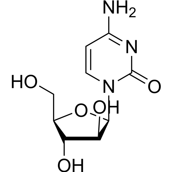
HY-13605
CytarabineDNA Synthesis Inhibitor -
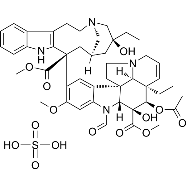
HY-N0488
Vincristine sulfateAntineoplastic Agent -
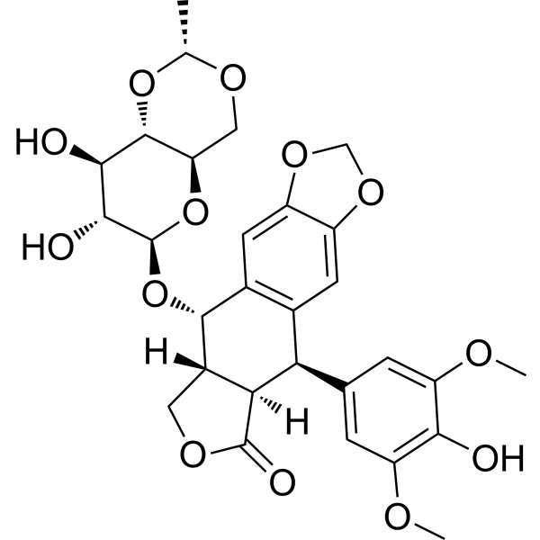
HY-13629
EtoposideTopoisomerase II Inhibitor -
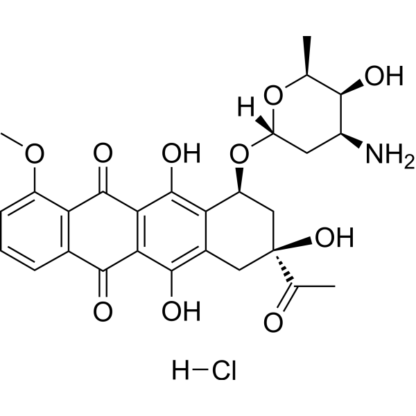
HY-13062
Daunorubicin hydrochlorideTopoisomerase II Inhibitor -
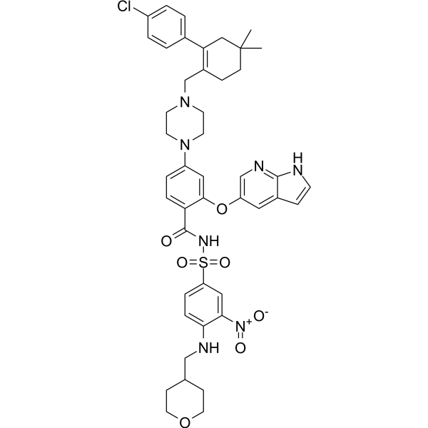
HY-15531
VenetoclaxBCL-2 Inhibitor -
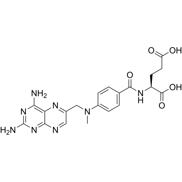
HY-14519
MethotrexateDihydrofolate Reductase Inhibitor -
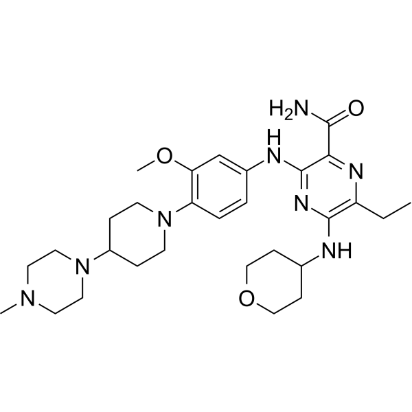
HY-12432
GilteritinibFLT3/AXL Inhibitor -
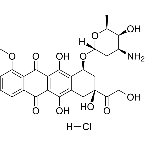
HY-15142
Doxorubicin hydrochlorideTopoisomerase Inhibitor -
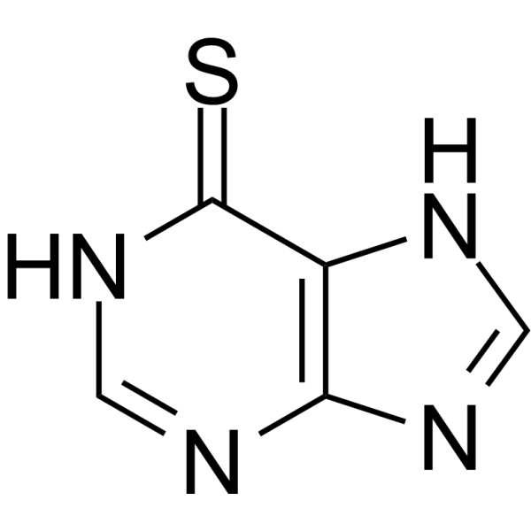
HY-13677
6-Mercaptopurine -
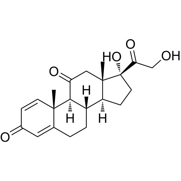
HY-B0214
PrednisoneGlucocorticoid Receptor Agonist -
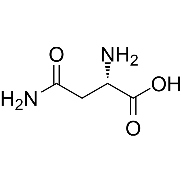
HY-N0667
L-AsparagineEndogenous Metabolite
MedChemExpress(MCE)の使用を引用している文献 L-Asparaginase
More- Nat Commun. 2025 Jul 1;16(1):5675. [Abstract]
- Adv Sci (Weinh). 2026 Mar 20:e16557. [Abstract]
- Cell Rep Med. 2025 May 20;6(5):102098. [Abstract]
- Sci Adv. 2025 Jun 20;11(25):eadt3075. [Abstract]
- Cell Death Dis. 2023 Aug 1;14(8):489. [Abstract]
- Mol Ther. 2025 Aug 12:S1525-0016(25)00650-1. [Abstract]
- EMBO Mol Med. 2023 Feb 8;15(2):e16554. [Abstract]
- Cell Rep. 2025 Nov 25;44(11):116496. [Abstract]
- Aging Cell. 2025 Oct;24(10):e70203. [Abstract]
- Biochem Pharmacol. 2023 Sep:215:115690. [Abstract]
- Cancers (Basel). 2022 Oct 19;14(20):5127. [Abstract]
- Front Mol Neurosci. 2020 Sep 30:13:563054. [Abstract]
- BMC Cancer. 2022 Jul 2;22(1):724. [Abstract]
- Front Oncol. 2024 Jul 19:14:1440650. [Abstract]
- Biochem Biophys Res Commun. 2026 Sep 24:832:154404.
- Blood Neoplasia. 2026 Apr 9;3(3):100230. [Abstract]
- bioRxiv. 2026 Mar 18.
- University of Montreal. 2026 Mar.
 Customer Validation & Images

-
Cell Migration/Invasion Assay
-
IHC
-
In Vivo Efficacy Study
-
Cell Proliferation/Viability Assay
-
Cell Imaging/Staining
DNA/RNA Synthesis アイソフォーム固有の製品をすべて表示
More

生物活性
製品説明
体外実験
L-Asparaginase has a cytotoxic effect on AML blasts and LSC subpopulations[1].
L-Asparaginase is effective on both CD34+CD38+ and CD34+CD38+ LSC fractions, MSC and monocytes/macrophages partially counteracted the effect[1].
Although lyophilized L-asparaginase is stable at room temperature for 3 weeks, it should be stored dry below -18°C. L-Asparaginase should Co-culture with cells at 37°C, 5% CO2 for 24, 48 h.
L-Asparaginase (100 IU/mL, 0-24 h) is co-cultured with L5178Y and B16 cells (5×10⁵/mL), and cells are arrested in the G1 phase and DNA strand breaks occurred[2].
Product Information
Optimal pH: 8.0-9.0
Agonists: Anaerobic conditions, amino acids
Inhibitors: 5-diazo-4-o-L-norvaline (DONV), dimethyl sulfoxide, glutaraldehyde, L-asparagine and L-aspartic acid, NH4+, trypsin
Instructions
Dissolve in 1 mg/ml 0.2 M Tris-HCl pH 8.6, store at 2-8°C after dissolution, and use as soon as possible within 1 week. Or dissolve according to specific experimental references.
MedChemExpress (MCE) has not independently confirmed the accuracy of these methods. They are for reference only.
臨床実験
| NCT Number | Sponsor | Condition | Start Date |
Phase

|
|---|---|---|---|---|
| NCT01329991 | Plexxikon| | 2011-05 | PHASE1 |
EC Number
3.5.1.1
Specific Activity
100 - 300 U/mg protein
Unit Definition
One unit will release 1.0μmol of ammonia from L-asparagine per minute at pH8.5 and 37°C.


化学情報
-
CAS 番号 9015-68-3
-
性状 Solid
-
分子量 34000 (monomer)
-
Color White to off-white
-
SMILES
[L-Asparaginase]
-
別名
L-Asparaginase; L-ASNase
-
輸送条件
Room temperature in continental US; may vary elsewhere.
-
保管条件
Please store the product under the recommended conditions in the Certificate of Analysis.


Publications (18)
-
Journal Impact Factor
-
Most Recent
-
Nat Commun
2025 Jul 1;16(1):5675. PMID: 40593710 -
Adv Sci (Weinh)
ETV4 Promotes Colorectal Cancer Progression by Reprogramming Asparagine Metabolism to Remodel the Stromal Microenvironment. [Abstract]2026 Mar 20:e16557. PMID: 41861091 -
Cell Rep Med
scRNA-seq reveals an immune microenvironment and JUN-mediated NK cell exhaustion in relapsed T-ALL. [Abstract]2025 May 20;6(5):102098. PMID: 40306275 -
Sci Adv
FAM50A drives breast cancer brain metastasis through interaction with C9ORF78 to enhance ʟ-asparagine production. [Abstract]2025 Jun 20;11(25):eadt3075. PMID: 40531994
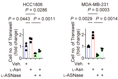
L-Asparaginase purchased from MedChemExpress. Usage Cited in: Sci Adv. 2025 Jun 20;11(25):eadt3075. [Abstract]
Transwell migration assay of indicated cells in the presence of ʟ-Asn(50 μg/ml) or ʟ-ASNase (L-Asparaginase; 10 μg/mL; 24-36 h).
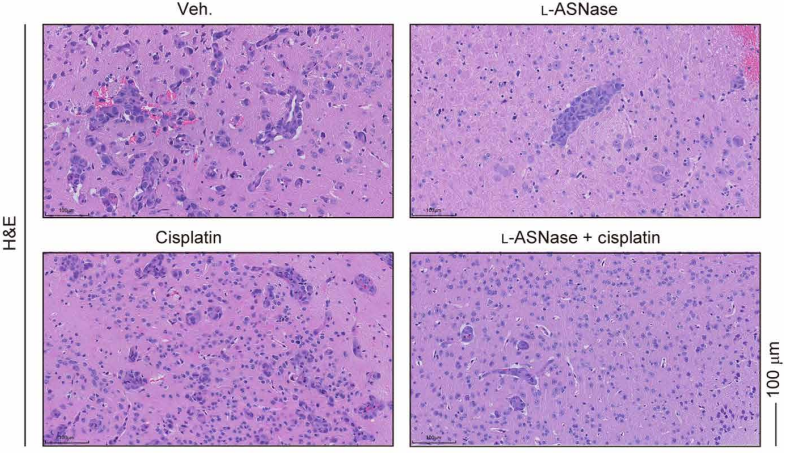
L-Asparaginase purchased from MedChemExpress. Usage Cited in: Sci Adv. 2025 Jun 20;11(25):eadt3075. [Abstract]
Brain slices of mice were stained with hematoxylin andeosin (H&E) at the endpoint. BCBM model mice, either cisplatin (4 mg/kg, intraperitoneally) or ʟ-ASNase (L-Asparaginase; 0.2 mg per mouse, intraperitoneally) was administered weekly or every 2 days for 2 weeks, respectively.
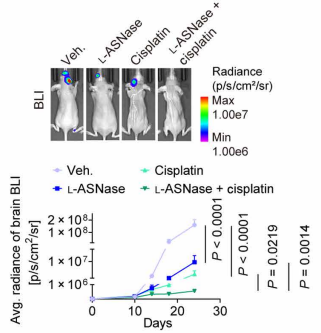
L-Asparaginase purchased from MedChemExpress. Usage Cited in: Sci Adv. 2025 Jun 20;11(25):eadt3075. [Abstract]
BLI was conducted at regular intervals. Representative BLI images and line chart of radiance. BCBM model, Mice were administered cisplatin (4 mg/kg, intraperitoneally) weekly or ʟ-ASNase (0.2 mg per mouse, intraperitoneally) every 2 days.
-
Cell Death Dis
NUCKS1, a LINC00629-upregulated gene, facilitated osteosarcoma progression and metastasis by elevating asparagine synthesis. [Abstract]2023 Aug 1;14(8):489. PMID: 37528150
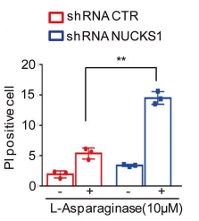
L-Asparaginase purchased from MedChemExpress. Usage Cited in: Cell Death Dis. 2023 Aug 1;14(8):489. [Abstract]
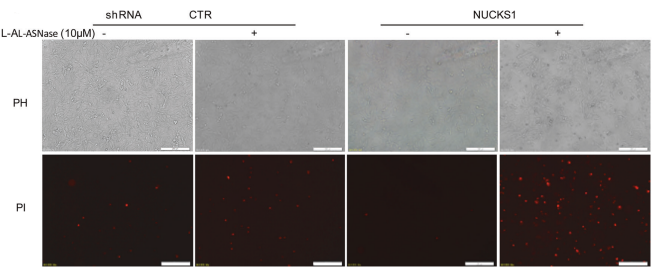
MNNG/HOS cells with or without NUCKS1 depletion were treated with 10 μM L-Asparaginase for 48 h. Cell death was measured by PI staining.
L-Asparaginase purchased from MedChemExpress. Usage Cited in: Cell Death Dis. 2023 Aug 1;14(8):489. [Abstract]
MNNG/HOS cells with or without NUCKS1 depletion were treated with 10 μM L-Asparaginase for 48 h. Cell death was measured by PI staining.
-
Mol Ther
Asparaginase enhances CAR-T cell antitumor immunity by asparagine metabolic reprogramming and central memory induction in ALL. [Abstract]2025 Aug 12:S1525-0016(25)00650-1. PMID: 40808257 -
EMBO Mol Med
β-Catenin activity induces an RNA biosynthesis program promoting therapy resistance in T-cell acute lymphoblastic leukemia. [Abstract]2023 Feb 8;15(2):e16554. PMID: 36597789 -
Cell Rep
A generalized strategy to kill leukemic cells by targeting the regulatory systems governing mitochondrial membrane potential. [Abstract]2025 Nov 25;44(11):116496. PMID: 41166305 -
Aging Cell
Asparaginase and Autophagy Inhibitors Effectively Remove Senescent Cells by Synergistically Limiting Asparagine Supply. [Abstract]2025 Oct;24(10):e70203. PMID: 40905331 -
Biochem Pharmacol
Methylseleninic acid overcomes gefitinib resistance through asparagine-MET-TOPK signaling axis in non-small cell lung cancer cells. [Abstract]2023 Sep:215:115690. PMID: 37481142 -
Cancers (Basel)
Association between Dysfunction of the Nucleolar Stress Response and Multidrug Resistance in Pediatric Acute Lymphoblastic Leukemia. [Abstract]2022 Oct 19;14(20):5127. PMID: 36291909 -
Front Mol Neurosci
L-Asparaginase Exerts Neuroprotective Effects in an SH-SY5Y-A53T Model of Parkinson's Disease by Regulating Glutamine Metabolism. [Abstract]2020 Sep 30:13:563054. PMID: 33117129 -
BMC Cancer
2022 Jul 2;22(1):724. PMID: 35778693 -
Front Oncol
Hypoxanthine in the microenvironment can enable thiopurine resistance in acute lymphoblastic leukemia. [Abstract]2024 Jul 19:14:1440650. PMID: 39099696 -
-
Blood Neoplasia
From cell lines to PDXs: in vivo confirmation of synergistic drug responses identified in leukemia cell line models. [Abstract]2026 Apr 9;3(3):100230. PMID: 42294111 -
-


溶剤 & 溶解度
体外:
H2O : ≥ 100 mg/mL
* "≥" means soluble, but saturation unknown.
体内:
For the following dissolution methods, please prepare the working solution directly:
It is recommended to prepare fresh solutions and use them promptly within a short period of time.
The percentages shown for the solvents indicate their volumetric ratio in the final prepared solution. If precipitation or phase separation occurs during preparation, heat and/or sonication can be used to aid dissolution.
Add each solvent one by one: PBS
Solubility: 25 mg/mL; Clear solution; Need ultrasonic


純度とドキュメンテーション
-
データシート (271 KB)
-
SDS (419 KB)
- English - EN (419 KB)
- Français - FR (419 KB)
- Deutsch - DE (419 KB)
- Norwegian - NO (419 KB)
- Español - ES (419 KB)
- Swedish - SV (419 KB)
- Italian - IT (419 KB)
- Korean - KR (419 KB)
- Portuguese - PT (419 KB)
-
取扱説明書 (2659 KB)
参考文献
[1]. Ilaria M Michelozzi, et al. Acute myeloid leukaemia niche regulates response to L-asparaginase. Br J Haematol. 2019 Aug;186(3):420-430. doi: 10.1111/bjh.15920. [Content Brief]
[2]. T Ueno, et al. Cell cycle arrest and apoptosis of leukemia cells induced by L-asparaginase. Leukemia. 1997 Nov;11(11):1858-61. [Content Brief]

Calculators
濃度 (開始) × 体積 (開始) = 濃度 (終了) × 体積 (終了)


L-Asparaginase 関連分類
HY-P1923 関連分類
 Powered by Bioz