



Lipase,Aspergillusniger (Immobilized)

Based on 1 Customer Validation
Lipase, Aspergillus niger (Immobilized) is an immobilized triacylglycerol acyl hydrolase and biocatalyst. Lipase, Aspergillus niger (Immobilized) broadly catalyzes the hydrolysis of glycerol-long-chain fatty acid esters, exhibits positional selectivity for the 1,3-positions of glycerol, but shows low hydrolysis efficiency for castor oil. Lipase, Aspergillus niger (Immobilized) displays optimal activity at 45°C and pH 7.0, and remains stable at temperatures up to 60°C and over a broad pH range of 2.0-9.0. Lipase, Aspergillus niger (Immobilized) is activated by Ca2+ and Mg2+, but inhibited by Mn2+, Fe2+ and Zn2+.
연구목적의 판매만을 진행합니다. 환자를 대상으로 한 판매는 하지 않습니다.
- CAS No.: 9001-62-1
-
보관:
Please store the product under the recommended conditions in the Certificate of Analysis.


To place orders, for customer services and technical support, please contact: MedChemExpress USA
Tel: 609-228-6898 E-mail: [email protected] [email protected]


-
 Biological Activity
-
Chemical Information
-
용액&용해도
- Protocol
- 순도&문서
- References
-
 Help & FAQs


-

HY-Y1422D
Lipase,Candida antarctica (Immobilized)Immobilized Lipase -

HY-Y1422I
Lipase,Pseudomonas fluorescens (Immobilized)Triacylglycerol Carboxylic Ester Bonds Catalyst, Short to Middle-Chain Fatty Acid Methyl-Ester Ester Bonds Catalyst, Triglyceride Ester Bonds Catalyst, Triolein 1,3-Ester Bonds Preferential Cleaver, EPA- and DHA-Containing Fats and Oils Decomposer -

HY-Y1422H
Lipase,Candida cylindracea (Immobilized)Biocatalyst -
HY-Y1422
Triacylglycerol lipaseTriglyceride Hydrolase -
HY-P2902
Glucose oxidaseOxidoreductase Enzyme -

HY-Y1422B
Lipase (MS grade)Lipase -

HY-Y1422J
Lipase,Burkholderiacepacia (Immobilized)Catalyzing Enzyme -
HY-14471
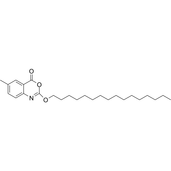
CetilistatLipase Inhibitor -

HY-14471R
Cetilistat (Standard)Lipase Inhibitor


Biological Activity
제품 설명
In Vitro
Lipase,Aspergillusniger (Immobilized) remains stable for 24 hours in 40% (V/V) methanol, 2-propanol, and methyl acetate. It tolerates high concentrations of water-miscible organic solvents, exhibits no relevant protease activity, and shows enhanced activity after pre-incubation at 37 or 40 °C in a medium supplemented with olive oil[1][2].
MedChemExpress (MCE) has not independently confirmed the accuracy of these methods. They are for reference only.


Chemical Information
-
CAS No. 9001-62-1
-
Appearance Solid
-
Color Yellow to brown
-
SMILES
[Lipase,Aspergillusniger (Immobilized)]
-
선적
Room temperature in continental US; may vary elsewhere.
-
보관
Please store the product under the recommended conditions in the Certificate of Analysis.


용액&용해도
In Vitro:
H2O : ≥ 3.33 mg/mL
* "≥" means soluble, but saturation unknown.


순도&문서
-
Data Sheet (265 KB)
-
SDS (251 KB)
- English - EN (251 KB)
- Français - FR (251 KB)
- Deutsch - DE (251 KB)
- Norwegian - NO (251 KB)
- Español - ES (251 KB)
- Swedish - SV (251 KB)
- Italian - IT (251 KB)
- Korean - KR (251 KB)
- Portuguese - PT (251 KB)
-
Handling Instructions (2659 KB)
References

Calculators
Concentration (start) × Volume (start) = Concentration (final) × Volume (final)


Lipase,Aspergillusniger (Immobilized) Related Classifications
HY-Y1422E Related Classifications
 Powered by Bioz