
Olopatadine
Based on 1 Customer Validation
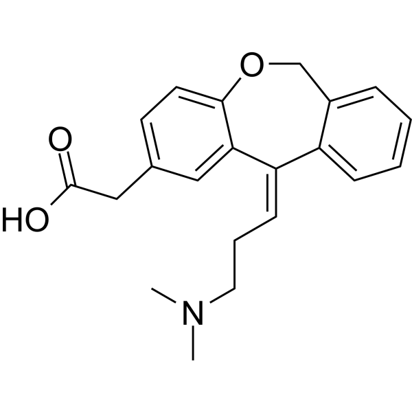
Olopatadine (ALO4943A; KW4679) is an orally active histamine H1 receptor antagonist and mast cell stabilizer. Olopatadine exerts antiallergic effects by blocking histamine H1 receptor-mediated activities. Olopatadine inhibits exocytosis, chemokine release, F-actin polymerization, CXCL10-induced calcium influx, and T cell chemotactic activity. Olopatadine also reduces the expression levels of CXCR3 on the surface of CD4+ and CD8+ T cells. Olopatadine inhibits scratching behavior, improves dermatitis scores, and suppresses intraepidermal neurite outgrowth. Olopatadine simultaneously decreases the levels of inflammatory markers, growth factors, histamine, and specific IgE, while increasing the expression of ErbB3A/HER3A. Olopatadine can be used in research related to seasonal pollinosis, chronic rhinitis, urticaria, allergic conjunctivitis, alopecia areata, and atopic dermatitis.
For research use only. We do not sell to patients.
- Purity : 98.94%
- CAS No.: 113806-05-6
- Formula: C21H23NO3
- Molecular Weight:337.41
-
Storage:
4°C, protect from light
* In solvent : -80°C, 6 months; -20°C, 1 month (protect from light)
All Histamine Receptor Isoforms
More
Biological Activity
Description
IC50 & Target
[1]|
H1 Receptor |
In Vitro
Olopatadine (1×10⁻⁶-1×10⁻⁵ M; 8 h) significantly inhibits the CXCL10-mediated chemotactic migration speed of CD4+ and CD8+ T cells isolated from patients with acute alopecia areata, with a stronger inhibitory effect at high concentrations than at low concentrations[2].
Olopatadine (1×10⁻⁶-1×10⁻⁵ M) significantly reduces the CXCR3 expression level and F-actin polymerization on the surface of CD4+ and CD8+ T cells isolated from patients with acute alopecia areata[2].
Olopatadine (1×10⁻⁶-1×10⁻⁵ M) significantly inhibits CXCL10-induced intracellular calcium influx in PBMCs isolated from one patient with acute alopecia areata, with a stronger inhibitory effect observed at the high concentration than at the low concentration[2].
MedChemExpress (MCE) has not independently confirmed the accuracy of these methods. They are for reference only. Further protocols information, click here.
In Vivo
MedChemExpress (MCE) has not independently confirmed the accuracy of these methods. They are for reference only.
Chemical Information
-
CAS No. 113806-05-6
-
Appearance Solid
-
Molecular Weight 337.41
-
Formula C21H23NO3
-
Color White to off-white
-
SMILES
O=C(O)CC1=CC=C(C/2=C1)OCC3=CC=CC=C3C2=C\CCN(C)C
-
Shipping
Room temperature in continental US; may vary elsewhere.
-
Storage
4°C, protect from light
* In solvent : -80°C, 6 months; -20°C, 1 month (protect from light)
Protocols
-
PC12 NGF-Induced Neuronal Differentiation Culture
PC12 cells are a rat adrenal pheochromocytoma clonal line that responds to NGF by stopping proliferation and extending branching neurite-like processes; after longer NGF exposure, cells develop long processes and neuronal-like ultrastructural and functional features. NGF-induced differentiation is read out mainly by neurite outgrowth, reduced proliferation, microtubule assembly, and neuronal differentiation-associated proteins such as MAPs, tau, GAP-43, and synapsin-1.
-
RNA extraction experimental
By lysing cells, releasing RNA, and removing impurities such as proteins and DNA, high-purity RNA products are finally obtained. The commonly used traditional method is the guanidine isothiocyanate/phenol/chloroform method (Trizol), which is suitable for a variety of animal materials including animal tissues, microorganisms, cultured cells, etc., and most plant materials.
-
SH-SY5Y Neuronal Differentiation Culture
SH-SY5Y neuronal differentiation culture uses sequential exposure to retinoic acid and neurotrophic factors to reduce proliferative neuroblastoma-like behavior and induce neuron-like morphology, including neurite extension, neuronal marker expression, and, in RA/BDNF protocols, greater synaptic-marker expression than undifferentiated culture. Retinoic acid is commonly used as the initiating differentiation cue, while BDNF in serum-reduced or serum-free medium supports later maturation and neurotrophic-factor-dependent neuron-like survival.
-
TPA/Croton Oil Ear Edema and Dermatitis
The TPA (12-O-tetradecanoylphorbol-13-acetate) and croton oil-induced mouse ear edema model is a well-established acute cutaneous inflammation system used to evaluate topical anti-inflammatory activity by measuring edema formation, neutrophil infiltration, vascular permeability, and cytokine-mediated skin responses in vivo. The inflammatory response is triggered by topical application of phorbol esters (TPA) or croton oil constituents, leading to rapid activation of protein kinase C signaling, leukocyte recruitment, and increased vascular permeability, which can be quantified by ear thickness, weight, dye extravasation, and biochemical markers such as myeloperoxidase (MPO) activity and pro-inflammatory mediators in ear tissue homogenates. This model is widely used for screening anti-inflammatory agents, where reductions in edema and inflammatory biomarkers reflect suppression of acute dermal inflammation and immune cell infiltration. Histological evaluation typically confirms epidermal
-
SH-SY5Y neuronal-like differentiation
SH-SY5Y neuronal-like differentiation uses defined culture conditions to shift proliferative human neuroblastoma cells toward a neuron-like state, mainly assessed by reduced proliferation, neurite extension, neuronal-marker expression, and, in some protocols, increased dependence on neurotrophic support. Retinoic acid (RA) is commonly used for the first differentiation phase, and sequential RA followed by brain-derived neurotrophic factor (BDNF) in serum-free medium is a well-characterized approach for generating neuron-like SH-SY5Y cultures with extensive neurite outgrowth. The primary readouts are morphology-based neurite outgrowth and marker-based confirmation using proteins such as βIII-tubulin, MAP2, GAP43, synaptophysin, NeuN, NSE, TH, or related neuronal/synaptic markers, depending on the study endpoint.
-
Cell differentiation
Cell differentiation refers to the process in which cells of the same origin gradually produce cell groups with different morphological structure and functional characteristics.
-
Ca2+ Staining Technique
Ca2+ staining is an experimental technique that utilizes specific fluorescent probes (such as Fluo-4 AM, Fura-2, etc.) to qualitatively or quantitatively detect dynamic changes in intracellular Ca2+ concentrations; this is achieved by monitoring the changes in fluorescent signals generated when these probes bind to free intracellular calcium ions. The underlying principle relies primarily on the presence of chelating groups within the probe's molecular structure that possess high affinity for calcium ions.
-
PC12 NGF-induced neuronal-like differentiation
PC12 cells are a rat adrenal pheochromocytoma-derived clonal cell line that responds to nerve growth factor by stopping proliferation and extending neurites, producing a sympathetic neuron-like phenotype used to study neuronal differentiation and neurite outgrowth. NGF acts through TrkA-dependent signaling, and neurite outgrowth is associated with ERK/Akt signaling, microtubule organization, neuronal-marker expression, and increased electrophysiological neuronal features such as sodium-channel density. The main assay readout is morphological differentiation, usually measured as the percentage of neurite-bearing cells, neurite length, neurite number, or total neurite length per cell. Additional readouts include GAP-43, tyrosine hydroxylase, βIII-tubulin, neurofilament, synapsin I, synaptophysin, ERK phosphorylation, Akt phosphorylation, and sodium-channel current density.
-
Research Protocol for Cancer Immunology
Cancer immunology studies how the immune system recognizes, suppresses, edits, or fails to eliminate malignant cells through tumor antigen release, antigen presentation, T-cell priming, immune trafficking, tumor-cell killing, and feedback inhibition in the tumor microenvironment. The cancer-immunity cycle links tumor antigenicity, dendritic-cell priming, CD8+ T-cell infiltration, cytotoxic function, and immune-checkpoint regulation to tumor rejection or immune escape. Immune-checkpoint pathways such as PD-1/PD-L1 and CTLA-4 suppress antitumor T-cell activity and can be therapeutically blocked, but many tumors remain resistant because of poor antigen presentation, weak T-cell infiltration, suppressive myeloid cells, regulatory T cells, and tumor-intrinsic immune-exclusion programs. Unresolved questions include which immune-cell states predict response, how tumor-intrinsic pathways exclude immune cells, how myeloid suppression limits checkpoint blockade, and which combination strategies
-
Phalloidin F-actin cytoskeleton staining
Phalloidin F-actin staining detects polymerized filamentous actin in fixed and permeabilized specimens by using fluorescent phalloidin or phalloidin-derived phallotoxins that bind actin filaments and generate a fluorescence microscopy readout corresponding to F-actin organization, including stress fibers, cortical actin, filament bundles, and tissue-specific actin networks. Phalloidin stabilizes F-actin by reducing actin subunit dissociation from filament ends, and fluorescent phallotoxins were established as tools for visualizing actin-containing structures in eukaryotic cells.
Purity & Documentation
-
Data Sheet (287 KB)
-
SDS (393 KB)
- English - EN (393 KB)
- Français - FR (393 KB)
- Deutsch - DE (393 KB)
- Norwegian - NO (393 KB)
- Español - ES (393 KB)
- Swedish - SV (393 KB)
- Italian - IT (393 KB)
- Korean - KR (393 KB)
- Portuguese - PT (393 KB)
-
Handling Instructions (2659 KB)
References
[1]. Baba A, et al. Olopatadine inhibits exocytosis in rat peritoneal mast cells by counteracting membrane surface deformation. Cell Physiol Biochem. 2015;35(1):386-396. [Content Brief]
[3]. Murota H, et al. Olopatadine hydrochloride improves dermatitis score and inhibits scratch behavior in NC/Nga mice. Int Arch Allergy Immunol. 2010;153(2):121-132. [Content Brief]
Calculators
Concentration (start) × Volume (start) = Concentration (final) × Volume (final)

