



SD-1029
Based on 1 publication(s) in Google Scholar
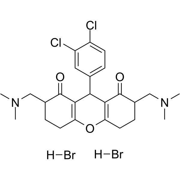
SD-1029 is a JAK2/STAT3 inhibitor. SD-1029 inhibits STAT3 nuclear translocation. SD-1029 is an inhibitor of STAT3 activation due to inhibition of JAK2 phosphorylation.
For research use only. We do not sell to patients.
- Purity : 88.8%
- CAS No.: 118372-34-2
- Formula: C25H32Br2Cl2N2O3
- Molecular Weight:639.25
-
Storage:
-20°C, sealed storage, away from moisture
* In solvent : -80°C, 6 months; -20°C, 1 month (sealed storage, away from moisture)

To place orders, for customer services and technical support, please contact: MedChemExpress USA
Tel: 609-228-6898 E-mail: [email protected] [email protected]


-
 Biological Activity
-
Chemical Information
- Protocols
- Purity & Documentation
- References
-
 Help & FAQs


Publications Citing Use of MedChemExpress (MCE) SD-1029
More

Biological Activity
Description
IC50 & Target
[1]|
STAT3 |
JAK2 |
In Vitro
SD-1029 (10 μM) inhibits EGFP-Stat3 nuclear translocation in BHK-21 and U-2OS cells[1].
SD-1029 (5 and 10 μM; 24 hours) inhibits cell growth and induces apoptosis in OVCAR8TR ovarian cancer cells[1].
SD-1029 (10 μM; 24 hours) suppresses p-Stat3 levels in human breast and ovarian cancer cell lines[1].
SD-1029 inhibits not only JAK2 phosphorylation, but also the phosphorylation of STAT1 and STAT3. SD-1029 strongly inhibits Tyk2 phosphorylation, implicating both JAK2 and Tyk2 as upstream requirements for IL-23-induced IL-23R expression[2].
MedChemExpress (MCE) has not independently confirmed the accuracy of these methods. They are for reference only.
-
Cell Line:OVCAR8TR ovarian cancer cells
-
Concentration:5 and 10 μM
-
Incubation Time:24 hours
-
Result:Down-regulated cell proliferation, and induced apoptotic cell death.
Treatment resulted in a marked, 20-fold induction of apoptosis in the OVCAR8TR cells that express constitutively activated Stat3.
-
Cell Line:MDA-MB-468 and MDA435 (breast cancer), OV1063 (ovarian cancer), and the paclitaxel-resistant ovarian cancer daughter lines, SKOV-3TR and OVCAR8TR.
-
Concentration:10 μM
-
Incubation Time:24 hours
-
Result:Led to reduced levels of pStat3.


Chemical Information
-
CAS No. 118372-34-2
-
Appearance Solid
-
Molecular Weight 639.25
-
Formula C25H32Br2Cl2N2O3
-
Color White to off-white
-
SMILES
O=C1C(C(C2=CC=C(Cl)C(Cl)=C2)C3=C(CCC(CN(C)C)C3=O)O4)=C4CCC1CN(C)C.[H]Br.[H]Br
-
Shipping
Room temperature in continental US; may vary elsewhere.
-
Storage
-20°C, sealed storage, away from moisture
* In solvent : -80°C, 6 months; -20°C, 1 month (sealed storage, away from moisture)


Publications (1)
-
Journal Impact Factor
-
Most Recent
-
Exp Ther Med
CX3CL1/CX3CR1 axis alleviates inflammation and apoptosis in human nucleus pulpous cells via M2 macrophage polarization. [Abstract]2023 Jun 7;26(1):359. PMID: 37324510


Purity & Documentation
-
Data Sheet (276 KB)
-
SDS (252 KB)
- English - EN (252 KB)
- Français - FR (252 KB)
- Deutsch - DE (252 KB)
- Norwegian - NO (252 KB)
- Español - ES (252 KB)
- Swedish - SV (252 KB)
- Italian - IT (252 KB)
- Korean - KR (252 KB)
- Portuguese - PT (252 KB)
-
Handling Instructions (2659 KB)
References
[1]. Zhenfeng Duan, et al. SD-1029 inhibits signal transducer and activator of transcription 3 nuclear translocation. Clin Cancer Res. 2006 Nov 15;12(22):6844-52. [Content Brief]
[2]. Nor Fazila Che Mat, et al. Interleukin-23-induced interleukin-23 receptor subunit expression is mediated by the Janus kinase/signal transducer and activation of transcription pathway in human CD4 T cells. J Interferon Cytokine Res. 2011 Apr;31(4):363-71. [Content Brief]

Calculators
Concentration (start) × Volume (start) = Concentration (final) × Volume (final)


SD-1029 Related Classifications
HY-112391 Related Classifications
 Powered by Bioz