



STAT3-IN-48
Based on 1 publication(s) in Google Scholar
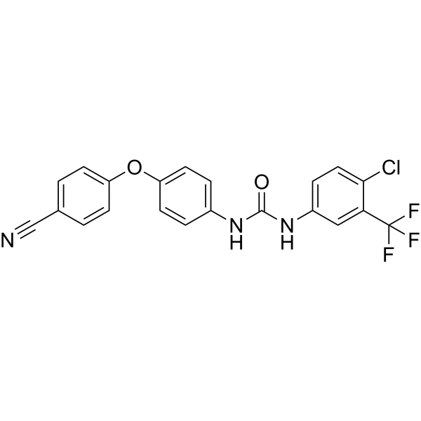
STAT3-IN-48 is a Sorafenib analogue and potently inhibits the phosphorylation of STAT3. STAT3-IN-48 induces cell apoptosis through SHP-1 dependent STAT3 inactivation. STAT3-IN-48 does not inhibit kinase activity and has anticancer effects.
For research use only. We do not sell to patients.
- Purity : 98.64%
- CAS No.: 1313019-65-6
- Formula: C21H13ClF3N3O2
- Molecular Weight:431.80
-
Storage:Powder -20°C, 3 years , 4°C, 2 years ; In solvent -80°C, 6 months , -20°C, 1 month

To place orders, for customer services and technical support, please contact: MedChemExpress USA
Tel: 609-228-6898 E-mail: [email protected] [email protected]


-
 Biological Activity
-
Chemical Information
-
Solvent & Solubility
- Protocols
- Purity & Documentation
- References
-
 Help & FAQs


-
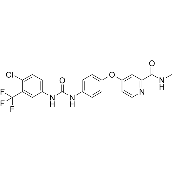
HY-10201
SorafenibMultikinase Inhibitor -
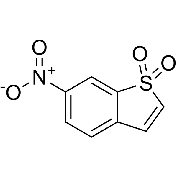
HY-13818
StatticSTAT3 Inhibitor -
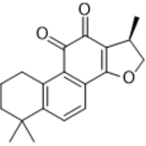
HY-N0174
CryptotanshinoneAutophagy Inducer -
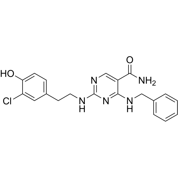
HY-100614
AS1517499STAT6 Inhibitor -
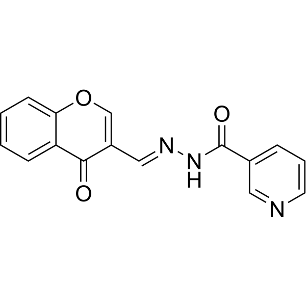
HY-101853
STAT5-IN-1STAT5 Inhibitor -
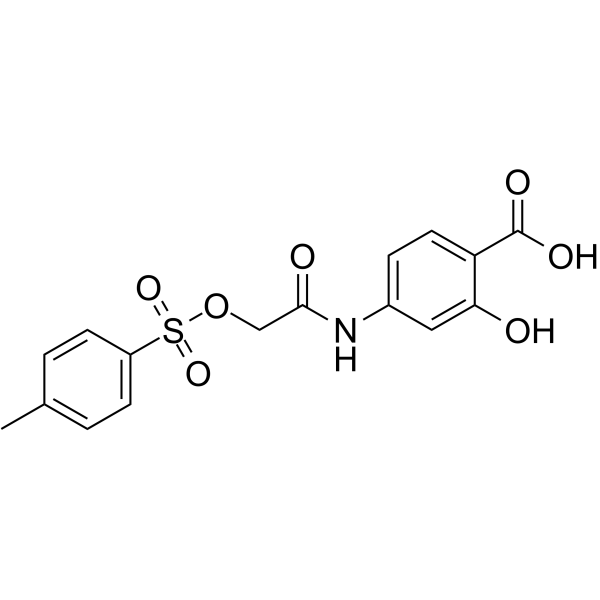
HY-15146
NSC 74859STAT3 Inhibitor -
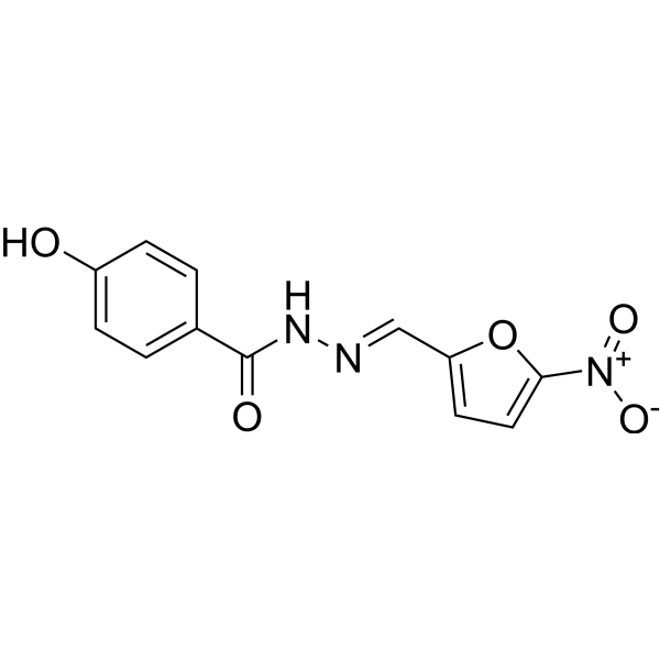
HY-B1436
NifuroxazideBacterial Inhibitor -
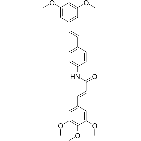
HY-100753
STAT3-IN-1STAT3 Inhibitor -
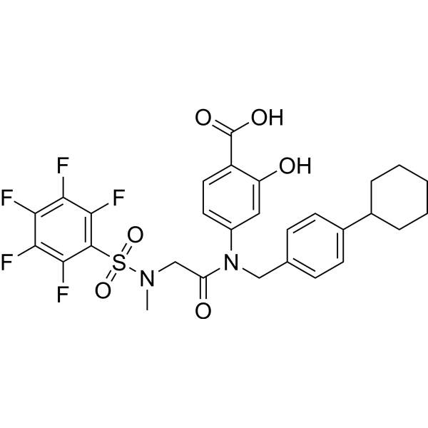
HY-100493
BP-1-102STAT3 Inhibitor -
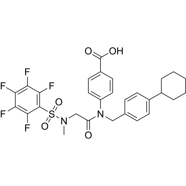
HY-16975
SH-4-54STAT3/5 Inhibitor -
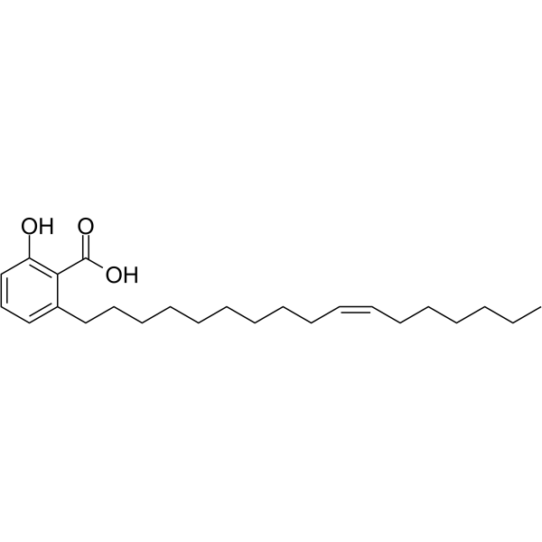
HY-N2116
Ginkgolic acid C17:1Antitumor Agent -
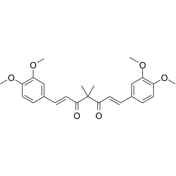
HY-N2521
TetramethylcurcuminSTAT3 Phosphorylation Inhibitor
Publications Citing Use of MedChemExpress (MCE) STAT3-IN-48
More

Biological Activity
Description
IC50 & Target
[1]|
p-STAT3 |
Cellular Effect
|
Cell Line

|
Type | Value | Description | References |
|---|---|---|---|---|
| PLC-PRF-5 | IC50 |
7.5 μM
Compound: 1
|
Cytotoxicity against human PLC/PRF/5 cells assessed as growth inhibition
Cytotoxicity against human PLC/PRF/5 cells assessed as growth inhibition
|
[PMID: 21531053] |
In Vitro
STAT3-IN-48 (SC-1; 1-10 μM; 48 hours; breast cancer cells) treatment demonstrates dose-dependent suppression of cell viability in all tested breast cancer cells[1].
STAT3-IN-48 (SC-1; 1-10 μM; 36 hours; breast cancer cells) treatment induces potent apoptotic activity[1].
STAT3-IN-48 (SC-1; 1-10 μM; 36 hours; breast cancer cells) treatment shows downregulation of p-STAT3 and its downstream proteins cyclin D1 and survivin in a dose-dependent manner in breast cancer cell lines[1].
MedChemExpress (MCE) has not independently confirmed the accuracy of these methods. They are for reference only.
-
Cell Line:HCC-1937, MDA-MB-231, MDA-MB-468, MDA-MB-453, SK-BR3 and MCF-7 cells
-
Concentration:1 μM, 2 μM, 5 μM, 7.5 μM, 10 μM
-
Incubation Time:48 hours
-
Result:Demonstrated dose-dependent suppression of cell viability in all tested breast cancer cells.
-
Cell Line:HCC-1937, MDA-MB-231, MDA-MB-468, MDA-MB-453, SK-BR3 and MCF-7 cells
-
Concentration:1 μM, 2 μM, 5 μM, 7.5 μM, 10 μM
-
Incubation Time:36 hours
-
Result:Induced potent apoptotic activity.
-
Cell Line:HCC-1937, MDA-MB-231, MDA-MB-468, MDA-MB-453, SK-BR3 and MCF-7 cells
-
Concentration:1 μM, 2 μM, 5 μM, 7.5 μM, 10 μM
-
Incubation Time:36 hours
-
Result:Showed downregulation of p-STAT3 and its downstream proteins cyclin D1 and survivin in a dose-dependent manner in breast cancer cell lines.
In Vivo
MedChemExpress (MCE) has not independently confirmed the accuracy of these methods. They are for reference only.
-
Animal Model:Female NCr athymic nude mice (4-6 weeks of age) injected with breast cancer cells [1]
-
Dosage:10 mg/kg
-
Administration:Oral gavage; daily; for 28 days
-
Result:Showed efficacious antitumor activity and p-STAT3 downregulation in MDA-MB-468 xenograft tumors.


Chemical Information
-
CAS No. 1313019-65-6
-
Appearance Solid
-
Molecular Weight 431.80
-
Formula C21H13ClF3N3O2
-
Color White to off-white
-
SMILES
O=C(NC1=CC=C(OC2=CC=C(C#N)C=C2)C=C1)NC3=CC=C(Cl)C(C(F)(F)F)=C3
-
Shipping
Room temperature in continental US; may vary elsewhere.
-
Storage
Powder -20°C 3 years 4°C 2 years In solvent -80°C 6 months -20°C 1 month


Publications (1)
-
Journal Impact Factor
-
Most Recent
-
Cancer Manag Res
Inhibition of microRNA-15b-5p Attenuates the Progression of Oral Squamous Cell Carcinoma via Modulating the PTPN4/STAT3 Axis. [Abstract]2020 Oct 28:12:10559-10572. PMID: 33149666


Solvent & Solubility
In Vitro:
DMSO : 250 mg/mL (578.97 mM; Need ultrasonic; Hygroscopic DMSO has a significant impact on the solubility of product, please use newly opened DMSO)
Please refer to the solubility information to select the appropriate solvent. Once prepared, please aliquot and store the solution to prevent product inactivation from repeated freeze-thaw cycles.
Storage method and period of stock solution: -80°C, 6 months; -20°C, 1 month. When stored at -80°C, please use it within 6 months. When stored at -20°C, please use it within 1 month.
Please refer to the solubility information to select the appropriate solvent. Once prepared, please aliquot and store the solution to prevent product inactivation from repeated freeze-thaw cycles.
Storage method and period of stock solution: -80°C, 6 months; -20°C, 1 month. When stored at -80°C, please use it within 6 months. When stored at -20°C, please use it within 1 month.
Concentration (start) × Volume (start) = Concentration (final) × Volume (final)


Purity & Documentation
-
Data Sheet (274 KB)
-
SDS (393 KB)
- English - EN (393 KB)
- Français - FR (393 KB)
- Deutsch - DE (393 KB)
- Norwegian - NO (393 KB)
- Español - ES (393 KB)
- Swedish - SV (393 KB)
- Italian - IT (393 KB)
- Korean - KR (393 KB)
- Portuguese - PT (393 KB)
-
Handling Instructions (2659 KB)
References


Complete Stock Solution Preparation Table
Please refer to the solubility information to select the appropriate solvent. Once prepared, please aliquot and store the solution to prevent product inactivation from repeated freeze-thaw cycles.
Storage method and period of stock solution: -80°C, 6 months; -20°C, 1 month. When stored at -80°C, please use it within 6 months. When stored at -20°C, please use it within 1 month.
| Optional Solvent | Concentration Solvent Mass | 1 mg | 5 mg | 10 mg | 25 mg |
|---|---|---|---|---|---|
| DMSO | 1 mM | 2.3159 mL | 11.5794 mL | 23.1589 mL | 57.8972 mL |
| 5 mM | 0.4632 mL | 2.3159 mL | 4.6318 mL | 11.5794 mL | |
| 10 mM | 0.2316 mL | 1.1579 mL | 2.3159 mL | 5.7897 mL | |
| 15 mM | 0.1544 mL | 0.7720 mL | 1.5439 mL | 3.8598 mL | |
| 20 mM | 0.1158 mL | 0.5790 mL | 1.1579 mL | 2.8949 mL | |
| 25 mM | 0.0926 mL | 0.4632 mL | 0.9264 mL | 2.3159 mL | |
| 30 mM | 0.0772 mL | 0.3860 mL | 0.7720 mL | 1.9299 mL | |
| 40 mM | 0.0579 mL | 0.2895 mL | 0.5790 mL | 1.4474 mL | |
| 50 mM | 0.0463 mL | 0.2316 mL | 0.4632 mL | 1.1579 mL | |
| 60 mM | 0.0386 mL | 0.1930 mL | 0.3860 mL | 0.9650 mL | |
| 80 mM | 0.0289 mL | 0.1447 mL | 0.2895 mL | 0.7237 mL | |
| 100 mM | 0.0232 mL | 0.1158 mL | 0.2316 mL | 0.5790 mL |


STAT3-IN-48 Related Classifications
HY-136658 Related Classifications
 Powered by Bioz