



Quadrol

Based on 1 Customer Validation
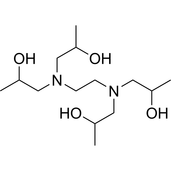
Quadrol is an immunostimulant that has been considered as a potentially useful active molecule for accelerating wound healing. Quadrol can form a complex with Ca2+, mediate the entry of Ca2+ into macrophages, and stimulate macrophage activation.
For research use only. We do not sell to patients.
- Purity : 99.96%
- CAS No.: 102-60-3
- Formula: C14H32N2O4
- Molecular Weight:292.41
-
Storage:Pure form -20°C, 3 years , 4°C, 2 years ; In solvent -80°C, 6 months , -20°C, 1 month

To place orders, for customer services and technical support, please contact: MedChemExpress USA
Tel: 609-228-6898 E-mail: [email protected] [email protected]


-
 Biological Activity
-
Chemical Information
-
Solvent & Solubility
- Protocols
- Purity & Documentation
- References
-
 Help & FAQs


-

HY-W047709
N,N,N',N'-Tetrakis(2-hydroxyethyl)ethylenediamineBiochemical Reagent -
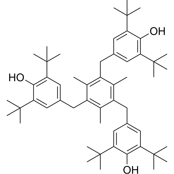
HY-120388
Ionox 330Antioxidant -

HY-Y1684
N,N'-Dimethyl-1,3-propylenediaminePROTAC Linker -

HY-W010653
Antioxidant 1010Antioxidant Agent -

HY-W012663
1,2,6-HexanetriolPolyol -

HY-W011696
Oleylamine, 80-90%Nanoparticle Modification Reagent -

HY-120388R
Ionox 330 (Standard)Standard of Ionox 330 -

HY-W235067
N,N-DimethyldodecylamineDynamin I Inhibitor -
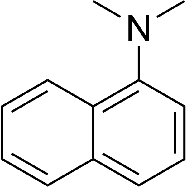
HY-D0222
N,N-Dimethyl-1-naphthylamineDye -

HY-W010653R
Antioxidant 1010 (Standard)Antioxidant Agent -

HY-W011588R
2-Ethylhexyl 4-(dimethylamino)benzoate (Standard) -

HY-125793
N,N-DimethylethylamineCatalyst
All Calcium Channel Isoforms
More

Biological Activity
Description
In Vitro
Results show that viability of macrophages incubated with Quadrol at concentrations of 0.5 mM, 1 mM and 4mM are identical to that for controls. Viability, however, is reduced to 50% of control at concentrations of 16 mM to 32 mM. After four hours, at concentrations of 1 mM and 4 mM, Quadrol produces enhanced spreading of 88% and 80%, respectively, as compared to the control of 28%. Quadrol, at a concentration of 16 mM, shows reduced percentage of spreading of macrophages after four hours. Exposure of macrophages to 1.0 mM or 4.0 mM Quadrol concentrations enhances phagocytic activity, 41% and 57%, respectively, over that of the cells exposed to media alone (34%)[1].
MedChemExpress (MCE) has not independently confirmed the accuracy of these methods. They are for reference only.
In Vivo
MedChemExpress (MCE) has not independently confirmed the accuracy of these methods. They are for reference only.


Chemical Information
-
CAS No. 102-60-3
-
Appearance Oil
-
Molecular Weight 292.41
-
Formula C14H32N2O4
-
Color Colorless to light yellow
-
SMILES
CC(O)CN(CC(O)C)CCN(CC(O)C)CC(O)C
-
Synonyms
N,N,N′,N′-Tetrakis(2-hydroxypropyl)ethylenediamine; EDTP
-
Shipping
Room temperature in continental US; may vary elsewhere.
-
Storage
Pure form -20°C 3 years 4°C 2 years In solvent -80°C 6 months -20°C 1 month


Solvent & Solubility
In Vitro:
Ethanol : 100 mg/mL (341.99 mM; Need ultrasonic)
DMSO : 100 mg/mL (341.99 mM; Need ultrasonic; Hygroscopic DMSO has a significant impact on the solubility of product, please use newly opened DMSO)
Please refer to the solubility information to select the appropriate solvent. Once prepared, please aliquot and store the solution to prevent product inactivation from repeated freeze-thaw cycles.
Storage method and period of stock solution: -80°C, 6 months; -20°C, 1 month. When stored at -80°C, please use it within 6 months. When stored at -20°C, please use it within 1 month.
Please refer to the solubility information to select the appropriate solvent. Once prepared, please aliquot and store the solution to prevent product inactivation from repeated freeze-thaw cycles.
Storage method and period of stock solution: -80°C, 6 months; -20°C, 1 month. When stored at -80°C, please use it within 6 months. When stored at -20°C, please use it within 1 month.
Concentration (start) × Volume (start) = Concentration (final) × Volume (final)
In Vivo:
Select the appropriate dissolution method based on your experimental animal and administration route.
- For the following dissolution methods, please ensure to first prepare a clear stock solution using an In Vitro approach and then sequentially add co-solvents:
- To ensure reliable experimental results, the clarified stock solution can be appropriately stored based on storage conditions. As for the working solution for In Vivo experiments, it is recommended to prepare freshly and use it on the same day.
- The percentages shown for the solvents indicate their volumetric ratio in the final prepared solution. If precipitation or phase separation occurs during preparation, heat and/or sonication can be used to aid dissolution.
Add each solvent one by one: 10% DMSO 40% PEG300 5% Tween-80 45% Saline
Solubility: ≥ 2.5 mg/mL (8.55 mM); Clear solution
This protocol yields a clear solution of ≥ 2.5 mg/mL (saturation unknown).
Taking 1 mL working solution as an example, add 100 μL DMSO stock solution (25.0 mg/mL) to 400 μL PEG300, and mix evenly; then add 50 μL Tween-80 and mix evenly; then add 450 μL Saline to adjust the volume to 1 mL.
Preparation of Saline: Dissolve 0.9 g sodium chloride in ddH₂O and dilute to 100 mL to obtain a clear Saline solution.
Add each solvent one by one: 10% DMSO 90% (20% SBE-β-CD in Saline)
Solubility: ≥ 2.5 mg/mL (8.55 mM); Clear solution
This protocol yields a clear solution of ≥ 2.5 mg/mL (saturation unknown).
Taking 1 mL working solution as an example, add 100 μL DMSO stock solution (25.0 mg/mL) to 900 μL 20% SBE-β-CD in Saline, and mix evenly.
Preparation of 20% SBE-β-CD in Saline (4°C, storage for one week): 2 g SBE-β-CD powder is dissolved in 10 mL Saline, completely dissolve until clear.
In Vivo Dissolution Calculator
Please enter the basic information of animal experiments:
-
-
-
-
Recommended: Prepare an additional quantity of animals to account for potential losses during experiments.
Please enter your animal formula composition:
-
%DMSO +
Recommended: Keep the proportion of DMSO in working solution below 2% if your animal is weak.
-
%+
-
+%Tween-80 + +
-
%Saline +
The co-solvents required include: DMSO, . All of co-solvents are available by MedChemExpress (MCE). , Tween 80. All of co-solvents are available by MedChemExpress (MCE).
Working solution concentration: 0.22 mg/mL
Method for preparing stock solution: mg drug dissolved in μL DMSO. Stock solution concentration: mg/mL.
1. Take μL DMSO stock solution;
2. Add μL .
μL , mix evenly;
3. Then add μL Tween 80, mix evenly;
4. Then add μL
Please ensure that the stock solution in the first step is dissolved to a clear state, and add co-solvents in sequence. You can use ultrasonic heating (ultrasonic cleaner, recommended frequency 20-40 kHz), vortexing, etc. to assist dissolution.


Purity & Documentation
-
Data Sheet (274 KB)
-
SDS (396 KB)
- English - EN (396 KB)
- Français - FR (396 KB)
- Deutsch - DE (396 KB)
- Norwegian - NO (396 KB)
- Español - ES (396 KB)
- Swedish - SV (396 KB)
- Italian - IT (396 KB)
- Korean - KR (396 KB)
- Portuguese - PT (396 KB)
-
Handling Instructions (2659 KB)
References
[1]. Bhide MV, et al. Investigation into the mechanism of stimulation of macrophages by Quadrol. Immunopharmacol Immunotoxicol. 1987;9(1):129-41. [Content Brief]
[2]. Bhide MV, et al. In vitro stimulation of macrophages by quadrol [N,N,N',N'-tetrakis(2-hydroxypropyl)ethylenediamine]. J Immunopharmacol. 1985;7(3):303-12. [Content Brief]
[3]. Bhide MV, et al. Promotion of wound collagen formation in normal and diabetic mice by quadrol. Immunopharmacol Immunotoxicol. 1988;10(4):513-22. [Content Brief]


Complete Stock Solution Preparation Table
Please refer to the solubility information to select the appropriate solvent. Once prepared, please aliquot and store the solution to prevent product inactivation from repeated freeze-thaw cycles.
Storage method and period of stock solution: -80°C, 6 months; -20°C, 1 month. When stored at -80°C, please use it within 6 months. When stored at -20°C, please use it within 1 month.
| Optional Solvent | Concentration Solvent Mass | 1 mg | 5 mg | 10 mg | 25 mg |
|---|---|---|---|---|---|
| Ethanol / DMSO | 1 mM | 3.4199 mL | 17.0993 mL | 34.1986 mL | 85.4964 mL |
| 5 mM | 0.6840 mL | 3.4199 mL | 6.8397 mL | 17.0993 mL | |
| 10 mM | 0.3420 mL | 1.7099 mL | 3.4199 mL | 8.5496 mL | |
| 15 mM | 0.2280 mL | 1.1400 mL | 2.2799 mL | 5.6998 mL | |
| 20 mM | 0.1710 mL | 0.8550 mL | 1.7099 mL | 4.2748 mL | |
| 25 mM | 0.1368 mL | 0.6840 mL | 1.3679 mL | 3.4199 mL | |
| 30 mM | 0.1140 mL | 0.5700 mL | 1.1400 mL | 2.8499 mL | |
| 40 mM | 0.0855 mL | 0.4275 mL | 0.8550 mL | 2.1374 mL | |
| 50 mM | 0.0684 mL | 0.3420 mL | 0.6840 mL | 1.7099 mL | |
| 60 mM | 0.0570 mL | 0.2850 mL | 0.5700 mL | 1.4249 mL | |
| 80 mM | 0.0427 mL | 0.2137 mL | 0.4275 mL | 1.0687 mL | |
| 100 mM | 0.0342 mL | 0.1710 mL | 0.3420 mL | 0.8550 mL |


Quadrol Related Classifications
HY-B2149 Related Classifications
 Powered by Bioz