



Antitumor agent-77
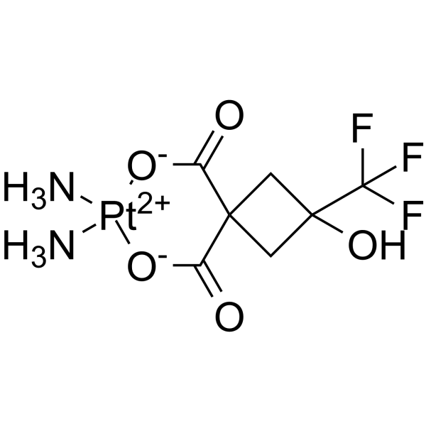
Antitumor agent-77 is an antitumor agent, inhibits cancer cells growth and migration. Antitumor agent-77 triggers ferroptosis by inhibiting GPx-4 and elevating COX2. Antitumor agent-77 also activates intrinsic apoptotic pathway (Bax-Bcl-2-caspase-3) and hinders Epithelial-mesenchymal transition (EMT) process of cancer cells.
商品は「研究用試薬」です。人や動物の医療用・臨床診断用・食品用の製品ではありません。
研究用途以外に使用した場合、当社は一切の責任を負いかねます。
- CAS 番号: 2870703-21-0
- 分子式: C7H11F3N2O5Pt
- 分子量:455.25
-
保管条件:
Please store the product under the recommended conditions in the Certificate of Analysis.

To place orders, for customer services and technical support, please contact: MedChemExpress USA
Tel: 609-228-6898 E-mail: [email protected] [email protected]


-
 生物活性
生物活性
-
化学情報
- プロトコル
- 純度とドキュメンテーション
- 参考文献
-
 Help & FAQs




生物活性
製品説明
IC50 & Target
|
COX-2 |
Bax |
Bcl-2 |
体外実験
Antitumor agent-77 (compound 2a) (30 μM; 4 h) exhibits better solubility and improved cellular uptake than Carboplatin (HY-17393) in A549 cells[1].
Antitumor agent-77 (20 μM; 36 h) produces cytotoxicity by inducing apoptosis of A549 cancer cells[1].
Antitumor agent-77 (20 μM; 24 h) results in significant down-regulation of Bcl-2 and upregulation of Bax, also leads to E-cadherin increase, Vimentin decrease[1].
Antitumor agent-77 (20 μM; 24 h) arrests cell cycle at S phase and G2/M phase[1].
Antitumor agent-77 (10 μM; 12 h) inhibits cells migration with inhibition rate of 52%[1].
MedChemExpress (MCE) has not independently confirmed the accuracy of these methods. They are for reference only. Further protocols information, click here.
-
Cell Line:A549 cells
-
Concentration:20 μM
-
Incubation Time:36 hours
-
Result:Resulte cell apopsotsis with average apoptotic values (including both early and late apoptotic states which were displayed in Q1-LR and Q1-UR, respectively) of 25.34%.
-
Cell Line:A549 cells
-
Concentration:20 μM
-
Incubation Time:24 hours
-
Result:Elevated the level of cleaved caspase-3 and reduced the level of caspase-3 in A549 cells.
Decreased anti-apoptotic protein Bcl-2 and increased pro-apoptotic protein Bax.
Elevated the expression of E-cadherin and on the other hand, lowered the protein level of Vimentin.
-
Cell Line:A549 cells
-
Concentration:20 μM
-
Incubation Time:24 hours
-
Result:Blocked cell cycle progression in S and G2/M phase with the values of 41.11% and 26.03%, respectively.
体内実験
MedChemExpress (MCE) has not independently confirmed the accuracy of these methods. They are for reference only.
-
Animal Model:A549 xenograft models in mouse[1]
-
Dosage:6 μg/kg
-
Administration:Intravenous injection; administration on day 8, 10, 12 after establishing xenograft models (A549 cells; s.c.)
-
Result:Significantly repressed tumor growth, and maintained normal kidney and liver architecture in mice.


化学情報
-
CAS 番号 2870703-21-0
-
分子量 455.25
-
分子式 C7H11F3N2O5Pt
-
SMILES
O=C1[O-][Pt+2]([NH3])([NH3])[O-]C(C12CC(C2)(C(F)(F)F)O)=O
-
輸送条件
Room temperature in continental US; may vary elsewhere.
-
保管条件
Please store the product under the recommended conditions in the Certificate of Analysis.

プロトコル
-
Apoptosis
Apoptosis, also called programmed cell death, is generally characterized by distinct morphological characteristics.
-
Cell migration
Cell migration is a method that plays an important role in wound healing, cell differentiation, embryonic development, etc.
-
Apoptosis Solutions
Apoptosis is a regulated, generally non-lytic cell-death pathway that removes unwanted, damaged, infected, or abnormal cells through coordinated morphological changes, caspase activation, DNA fragmentation, and membrane remodeling. The intrinsic apoptosis pathway is controlled mainly by mitochondrial outer membrane permeabilization, BCL-2 family proteins, cytochrome c release, apoptosome formation, caspase-9 activation, and downstream executioner caspase-3/7 activation. The extrinsic apoptosis pathway is initiated by death receptors such as Fas, TNFR, and TRAIL receptors, which recruit adaptor proteins and activate caspase-8 before engaging executioner caspases or mitochondrial amplification through BID cleavage. Apoptosis is linked to many phenotypes, including cancer cell killing, tissue homeostasis, immune regulation, neurodegeneration, infection response, and treatment-induced cytotoxicity; unresolved questions include how apoptosis interacts with necroptosis, pyroptosis, ferroptos
-
Ferroptosis Solutions
Ferroptosis is an iron-dependent, non-apoptotic form of regulated cell death characterized by lethal lipid peroxidation and sensitivity to suppression by iron chelators or lipophilic radical-trapping antioxidants. The core pathway links cystine uptake through system Xc−, glutathione availability, GPX4-dependent detoxification of phospholipid hydroperoxides, iron-dependent oxidative reactions, and polyunsaturated-phospholipid metabolism into a cell-death program that is biochemically and morphologically distinct from apoptosis, necrosis, and autophagy. The ferroptosis pathway is experimentally linked to phenotype through chemical and genetic perturbation. Erastin induces ferroptosis by inhibiting cystine uptake through system Xc− and weakening antioxidant defenses, while GPX4 inhibition or depletion causes lipid peroxide accumulation and ferroptotic cancer-cell death. ACSL4 and oxidizable arachidonoyl- or adrenoyl-containing phosphatidylethanolamines shape ferroptosis sensitivity by con


純度とドキュメンテーション
-
データシート (278 KB)
-
SDS (252 KB)
- Français - FR (252 KB)
- Deutsch - DE (252 KB)
- Norwegian - NO (252 KB)
- Español - ES (252 KB)
- Swedish - SV (252 KB)
- Italian - IT (252 KB)
- Korean - KR (252 KB)
- Portuguese - PT (252 KB)
-
取扱説明書 (2659 KB)
参考文献

Calculators
濃度 (開始) × 体積 (開始) = 濃度 (終了) × 体積 (終了)


Antitumor agent-77 関連分類
HY-151429 関連分類
 Powered by Bioz