



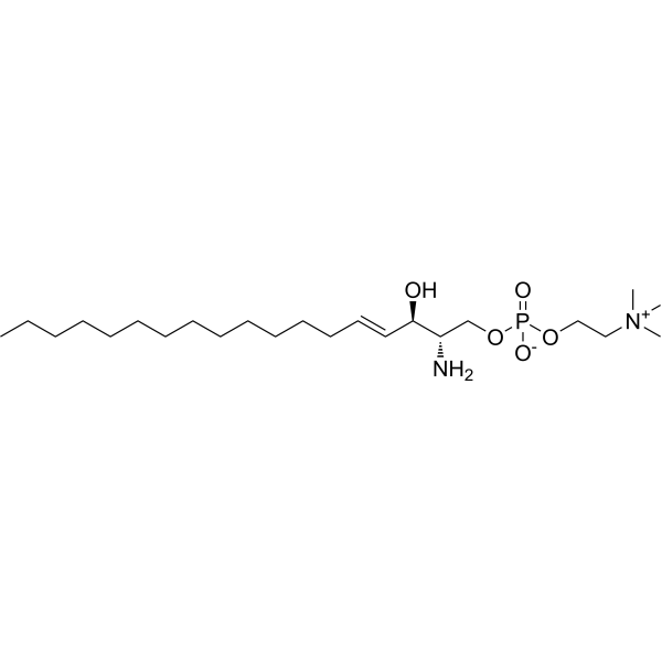
Sphingosylphosphorylcholine

Based on 1 Customer Validation
Sphingosylphosphorylcholine is a bioactive lipid and a major component of plasma high-density lipoprotein that binds to OGR1 with a Kd of 33.3 nM. Sphingosylphosphorylcholine triggers delayed phosphorylation of Smad2, upregulates α-SMA expression, and activates TRPM3. Sphingosylphosphorylcholine reduces Apoptosis and upregulates the expression of uPA and its receptor uPA-R. Sphingosylphosphorylcholine exerts anti-apoptotic, anti-cardiac hypertrophy and pro-wound healing effects. Sphingosylphosphorylcholine induces scratching behavior in mice. Sphingosylphosphorylcholine is used in studies related to atopic dermatitis, promyelocytic leukemia, heart failure, myocardial ischemia/reperfusion injury, ovarian cancer, breast cancer, pancreatic cancer, and skin wound healing disorders in genetically impaired healing diabetes.
Nur für Forschungszwecke. Wir verkaufen nicht an Patienten.
- Reinheit: 99.50%
- CAS. Nr.: 1670-26-4
- Formel: C23H49N2O5P
- Molecular Weight:464.62
-
Speicherung:
-20°C, protect from light, stored under nitrogen
* In solvent : -80°C, 6 months; -20°C, 1 month (protect from light, stored under nitrogen)


-
 Biologische Aktivität
-
Chemical Information
-
Lösungsmittel & Löslichkeit
- Reinheit & Dokumentation
- Verweise
-
 Help & FAQs





Biologische Aktivität
|
TRPM3 |
Sphingosylphosphorylcholine (10 nM-50 μM; 24 h post-transfection incubation) activates TRPM3 in HEK293T cells to induce intracellular calcium elevation primarily via extracellular calcium influx, with a minor contribution from endoplasmic reticulum calcium stores, and this activation is independent of Gβγ signaling[2].
Sphingosylphosphorylcholine (15-20 μM; 15 min-4 h) potently induces actin stress fiber formation and focal adhesion assembly in mouse ES cell-derived EB outgrowths in a concentration- and time-dependent manner[3].
Sphingosylphosphorylcholine (10 μM) inhibits Ang II-induced hypertrophy in neonatal mouse cardiomyocytes, reducing cell surface area and lowering ANP and BNP protein levels[4].
Sphingosylphosphorylcholine (10 μM; 210 min hypoxia, 150 min reoxygenation) protects rat neonatal cardiomyocytes from apoptosis induced by 210 min of hypoxia followed by 150 min of reoxygenation[5].
Sphingosylphosphorylcholine (0.5-100 nM; 0-140 min) binds to OGR1 in OGR1-transfected HEK293 cells with high affinity (Kd = 33.3 nM) and high specificity[6].
Sphingosylphosphorylcholine (0.1-5 μM; 24 h) upregulates cell surface expression of uPA and its receptor uPA-R in normal human foreskin keratinocytes in a dose-dependent manner over 24 h[7].
MedChemExpress (MCE) has not independently confirmed the accuracy of these methods. They are for reference only.
-
Cell Line:human NB4 promyelocytic leukemia cells
-
Concentration:12.5-15 μM
-
Incubation Time:48-96 h
-
Result:Caused a 45% decrease in NB4 cell numbers after 96 h compared to untreated controls.
Significantly increased relative CD11c mRNA expression at 48 h and 96 h, reaching ~10% of the effect of all-trans retinoic acid (ATRA).
Significantly increased relative CD18 mRNA expression at 48 h and 96 h, reaching 8-18% of the effect of ATRA.
Substantially increased cell surface CD11c protein expression, reaching ~20% of the effect of ATRA.
Sphingosylphosphorylcholine (10 μM/kg/day; subcutaneous injection; daily, for 4 consecutive weeks) increases the survival rate of mice to 63%, and alleviates pressure overload-induced myocardial hypertrophy, fibrosis and apoptosis by inhibiting the CaM-JNK/p38 signaling pathway[4].
Sphingosylphosphorylcholine (0.625-2.5 μg/g body weight; intravenous injection; prophylactic administration 30 minutes before coronary artery ligation, or administration at the onset of reperfusion) dose-dependently reduces myocardial infarct size via a mechanism dependent on the S1P3 receptor: the reduction reaches up to 50% with prophylactic administration and 40% with administration upon reperfusion[5].
Topical application of 2 μM Sphingosylphosphorylcholine statistically significantly promotes skin wound healing in diabetic db/db mice with genetically impaired healing capacity, as evidenced by reduced wound area, increased granulation tissue volume, and shortened length of non-re-epithelialized wound surface[7].
MedChemExpress (MCE) has not independently confirmed the accuracy of these methods. They are for reference only.
-
Animal Model:C57BL/6 (male, 10- to 14-week-old)[2]
-
Dosage:100 nmol/site
-
Administration:i.d.; single administration
-
Result:Induced a mean of 74.83 ± 27.46 scratching bouts per 30 min, compared to 12 ± 4.374 scratching bouts in vehicle-treated mice.
Did not induce a significant change in wiping behavior, with a mean of 4.000 ± 0.578 wiping bouts per 30 min, compared to 6.333 ± 0.989 in vehicle-treated mice.\nInduced a mean of 89.67 ± 7.756 scratching bouts per 30 min, compared to 14.33 ± 3.303 scratching bouts in vehicle-treated mice.
-
Animal Model:C57BL/6 (male, 6-8 weeks old; pressure overload induced via trans-aortic constriction surgery)[4]
-
Dosage:10 μM/kg/day
-
Administration:s.c.; daily; 4 weeks
-
Result:Increased survival rate to 63% in TAC mice.
Significantly increased left ventricular ejection fraction (LVEF) and left ventricular fractional shortening (LVFS) relative to untreated TAC mice.
Significantly reduced the ratios of heart weight to body weight (HW/BW) and left ventricular weight to body weight (LW/BW) relative to untreated TAC mice.
Reduced cardiomyocyte cross-sectional area relative to untreated TAC mice.
Decreased the mRNA and protein expression of hypertrophy markers ANP, BNP, and β-MHC compared to untreated TAC mice.
Reduced left ventricular collagen volume fraction relative to untreated TAC mice.
Decreased protein and mRNA expression of fibrosis markers collagen I, collagen III, α-SMA, TGF-β, and fibronectin relative to untreated TAC mice.
Reduced the number of TUNEL-positive apoptotic cardiomyocytes relative to untreated TAC mice.
Decreased protein expression of Bax in TAC mouse hearts compared to untreated TAC mice.
Increased protein expression of Bcl-2 in TAC mouse hearts compared to untreated TAC mice.
Reduced the levels of CaM, phosphorylated JNK (p-JNK), and phosphorylated p38 (p-p38) in TAC mouse hearts compared to untreated TAC mice.
-
Animal Model:Outbred Swiss mice (strain-matched, age-matched, sex-matched); C57BL/6 wild-type mice; S1P3-deficient (S1P3-/-) mice on C57BL/6 background (strain-matched, age-matched, sex-matched)[5]
-
Dosage:0.625 μg/g body weight; 1.25 μg/g body weight; 2.5 μg/g body weight
-
Administration:i.v.; 30 minutes before coronary ligation; single dose; i.v. (therapeutically at reperfusion onset; single dose)
-
Result:Reduced infarct size by 23% (0.625 μg/g preventive).
Reduced infarct size by 36% (1.25 μg/g preventive).
Reduced infarct size by 50% (2.5 μg/g preventive).
Reduced infarct size by 40% (1.25 μg/g at reperfusion).
Reduced polymorphonuclear neutrophil recruitment to infarcted area from 629 ± 45 PMN/mm2 to 332 ± 43 PMN/mm2 (preventive treatment).
Reduced TUNEL-positive apoptotic cells in area at risk from 920 ± 225 cells/mm2 to 643 ± 66 cells/mm2 (preventive treatment).
Reduced infarction/area at risk to 29 ± 3.8% vs control 34 ± 2% (1.25 μg/g preventive in C57BL/6 wild-type mice).
Showed no effect on infarct size (105 ± 9% of vehicle control, no significant difference) in S1P3-/- mice.


Chemical Information
-
CAS. Nr. 1670-26-4
-
Appearance Solid
-
Molecular Weight 464.62
-
Formel C23H49N2O5P
-
Color White to off-white
-
SMILES
CCCCCCCCCCCCC/C=C/[C@@H](O)[C@@H](N)COP(OCC[N+](C)(C)C)([O-])=O
-
Structure Classification
-
Initial Source
-
Versand
Room temperature in continental US; may vary elsewhere.
-
Speicherung
-20°C, protect from light, stored under nitrogen
* In solvent : -80°C, 6 months; -20°C, 1 month (protect from light, stored under nitrogen)


Lösungsmittel & Löslichkeit
Ethanol : 2.5 mg/mL (5.38 mM; ultrasonic and warming and heat to 60°C)
Please refer to the solubility information to select the appropriate solvent. Once prepared, please aliquot and store the solution to prevent product inactivation from repeated freeze-thaw cycles.
Storage method and period of stock solution: -80°C, 6 months; -20°C, 1 month (protect from light, stored under nitrogen). When stored at -80°C, please use it within 6 months. When stored at -20°C, please use it within 1 month.
Please refer to the solubility information to select the appropriate solvent. Once prepared, please aliquot and store the solution to prevent product inactivation from repeated freeze-thaw cycles.
Storage method and period of stock solution: -80°C, 6 months; -20°C, 1 month (protect from light, stored under nitrogen). When stored at -80°C, please use it within 6 months. When stored at -20°C, please use it within 1 month.
Konzentration (Stammlösung) × Volumen (Stammlösung) = Konzentration (Ziellösung) × Volumen (Ziellösung)
Please enter the basic information of animal experiments:
-
-
-
-
Recommended: Prepare an additional quantity of animals to account for potential losses during experiments.
Working solution concentration: 0.22 mg/mL


Reinheit & Dokumentation
-
Data Sheet (280 KB)
-
SDS (394 KB)
- English - EN (394 KB)
- Français - FR (394 KB)
- Deutsch - DE (394 KB)
- Norwegian - NO (394 KB)
- Español - ES (394 KB)
- Swedish - SV (394 KB)
- Italian - IT (394 KB)
- Portuguese - PT (394 KB)
-
Handling Instructions (2659 KB)
Verweise
[1]. Jeon ES, et al. Sphingosylphosphorylcholine induces differentiation of human mesenchymal stem cells into smooth-muscle-like cells through a TGF-beta-dependent mechanism. J Cell Sci. 2006;119(Pt 23):4994-5005. [Content Brief]
[2]. Song DE, et al. Sphingosylphosphorylcholine induces itch via activation of TRPM3 and TRPA1 in mice. Biochem Pharmacol. 2025;237:116952. [Content Brief]
[3]. Kleger A, et al. The bioactive lipid sphingosylphosphorylcholine induces differentiation of mouse embryonic stem cells and human promyelocytic leukaemia cells. Cell Signal. 2007;19(2):367-377. [Content Brief]
[4]. Ren FF, et al. Sphingosylphosphorylcholine alleviates pressure overload-induced myocardial remodeling in mice via inhibiting CaM-JNK/p38 signaling pathway. Acta Pharmacol Sin. 2024;45(2):312-326. [Content Brief]
[5]. Herzog C, et al. Intravenous sphingosylphosphorylcholine protects ischemic and postischemic myocardial tissue in a mouse model of myocardial ischemia/reperfusion injury. Mediators Inflamm. 2010;2010:425191. [Content Brief]
[6]. Xu Y, et al. Sphingosylphosphorylcholine is a ligand for ovarian cancer G-protein-coupled receptor 1. Nat Cell Biol. 2000 May;2(5):261-7. doi: 10.1038/35010529. Retraction in: Nat Cell Biol. 2006 Mar;8(3):299. [Content Brief]
[7]. Wakita H, et al. Sphingosylphosphorylcholine stimulates proliferation and upregulates cell surface-associated plasminogen activator activity in cultured human keratinocytes. J Invest Dermatol. 1998;110(3):253-258. [Content Brief]


Complete Stock Solution Preparation Table
Please refer to the solubility information to select the appropriate solvent. Once prepared, please aliquot and store the solution to prevent product inactivation from repeated freeze-thaw cycles.
Storage method and period of stock solution: -80°C, 6 months; -20°C, 1 month (protect from light, stored under nitrogen). When stored at -80°C, please use it within 6 months. When stored at -20°C, please use it within 1 month.
| Optional Solvent | Concentration Solvent Mass | 1 mg | 5 mg | 10 mg | 25 mg |
|---|---|---|---|---|---|
| Ethanol | 1 mM | 2.1523 mL | 10.7615 mL | 21.5230 mL | 53.8074 mL |
| 5 mM | 0.4305 mL | 2.1523 mL | 4.3046 mL | 10.7615 mL |


 Powered by Bioz