



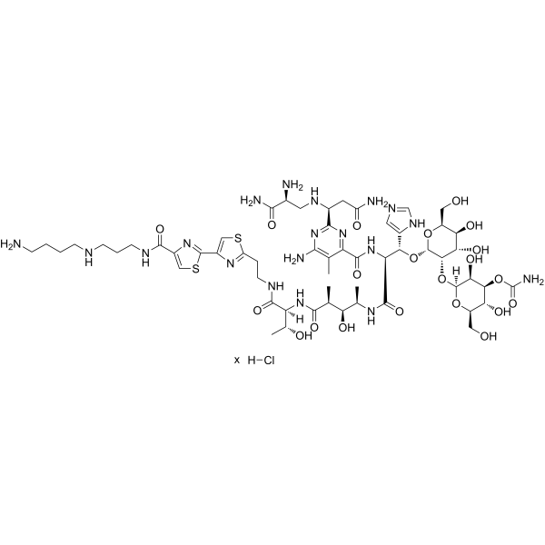
Bleomycin A5 hydrochloride
Based on 1 publication(s) in Google Scholar
Bleomycin A5 (Pingyangmycin) hydrochloride is a glycopeptide antibiotic with multiple biological activities, which can be isolated from Streptomyces. Bleomycin A5 hydrochloride exerts cytotoxic effects by binding to Fe2+ to form a complex, inducing single-strand and double-strand DNA breaks, and inhibiting DNA replication. Bleomycin A5 hydrochloride inhibits Drp1-mediated mitochondrial fission and suppresses PINK1/Parkin pathway-mediated mitophagy, ultimately triggering mitochondria-mediated cellular apoptosis. Bleomycin A5 hydrochloride can be used in cancer research.
For research use only. We do not sell to patients.
- CAS No.: 55658-47-4
- Formula: C57H90ClN19O21S2
- Molecular Weight:1477.02
-
Storage:
Please store the product under the recommended conditions in the Certificate of Analysis.

To place orders, for customer services and technical support, please contact: MedChemExpress USA
Tel: 609-228-6898 E-mail: [email protected] [email protected]


-
 Biological Activity
-
Chemical Information
- Protocols
- Purity & Documentation
- References
-
 Help & FAQs


Publications Citing Use of MedChemExpress (MCE) Bleomycin A5 hydrochloride
MoreAll Antibiotic Isoforms
MoreAll DNA/RNA Synthesis Isoforms
More

Biological Activity
Description
In Vitro
Bleomycin A5 (25-400 μM; 6-72 h) hydrochloride induces mitochondria-mediated apoptosis in primary human nasal polyp-derived fibroblasts (NPDFs) in a time- and dose-dependent manner[2].
Bleomycin A5 (200 μM; 48 h) hydrochloride inhibits Drp1-mediated mitochondrial fission in primary human nasal polyp-derived fibroblasts (NPDFs), leading to the formation of a fused, long tubular mitochondrial network[2].
Bleomycin A5 (50-200 μM; 48 h) hydrochloride induces mitochondrial dysfunction in primary human nasal polyp-derived fibroblasts (NPDFs), and knockdown of Drp1 exacerbates this dysfunction, including increased superoxide accumulation, elevated mtDNA copy number, reduced ATP production, and aggravated mitochondrial membrane potential depletion[2].
Bleomycin A5 (200 μM; 48 h) hydrochloride inhibits Drp1-mediated mitophagy in primary human nasal polyp-derived fibroblasts (NPDFs), and knockdown of Drp1 enhances this inhibitory effect, which is confirmed by the decreased levels of mitochondrial PINK1, Parkin, and LC3B[2].
Bleomycin A5 hydrochloride is taken up by human cancer cells via mediation of the human transporter hCT2; specifically, the NT2/D1 testicular cancer cells with high hCT2 expression are 333 times more sensitive to it (IC50 = 0.01 μM) than the HCT116 colon cancer cells with undetectable hCT2 expression (IC50 = 3.33 μM)[3].
Bleomycin A5 (10-160 μM; 48 h) hydrochloride induces concentration-dependent cytotoxicity in HCT116, HT29, H1299, A549, CHOK1, CHO745 and CHO3.1 cells, with the strongest activity observed in HCT116 cells (IC50 = 9.9 μM) and reduced activity in GAG-deficient CHO745 cells (IC50 = 327.7 μM)[5].
Bleomycin A5 (10-80 μM; 4-48 h) hydrochloride significantly reduces the sulfation level of heparan sulfate in A549 and HCT116 cells (characterized by an increase in non-sulfated disaccharides and a decrease in sulfated disaccharides), and alters the disaccharide composition of chondroitin sulfate[5].
MedChemExpress (MCE) has not independently confirmed the accuracy of these methods. They are for reference only.
-
Cell Line:HCT116, HT29, H1299, A549, CHOK1, CHO745, CHO3.1
-
Concentration:10, 20, 40, 80, 160 μM
-
Incubation Time:48 h
-
Result:Reduced cell survival rate in all tested cell lines in a concentration-dependent manner.
Exhibited the strongest cytotoxicity in HCT116 cells, with an IC50 of 9.9 μM.
Had IC50 values of 55.4 μM (HT29), 11.6 μM (A549), 71.7 μM (H1299), 126.4 μM (CHOK1), 327.7 μM (CHO745), and 122.8 μM (CHO3.1).
Showed higher cytotoxicity at 160 μM in GAG-sufficient CHOK1 and CHO3.1 cells than in GAG-deficient CHO745 cells.
In Vivo
Bleomycin A5 (1.2 mg/kg; intramuscular injection; once every 3 days; for 28 consecutive days) hydrochloride inhibits tumor growth of Lewis lung carcinoma and significantly reduces the sulfation levels of chondroitin sulfate (CS) and heparan sulfate (HS) in murine lung cancer[5].
MedChemExpress (MCE) has not independently confirmed the accuracy of these methods. They are for reference only.
-
Animal Model:Wistar (male, 200-220 g)[4]
-
Dosage:5 mg/kg
-
Administration:i.t.; single dose
-
Result:Induced lung inflammation score of ~2.7, lung index of ~19 mg wet weight/g body weight, BAL fluid total cell count of 47.81 × 10⁴/mL, lung hydroxyproline content of ~1.3 mg/g lung tissue, lung malondialdehyde (MDA) levels of ~61 nmol/mg protein, and serum tumor necrosis factor-α (TNF-α) levels of ~1.012 ng/mL serum at day 7, all significantly elevated compared to sham-treated rats.
Induced lung fibrosis score of ~2.5, lung index of ~8 mg wet weight/g body weight, BAL fluid total cell count of 24.13 × 10⁴/mL, lung hydroxyproline content of ~2.4 mg/g lung tissue, and lung MDA levels of ~27 nmol/mg protein at day 28, all significantly elevated compared to sham-treated rats.
-
Animal Model:C57BL/6 (female, 6-8 weeks old, 18.6 g weight, subcutaneous Lewis lung carcinoma cell injection)[5]
-
Dosage:1.2 mg/kg
-
Administration:i.m.; every 3rd day; 28 days
-
Result:Reduced lung tumor weight to 0.22 g.
Significantly reduced sulphated chondroitin sulfate (CS) disaccharides (D0a6 and D0a10) and overall heparan sulfate (HS) disaccharide sulphation in tumor tissues.
Increased the proportion of non-sulphated HS disaccharides in tumor tissues.
Clinical Trial
| NCT Number | Sponsor | Condition | Start Date |
Phase

|
|---|---|---|---|---|
| NCT01329991 | 2011-05 | PHASE1 |


Chemical Information
-
CAS No. 55658-47-4
-
Molecular Weight 1477.02
-
Formula C57H90ClN19O21S2
-
SMILES
[H]Cl.O=C(C1=NC([C@H](CC(N)=O)NC[C@H](N)C(N)=O)=NC(N)=C1C)N[C@H](C(N[C@H](C)[C@@H](O)[C@H](C)C(N[C@]([C@H](O)C)([H])C(NCCC2=NC(C3=NC(C(NCCCNCCCCN)=O)=CS3)=CS2)=O)=O)=O)[C@H](C4=CN=CN4)O[C@H]5[C@H]([C@H]([C@H](O)[C@H](CO)O5)O)O[C@@]6([H])[C@H]([C@H]([C@H](O)[C@@H](CO)O6)OC(N)=O)O.[x]
-
Synonyms
Pingyangmycin hydrochloride
-
Structure Classification
-
Initial Source
Streptomyces verticillus var
-
Shipping
Room temperature in continental US; may vary elsewhere.
-
Storage
Please store the product under the recommended conditions in the Certificate of Analysis.


Publications (1)
-
Journal Impact Factor
-
Most Recent
-
Exp Ther Med
MicroRNA‑203a‑3p improves bleomycin and pingyangmycin sensitivity by inactivating the PI3K/AKT pathway in hemangioma. [Abstract]2024 Jan 4;27(2):80. PMID: 38274341


Purity & Documentation
References
[1]. Yang Y, et al. Bleomycin A5 sclerotherapy for cervicofacial lymphatic malformations. J Vasc Surg. 2011;53(1):150-155. [Content Brief]
[2]. Wu F, et al. Bleomycin A5 suppresses Drp1‑mediated mitochondrial fission and induces apoptosis in human nasal polyp‑derived fibroblasts. Int J Mol Med. 2021;47(1):346-360. [Content Brief]
[3]. Aouida M, et al. A new twist in cellular resistance to the anticancer drug bleomycin-A5. Curr Drug Metab. 2010;11(7):595-602. [Content Brief]
[4]. Pan JB, et al. Rolipram attenuates bleomycin A5-induced pulmonary fibrosis in rats. Respirology. 2009;14(7):975-982. [Content Brief]
[5]. Lan Y, et al. Pingyangmycin inhibits glycosaminoglycan sulphation in both cancer cells and tumour tissues. J Cell Mol Med. 2020;24(6):3419-3430. [Content Brief]

Calculators
Concentration (start) × Volume (start) = Concentration (final) × Volume (final)


Bleomycin A5 hydrochloride Related Classifications
HY-125918 Related Classifications
 Powered by Bioz
Keywords
- Bleomycin A5
- 55658-47-4
- Pingyangmycin
- Bleomycin A 5
- Bleomycin A-5
- Antibiotic
- DNA/RNA Synthesis
- Apoptosis
- Dynamin
- PINK1/Parkin
- Mitophagy
- DNA
- NT2/D1 testicular carcinoma cells
- HCT116 colon carcinoma cells
- endothelial cells
- mitochondrial fission
- Saccharomyces cerevisiae
- glycosaminoglycans
- mitophagy
- apoptosis
- Wistar rats
- Inhibitor
- inhibitor
- inhibit