



KMI169

Based on 1 Customer Validation
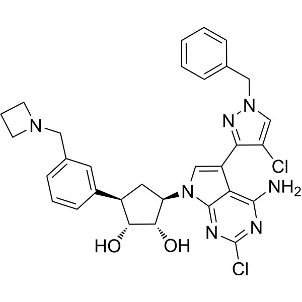
KMI169 is a potent and selective inhibitor targeting lysine methyl-transferase (KMT9) (IC50 = 0.05 μM, Kd = 0.025 μM). KMI169 functions as a bi-substrate inhibitor targeting the cofactor S-5’-adenosyl-L-methionine (SAM) and substrate binding pockets of KMT9. KMI169 can downregulate target genes involved in cell cycle regulation and impair proliferation of tumor cells by inhibiting KMT9. KMI169 is a valuable tool to probe cellular KMT9 functions and can be research for combating diseases including prostate, lung, colon, and invasive bladder cancer.
Nos produits utilisent uniquement pour la recherche. Nous ne vendons pas aux patients.
- Pureté : 98.00%
- Formule: C31H31Cl2N7O2
- Masse moléculaire:604.53
-
Stockage:
4°C, protect from light, stored under nitrogen
* In solvent : -80°C, 6 months; -20°C, 1 month (protect from light, stored under nitrogen)

To place orders, for customer services and technical support, please contact: MedChemExpress USA
Tel: 609-228-6898 E-mail: [email protected] [email protected]


-
 Activité biologique
-
Chemical Information
-
Solvant et solubilité
- Protocole
- Pureté et documentation
- Références
-
 Help & FAQs




Activité biologique
Description
IC50 & Target
KMT9[1]
In Vitro
KMI169 (30 μM) only results in a 60% maximum inhibition against PRMT5, suggesting KMI169 does not target cellular PRMT5[1].
KMI169 (500 nM, 3 d) reduces the levels of H4K12mel, but does not alter histone markers such as H3K4me2, H3K9me2, and H4K20mel in PC-3M prostate tumor cells[1].
KMI169 (0.2-1 μM, 7 d) impairs proliferation of castration- and Enzalutamide (HY-70002)-resistant prostate cancer cell lines such as PC-3M, DU145, LNCaP-abl, and LNCaP-abl EnzaR, whereas proliferation of HepG2 and PANC-1 cells are not affected[1].
KMI169 (360 nM, 4 d) reduces mRNA levels for a selection of genes including MYB, AURKB, FOXA1, CDK2, BIRC5, E2F1, E2F8, CDC6, and LIG1[1].
KMI169 strongly reduces the proliferation of various cell lines such as J82 cells with a GI50 of 371 nM and RT-112 cells with a GI50 of 320 nM, as well as impairing proliferation of 5637, CAL-29, and UM-UC-3 cells[2].
MedChemExpress (MCE) has not independently confirmed the accuracy of these methods. They are for reference only. Further protocols information, click here.
-
Cell Line:Prostate cancer cell lines PC-3M and LNCaP-abl
-
Concentration:0.2-1 μM
-
Incubation Time:7 d
-
Result:Revealed strong stabilization of endogenous KMT9.
Did not stabilize PRMT5 under the same conditions.
Strongly suppressed the proliferation of PC-3M cells with a GI50 of 150 nM.


Chemical Information
-
Appearance Solid
-
Masse moléculaire 604.53
-
Formule C31H31Cl2N7O2
-
Color White to off-white
-
SMILES
NC1=C2C(C3=NN(C=C3Cl)CC4=CC=CC=C4)=CN([C@@H]5C[C@H](C6=CC(CN7CCC7)=CC=C6)[C@@H](O)[C@H]5O)C2=NC(Cl)=N1
-
Livraison
Room temperature in continental US; may vary elsewhere.
-
Stockage
4°C, protect from light, stored under nitrogen
* In solvent : -80°C, 6 months; -20°C, 1 month (protect from light, stored under nitrogen)


Solvant et solubilité
In Vitro:
DMSO : 100 mg/mL (165.42 mM; Need ultrasonic; Hygroscopic DMSO has a significant impact on the solubility of product, please use newly opened DMSO)
Please refer to the solubility information to select the appropriate solvent. Once prepared, please aliquot and store the solution to prevent product inactivation from repeated freeze-thaw cycles.
Storage method and period of stock solution: -80°C, 6 months; -20°C, 1 month (protect from light, stored under nitrogen). When stored at -80°C, please use it within 6 months. When stored at -20°C, please use it within 1 month.
Please refer to the solubility information to select the appropriate solvent. Once prepared, please aliquot and store the solution to prevent product inactivation from repeated freeze-thaw cycles.
Storage method and period of stock solution: -80°C, 6 months; -20°C, 1 month (protect from light, stored under nitrogen). When stored at -80°C, please use it within 6 months. When stored at -20°C, please use it within 1 month.
Concentration (start) × Volume (start) = Concentration (final) × Volume (final)
In Vivo:
Select the appropriate dissolution method based on your experimental animal and administration route.
- For the following dissolution methods, please ensure to first prepare a clear stock solution using an In Vitro approach and then sequentially add co-solvents:
- To ensure reliable experimental results, the clarified stock solution can be appropriately stored based on storage conditions. As for the working solution for In Vivo experiments, it is recommended to prepare freshly and use it on the same day.
- The percentages shown for the solvents indicate their volumetric ratio in the final prepared solution. If precipitation or phase separation occurs during preparation, heat and/or sonication can be used to aid dissolution.
Add each solvent one by one: 10% DMSO 40% PEG300 5% Tween-80 45% Saline
Solubility: ≥ 2.5 mg/mL (4.14 mM); Clear solution
This protocol yields a clear solution of ≥ 2.5 mg/mL (saturation unknown).
Taking 1 mL working solution as an example, add 100 μL DMSO stock solution (25.0 mg/mL) to 400 μL PEG300, and mix evenly; then add 50 μL Tween-80 and mix evenly; then add 450 μL Saline to adjust the volume to 1 mL.
Preparation of Saline: Dissolve 0.9 g sodium chloride in ddH₂O and dilute to 100 mL to obtain a clear Saline solution.
In Vivo Dissolution Calculator
Please enter the basic information of animal experiments:
-
-
-
-
Recommended: Prepare an additional quantity of animals to account for potential losses during experiments.
Please enter your animal formula composition:
-
%DMSO +
Recommended: Keep the proportion of DMSO in working solution below 2% if your animal is weak.
-
%+
-
+%Tween-80 + +
-
%Saline +
The co-solvents required include: DMSO, . All of co-solvents are available by MedChemExpress (MCE). , Tween 80. All of co-solvents are available by MedChemExpress (MCE).
Working solution concentration: 0.22 mg/mL
Method for preparing stock solution: mg drug dissolved in μL DMSO. Stock solution concentration: mg/mL. * In solvent : -80°C, 6 months; -20°C, 1 month (protect from light, stored under nitrogen)
1. Take μL DMSO stock solution;
2. Add μL .
μL , mix evenly;
3. Then add μL Tween 80, mix evenly;
4. Then add μL
Please ensure that the stock solution in the first step is dissolved to a clear state, and add co-solvents in sequence. You can use ultrasonic heating (ultrasonic cleaner, recommended frequency 20-40 kHz), vortexing, etc. to assist dissolution.

Protocole
-
Flow cytometric DNA-content cell-cycle staining
Flow cytometric DNA-content cell-cycle staining measures the fluorescence intensity of DNA-bound fluorochromes in single cells or nuclei to estimate DNA content distributions, allowing assignment of populations to G0/G1, S, and G2/M phases by DNA histogram deconvolution. Propidium iodide (PI) intercalates into DNA, and PI fluorescence is proportional to cellular DNA content when staining is performed under conditions that make DNA accessible and minimize non-DNA signal. Cells with G2/M DNA content are expected to show approximately twice the fluorescence intensity of G0/G1 cells, while S-phase cells occupy intermediate fluorescence values. PI-based DNA-content analysis can also detect cells with fractional DNA content, often reported as sub-G1, when DNA fragmentation and extraction during staining reduce retained DNA signal in apoptotic cells. DAPI is an alternative DNA fluorochrome for univariate DNA-content analysis, while bivariate approaches combining DNA content with proliferation
-
BrdU Incorporation Assay
Bromodeoxyuridine (BrdU) incorporation assay is based on the principle that BrdU, a thymidine analog, is incorporated into newly synthesized DNA during the S phase of the cell cycle, thereby serving as a marker of DNA replication and cellular proliferation. Incorporated BrdU can be detected using anti-BrdU antibodies following DNA denaturation, enabling visualization or quantification of proliferating cells through immunochemical detection methods such as immunofluorescence or immunohistochemistry.
-
Protocol for Cell Cycle
Cell-cycle analysis by flow cytometry measures DNA content in single cells to estimate the fraction of cells in G0/G1, S, and G2/M phases. Propidium iodide intercalates into DNA, and after RNA removal with RNase, fluorescence intensity reflects cellular DNA content: 2N cells are assigned to G0/G1, cells between 2N and 4N to S phase, and 4N cells to G2/M. DNA-content analysis alone cannot reliably separate G0 from G1 or G2 from M. Ki-67 can distinguish quiescent G0 cells from cycling cells, EdU or BrdU incorporation marks active DNA synthesis in S phase, and phospho-histone H3 staining identifies mitotic cells within the 4N population.
-
Directly Induced Neuron Culture
Directly induced neuron culture converts somatic cells, most commonly fibroblasts, into induced neurons without passing through a pluripotent or neural progenitor stage; classic evidence shows that mouse fibroblasts can be converted by Ascl1, Brn2/Pou3f2, and Myt1l, human fibroblasts can be converted by defined neuronal transcription factors, and human fibroblasts can also be converted by miR-9/9-124 with neurogenic or subtype-specifying transcription factors. The readout is acquisition of neuronal identity and function, assessed by neuronal morphology, neuronal markers such as Tuj1/βIII-tubulin, MAP2, synapsin, and subtype markers when relevant, together with functional assays such as action-potential firing, synaptic activity, and electrophysiology.
-
Cell Viability Determination by MTT Colorimetric Assay
The following protocol uses the MTT colorimetric assay as a classic literature-established method for assessing cell viability/metabolic activity in cultured mammalian cells. MTT[3-(4,5-dimethylthiazol-2-yl)-2,5-diphenyltetrazolium bromide] is reduced by metabolically active cells to a colored formazan product; the amount of formazan is quantified spectrophotometrically and provides an indirect measure of metabolically active viable cells. Importantly, MTT reduction reflects cellular oxidoreductase/metabolic activity rather than an absolute direct count of living cells, so changes in cellular metabolism can alter the signal independently of cell number.


Pureté et documentation
-
Fiche technique (281 KB)
-
SDS (251 KB)
- English - EN (251 KB)
- Français - FR (251 KB)
- Deutsch - DE (251 KB)
- Norwegian - NO (251 KB)
- Español - ES (251 KB)
- Swedish - SV (251 KB)
- Italian - IT (251 KB)
- Korean - KR (251 KB)
- Portuguese - PT (251 KB)
-
Instruction de manipulation (2659 KB)
Références
[1]. Wang S, et al. Structure-guided design of a selective inhibitor of the methyltransferase KMT9 with cellular activity. Nat Commun. 2024 Jan 2;15(1):43. [Content Brief]
[2]. Totonji, S., et., (2024). Lysine Methyltransferase 9 (KMT9) Is an Actionable Target in Muscle-Invasive Bladder Cancer. Cancers, 16(8), 1532. [Content Brief]


Complete Stock Solution Preparation Table
Please refer to the solubility information to select the appropriate solvent. Once prepared, please aliquot and store the solution to prevent product inactivation from repeated freeze-thaw cycles.
Storage method and period of stock solution: -80°C, 6 months; -20°C, 1 month (protect from light, stored under nitrogen). When stored at -80°C, please use it within 6 months. When stored at -20°C, please use it within 1 month.
| Optional Solvent | Concentration Solvent Mass | 1 mg | 5 mg | 10 mg | 25 mg |
|---|---|---|---|---|---|
| DMSO | 1 mM | 1.6542 mL | 8.2709 mL | 16.5418 mL | 41.3544 mL |
| 5 mM | 0.3308 mL | 1.6542 mL | 3.3084 mL | 8.2709 mL | |
| 10 mM | 0.1654 mL | 0.8271 mL | 1.6542 mL | 4.1354 mL | |
| 15 mM | 0.1103 mL | 0.5514 mL | 1.1028 mL | 2.7570 mL | |
| 20 mM | 0.0827 mL | 0.4135 mL | 0.8271 mL | 2.0677 mL | |
| 25 mM | 0.0662 mL | 0.3308 mL | 0.6617 mL | 1.6542 mL | |
| 30 mM | 0.0551 mL | 0.2757 mL | 0.5514 mL | 1.3785 mL | |
| 40 mM | 0.0414 mL | 0.2068 mL | 0.4135 mL | 1.0339 mL | |
| 50 mM | 0.0331 mL | 0.1654 mL | 0.3308 mL | 0.8271 mL | |
| 60 mM | 0.0276 mL | 0.1378 mL | 0.2757 mL | 0.6892 mL | |
| 80 mM | 0.0207 mL | 0.1034 mL | 0.2068 mL | 0.5169 mL | |
| 100 mM | 0.0165 mL | 0.0827 mL | 0.1654 mL | 0.4135 mL |


KMI169 Related Classifications
HY-157486 Related Classifications
 Powered by Bioz